Use the labels in the right column to find what you want. Or you can go thru them one by one, there are only 33,991 posts. Searching is done in the search box in upper left corner. I blog on anything to do with stroke. DO NOT DO ANYTHING SUGGESTED HERE AS I AM NOT MEDICALLY TRAINED, YOUR DOCTOR IS, LISTEN TO THEM. BUT I BET THEY DON'T KNOW HOW TO GET YOU 100% RECOVERED. I DON'T EITHER BUT HAVE PLENTY OF QUESTIONS FOR YOUR DOCTOR TO ANSWER.
Changing stroke rehab and research worldwide now.Time is Brain!trillions and trillions of neuronsthatDIEeach day because there areNOeffective hyperacute therapies besides tPA(only 12% effective). I have 523 posts on hyperacute therapy, enough for researchers to spend decades proving them out. These are my personal ideas and blog on stroke rehabilitation and stroke research. Do not attempt any of these without checking with your medical provider. Unless you join me in agitating, when you need these therapies they won't be there.
What this blog is for:
My blog is not to help survivors recover, it is to have the 10 million yearly stroke survivors light fires underneath their doctors, stroke hospitals and stroke researchers to get stroke solved. 100% recovery. The stroke medical world is completely failing at that goal, they don't even have it as a goal. Shortly after getting out of the hospital and getting NO information on the process or protocols of stroke rehabilitation and recovery I started searching on the internet and found that no other survivor received useful information. This is an attempt to cover all stroke rehabilitation information that should be readily available to survivors so they can talk with informed knowledge to their medical staff. It lays out what needs to be done to get stroke survivors closer to 100% recovery. It's quite disgusting that this information is not available from every stroke association and doctors group.
1Laboratory of Experimental Stroke Research,
Institute for Stroke and Dementia Research (ISD), University of Munich
Medical Center, Munich, Germany
2Munich Cluster of Systems Neurology (Synergy), Munich, Germany
3Department of Neurosurgery, Graduate School of Medicine, Kyoto University, Kyoto, Japan
4Department of Neurosurgery, University of Giessen, Giessen, Germany
Background: Leukocytes contribute to
tissue damage after cerebral ischemia; however, the mechanisms
underlying this process are still unclear. This study investigates the
temporal and spatial relationship between vascular leukocyte recruitment
and tissue damage and aims to uncover which step of the leukocyte
recruitment cascade is involved in ischemic brain injury.
Methods: Male wild-type, ICAM-1-deficient, anti-CD18 antibody treated, or selectin-deficient [fucusyltransferase (FucT IV/VII−/−)]
mice were subjected to 60 min of middle cerebral artery occlusion
(MCAo). The interaction between leukocytes and the cerebrovascular
endothelium was quantified by in vivo fluorescence microscopy up
to 15 h thereafter. Temporal dynamics of neuronal cell death and
leukocyte migration were assessed at the same time points and in the
same tissue volume by histology.
Results: In wild-type mice, leukocytes
started to firmly adhere to the wall of pial postcapillary venules two
hours after reperfusion. Three hours later, neuronal loss started and 13
h later, leukocytes transmigrated into brain tissue. Loss of selectin
function did not influence this process. Application of an anti-CD18
antibody or genetic deletion of ICAM-1, however, significantly reduced
tight adhesion of leukocytes to the cerebrovascular endothelium (-60%; p < 0.01) and increased the number of viable neurons in the ischemic penumbra by 5-fold (p < 0.01); the number of intraparenchymal leukocytes was not affected.
Conclusions: Our findings suggest that
ischemia triggers only a transient adhesion of leukocytes to the venous
endothelium and that inhibition of this process is sufficient to partly
prevent ischemic tissue damage.
Introduction
Ischemic stroke is one of the most frequent causes of death and disability worldwide (1, 2). Current therapies include thrombolysis with rtPA or rtPA in combination with mechanical thrombectomy (3–5); however, only up to 30% of stroke patients are eligible for these interventions (5).
Hence, there is an ongoing and urgent need for the development of novel
therapeutic options for the 70% of stroke patients who do not receive
any causal treatment.
For more than four decades, inflammation has been
recognized as a major pathomechanism, which is responsible for brain
injury following ischemic stroke (6–11).
A plethora of elegant experimental and clinical studies discovered that
ischemia triggers an acute innate immune response within the brain
parenchyma which results in the production of inflammatory cytokines,
the upregulation of adhesion molecules on endothelial cells, and the
subsequent recruitment of granulocytes and monocytes into ischemic
tissue within the first few hours and days after vessel occlusion (12–19). Later on, T-lymphocytes invade the infarcted tissue and may cause further damage (20–22).
Despite these impressive steps forward in our understanding of
postischemic inflammation, none of the above-mentioned mechanisms
translated into a viable therapeutic approach for stroke patients (14).
Hence, reevaluation of previous experimental findings and
identification of significant knowledge gaps may help identify so far
unexplored or neglected therapeutic principles related to postischemic
leukocyte recruitment.
Histopathological studies in human tissue, nonhuman
primates, and rodents and investigations using radioactively labeled
leukocytes in humans and experimental animal models univocally
demonstrate that cerebral ischemia is associated with accumulation of
polymorphonuclear leukocytes (PMNs) or granulocytes in the brain (10, 11, 23–31).
According to experimental studies using radioactively labeled
leukocytes or direct visualization of leukocytes by intravital
microscopy, recruitment of leukocytes to the ischemic brain starts
within the first 2 h after the onset of ischemia (9, 32–35), a time course also supported by investigations in stroke patients (30, 36).
The nature of this accumulation seems to follow two different
mechanisms: leukocytes may plug capillaries and arterioles during
ischemia, thus participating in the so-called no reflow phenomenon (33, 37, 38), and/or they may adhere to the endothelium of postcapillary venules due to upregulation of adhesion molecules (27, 32, 34, 35).
No matter which concept of accumulation is followed, most laboratories
report that depletion of granulocytes or inhibition or deletion of
adhesion molecules reduces ischemic brain damage and improves outcome
following experimental stroke (27, 33, 37, 39–50).
Hence, there is general agreement that leukocytes accumulate in the
brain within the first few hours after cerebral ischemia and inhibition
or deletion of adhesion molecules reduce ischemic brain damage. Beyond
this generally accepted concept, however, many crucial issues on the
role of leukocytes for ischemic tissue damage are still unsolved or
highly debated (17, 27, 28).
One of the main reasons for this discussion is that most of the
above-cited studies used static, histopathological techniques to
investigate leukocytes after stroke. Therefore, our knowledge about the
dynamics of adhesion, transmigration, and accumulation of leukocytes in
the brain after a stroke and how these processes are related to tissue
damage is still surprisingly limited.
Technically, leukocyte dynamics after stroke can be addressed by longitudinal in vivo
imaging; however, the few studies using this approach either focused on
the very first hours after cerebral ischemia, a time when the neuronal
injury was not yet present, or on time points later than 24 h after
stroke, when the ischemic injury had already occurred (27, 32–35, 51).
Consequently, we still do not know whether leukocytes are present in
the brain when ischemic damage occurs or whether leukocytes are present
at the site of injury (6, 7, 17, 19–21, 52–57).
To answer these two important issues, we investigated the full-time
course and sequence of leukocyte accumulation to the ischemic brain in
parallel with neuronal cell death and tried to decipher which part of
the leukocyte adhesion cascade may be involved in ischemic tissue
damage.
And just WHY THE FUCK are you predicting recovery rather than producing recovery? Your mentors and senior researchers incompetently didn't tell you that the only goal in stroke is 100% recovery? Current predictions are totally useless since they are based on the 10% full recovery rate. Of course your doctor doesn't bother telling you how fucking bad the failures are in stroke recovery, that might lead to firings.
Predicting to a 10% full recovery rate
is the height of stupidity. So first you need to create 100% recovery
protocols, THEN you can do predictions. You are doing the order of
research wrong, If we had stroke leadership we could fix this problem.
White blood cell (WBC) and neutrophil counts (NC) are common markers of
inflammation and neurological stroke damage and could be expected to
predict poststroke outcomes.
Objective.
The aim of this study was
to explore the prognostic value of early poststroke WBC and NC to
predict cognition, mood, and disability outcomes at 3 and 12 months
poststroke.
Methods.
Routine clinical analyses WBC and NC were
collected at 3 time points in the first 4 days of hospitalization from
156 acute stroke patients. Correlations using hierarchical or ordinal
regressions were explored between acute WBC and NC and functional
recovery, depression, and cognition at 3 and 12 months poststroke, after
covarying for age and baseline stroke severity.
Results.
We found significant increases in NC between <12 hours and 24 to 48 hours time points (P = .05). Hierarchical regressions, covaried for age and baseline stroke severity, found that 24 to 48 hours WBC (P = .05) and NC (P = .04) significantly predicted 3-month cognition scores. Similarly, 24 to 48 hours WBC (P = .05) and NC (P
= .02) predicted cognition scores at 12 months. Increases in WBC and NC
were predictive of increased cognition scores at both 3 and 12 months
(positive recovery) though there were no significant associations
between WBC and NC and disability or depression scores.
Conclusions.
Routine acute stroke clinical laboratory tests such as WBC and NC taken
between 24 and 48 hours poststroke are predictive of cognition
poststroke. It is interpreted that higher rapid immunological activation
in the acute phase is an indicator for the trajectory of positive
stroke recovery.
Sounds like this is describing the neuronal cascade of death. But I see nothing here that suggests that the stroke strategy will be updated and followup occur.
1Sanders–Brown Center on Aging, University of Kentucky, Lexington, KY, United States
2Department of Neuroscience, University of Kentucky, Lexington, KY, United States
3Department of Neurology, University of Kentucky, Lexington, KY, United States
4Department of Neurosurgery, University of Kentucky, Lexington, KY, United States
Ischemic stroke is a leading cause of death and
disability with limited therapeutic options. Resulting inflammatory
mechanisms after reperfusion (removal of the thrombus) result in
cytokine activation, calcium influx, and leukocytic infiltration to the
area of ischemia. In particular, leukocytes migrate toward areas of
inflammation by use of integrins, particularly integrins β1 and β2.
Integrins have been shown to be necessary for leukocyte adhesion and
migration, and thus are of immediate interest in many inflammatory
diseases, including ischemic stroke. In this review, we identify the
main integrins involved in leukocytic migration following stroke (αLβ2, αDβ2, α4β1, and α5β1) and targeted clinical therapeutic interventions.
Introduction
Ischemic stroke is a leading cause of death and
disability in the United States with limited therapeutic interventions
available, including tissue plasminogen activator (t-PA) and
endovascular mechanical thrombectomy (Rao et al., 2014; Benjamin et al., 2017; Rai et al., 2017).
These interventions are focused on the removal of the thrombus,
restoring blood flow, oxygen and glucose to hypoperfused areas, but are
unable to affect the inflammatory, necrotic, and blood-brain barrier
(BBB) mechanism that follow. In particular, the initial inflammatory
cascade is initiated by the decrease in ATP production, release of
cytokines, influx of intracellular calcium, reactive oxygen species,
etc., that develops during occlusion and continues for days afterward (Sandoval and Witt, 2008).
Using shear forces from cerebral blood flow, marrow-derived leukocytes
(including polymorphonuclear leukocytes (PMNs), neutrophils, lymphocytes
and monocytes) are recruited to the site of injury (Dereski et al., 1993; del Zoppo, 1994; Stefanidakis and Koivunen, 2006).
For the purpose of this review, we will focus on the
recruitment and rolling of leukocytes under the direction of integrins,
as well as some of their ligands following reperfusion after ischemic
stroke. We will then introduce recent β2 and β1 integrin-specific stroke clinical trials, and, finally, discuss potential future directions for the field.
Role of Integrins Post-Stroke: an Overview
Integrins are a diverse group of heterodimers composed of
18 different α and β subunits, creating 24 unique combinations.
Integrins exist on every cell type, while exhibiting a high diversity of
ligands and grouped into four different receptor groups: RGD
(Arg-Gly-Asp), laminin receptors, collagen receptors, and
leukocyte-specific receptors. Within these groups, integrins can have a
variety of ligands and roles following ischemic stroke (reviewed in Edwards and Bix, 2019). Under normal cerebrovascular conditions, integrins are in a highly inactive state, typically in a bent conformation (Takagi et al., 2002; Nishida et al., 2006).
Following ischemic stroke, activation signals are sent. Chemokines are
translocated to the lumen, on the apical side of endothelial cells, to
induce “inside-out” signaling (Chavakis, 2012).
Integrins then undergo a conformational change to increase integrin
affinity for potential ligands while enhancing detection by localizing
to the leading or rear-facing edge of the leukocyte’s cell wall for
ligand detection (Ridley et al., 2003; Hyun et al., 2009).
Activated integrins then bind to available ligands, permitting
leukocytic rolling and intracellular signaling. This is termed
“outside-in” signaling (Hato et al., 1998; Tominaga et al., 1998; Ley et al., 2007).
Leukocytes continue movement to the site of injury, looking for areas
to cross the endothelial cell barrier, and eventually coming to a halt.
Aggregation/clustering of integrins increases binding avidity (strength
of binding), preventing flow conditions from detaching leukocytes from
the endothelial cells (Ley et al., 2007). Using transmigration, leukocytes will infiltrate into the cerebral parenchyma using these integrin-ligand connections.
β2 Integrins
β2 integrins are the only group of integrins exclusively expressed on leukocytes (derived from hematopoietic cells) (Schenkel et al., 2004), and like most integrins, are highly conserved across species (Schittenhelm et al., 2017).
They are also the most highly expressed integrin on circulating blood
leukocytes, tending to cluster at the retraction area of the cell (the
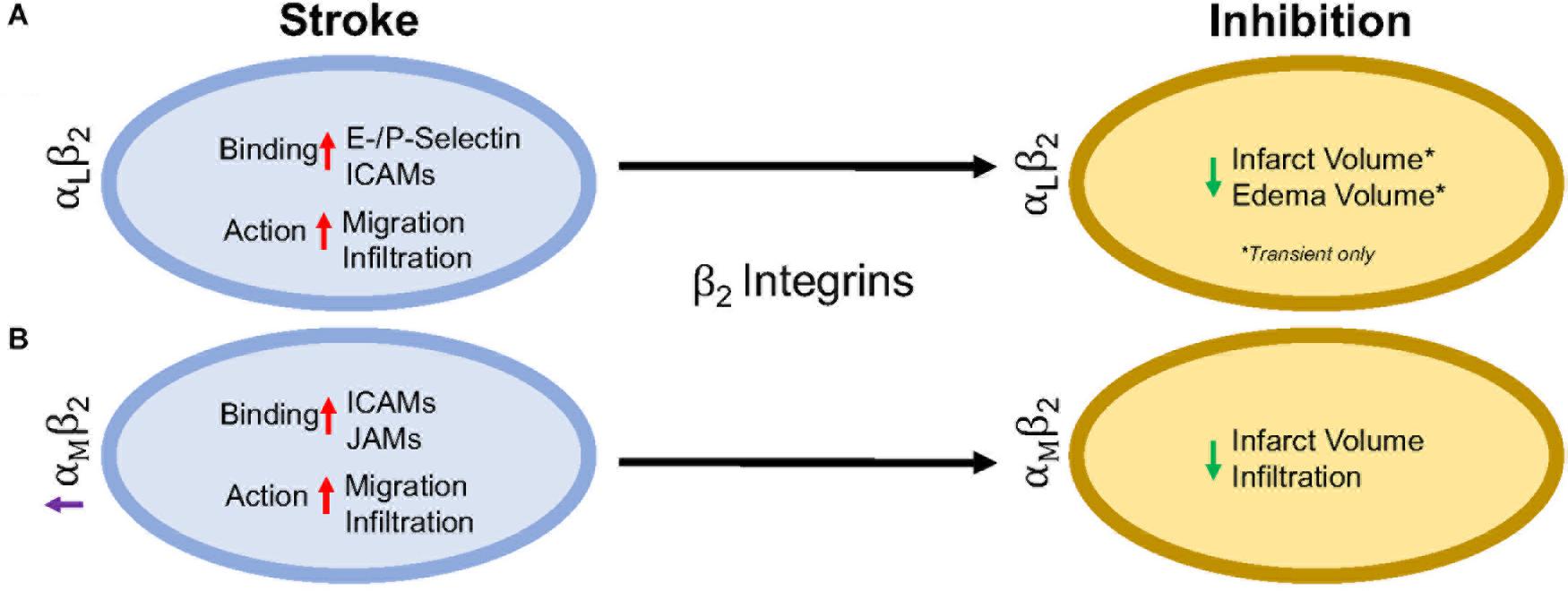
rear), in both an active and inactive state, compared to other β1, β4, β3, and β7 integrins found on circulating leukocytes (Pierini et al., 2000; Lindbom and Werr, 2002). Genetic leukocyte adhesion changes (LAD-1, as discussed above) has been attributed to mutations in the β2 subunit, reducing β2 expression. Thus, leukocytic movement is reduced on the cell surface with less movement toward the site of inflammation (Arnaout, 1990; Scharffetter-Kochanek et al., 1998). Importantly, in β2 inhibited mice, there is not total arrest of leukocytic recruitment or infiltration (Pierini et al., 2000), suggesting that other factors likely play a role. There are 4 identified heterodimers of β2 integrins, and of these, the most highly studied are αLβ2 and αMβ2 in ischemic stroke, and will be reviewed in more detail below. The other β2 integrins, αXβ2 and αDβ2, have not been individually studied in the context of stroke as have αLβ2 and αMβ2 integrins, though CD18 (β2)
inhibition in addition to t-PA has been shown to increase the time
window of t-PA administration without an increase in hemorrhagic
transformation in a rat embolic stroke model (Zhang et al., 1999). Furthermore, Figure 2 summarizes the results in this section.
FIGURE 2
Figure 2. Representative image of the β2 integrin response following experimental stroke and inhibitory antibody treatment in preclinical trials. Inhibition of (A) αLβ2 and (B) αMβ2 integrins post-stroke responses and effects.
αLβ2 Integrin
Integrin αLβ2 is also referred to as CD11a/CD18 and LFA-1 (lymphocyte functional-associating antigen-1). αLβ2
integrin acutely increases in ischemic stroke patients, with detectable
amounts through 72 h associated around the area of ischemia (Gerhard et al., 2000; Zhao et al., 2002). This suggests a correlation between αLβ2 integrin expression and inflammatory damage following ischemia. αLβ2 is expressed on all leukocytes (Soriano et al., 1999), though at particularly high levels on T-lymphocytes (Hammond et al., 2014; Walling and Kim, 2018). In healthy individuals, extracted blood analysis revealed that αLβ2 activation requires leukocytic rolling on P- or E-selectins, inducing an active conformational change (Kuwano et al., 2010),
but it is the binding of chemokines g-protein coupled receptors (GPCR)
and Rap-1 activation that induces the high-affinity conformational state
of αLβ2 (Steffen et al., 1994; Greenwood et al., 1995; Ghandour et al., 2007). In this state, αLβ2 has many possible ligands, ICAM-1, ICAM-2, ICAM-3, ICAM-4, ICAM-5, and junctional adhesion molecule-1 (JAM-1) (Marlin and Springer, 1987; de Fougerolles et al., 1991, 1994; Tian et al., 2000), though ICAM-1 is preferentially bound (Walling and Kim, 2018). The high avidity αLβ2-ICAM-1
complex, once formed, allows t-lymphocytes to move against circulatory
flow and the shear forces, resulting in the high-speed movement of
leukocytes (Katakai et al., 2013; Dominguez et al., 2015).
In an intraluminal model of experimental ischemic stroke, αLβ2
inhibition with the use of transgenic mice results in reduced infarct
volume, edema volume and mortality. However, this phenomenon is evident
in transient, but not permanent middle cerebral artery occlusion (Arumugam et al., 2004). This may be due to the previously mentioned high avidity of αLβ2-ICAM-1 bonds, and is evident in an in vitro study using αLβ2
(LFA-1) knock-in mice that experience high avidity through binding of
lymphocytes mediated through ICAM-1 binding, but are unable to continue
movement due to a non-polarized uropod (Park et al., 2010). An explanation for this phenomenon may be that the recycling process within the leukocyte is overwhelmed (Shaw et al., 2004). By enhancing αLβ2 expression, recycling may not be able to allow for dislocation of αLβ2-ICAM-1 complexes, preventing movement from the loss of high adhesion bonds. Enhanced αLβ2 expression could be a potential new avenue for therapy, especially if no enhanced mortality, infection, etc., are observed.
Independently, ICAMs play a significant role in
inflammation following ischemic stroke. ICAM-1, in particular, is
acutely increased in both cultured human endothelial cells undergoing
hypoxia and following intraluminal suture middle cerebral artery
occlusion, while expression remains sustained for up to a week
post-injury (Hess et al., 1994a,b; Zhang et al., 1995). ICAM-2, another possible ligand, does not change in expression following cytokine stimulation (de Fougerolles et al., 1991; Nortamo et al., 1991a,b). Furthermore, serum of ischemic stroke patients contains soluble ICAM-1, but not ICAM-2 in addition to being a risk factor (Kaplanski et al., 1994; Shyu et al., 1997).
Antibodies targeting ICAM-1 in rodents and humans have shown
contradictory results. An intraluminal suture middle cerebral artery
occlusion model in mice and rats showed a decrease in leukocyte
infiltration and infarct volume (Connolly et al., 1996; Kitagawa et al., 1998; Vemuganti et al., 2004), while one study reported opposing effects (Furuya et al., 2001).
ICAM-1 inhibition was translated to the clinic through testing of the
murine ICAM-1 antibody, Enlimomab in ischemic stroke. Unfortunately, the
study was halted early due to increased rate of infection, infarct
volumes, neurological scores and mortality for patients (Furuya et al., 2001).
αMβ2 Integrin
Integrin αMβ2, also known as CD11b/CD18 and Mac-1 (macrophage-1 antigen), exhibits many similarities to αLβ2 through its expression on all leukocytes (Springer et al., 1979), and common ligand binding partners such as the family of ICAMs and JAMs (von Andrian et al., 1991). Additional ligands are fibrinogen, heparin (von Andrian et al., 1991), elastase (Cai and Wright, 1996), complement C3 fragment (C3bi) (Micklem and Sim, 1985), kinogen components, and urokinase and its receptor (Chavakis et al., 1999). Just as αLβ2, hypoxia induced factors (cytokines, chemokines, etc.) induce conformational change of αMβ2 to a high affinity ligand-binding state (Stanimirovic et al., 1997). Binding assays with ICAM-1 as a ligand and both αLβ2 and αMβ2 as receptors show αLβ2 integrin is preferably bound (Lub et al., 1996). This suggests that the binding sites on both αLβ2 and αMβ2 compete for ICAM-1 binding.
Following experimental ischemic stroke in rats, integrin αMβ2 is upregulated (Campanella et al., 2002),
and has shown benefit when inhibited. Antibodies against both
CD11b/CD18 reduce infarct volume and reestablish cerebral blood flow as a
result of decreased neutrophil infiltration following intraluminal
stroke surgery (Chen et al., 1994; Bowes et al., 1995; Zhang et al., 1995).
In a different approach, the addition of recombinant neutrophil
inhibitory factor (rNIF) inhibits a binding domain on Mac-1 and yields
similar results in the same intraluminal occlusion model (Jiang et al., 1998). Furthermore, and similarly to αLβ2 integrin inhibition, inhibition of αMβ2 is also effective in transient, but not permanent experimental ischemic stroke in an embolic occlusion model (Zhang et al., 2003).
β1 Integrins
β1 integrins are a diverse set of integrins, with laminin-binding, collagen-binding, RGD-binding and leukocyte heterodimers. β1 integrins are not as highly expressed on leukocytes as β2 integrins, but they do play a major role in leukocyte adhesion and migration following ischemic stroke. The activity of β1 integrins is similar to β2 integrins. They undergo a conformational change to induce “inside-out” and “outside-in” cellular signaling (Campanero et al., 1994). As the cells migrate, the β1
integrins are most commonly clustered around the uropod, but will be
located in any area of the leukocyte that is in contact with the
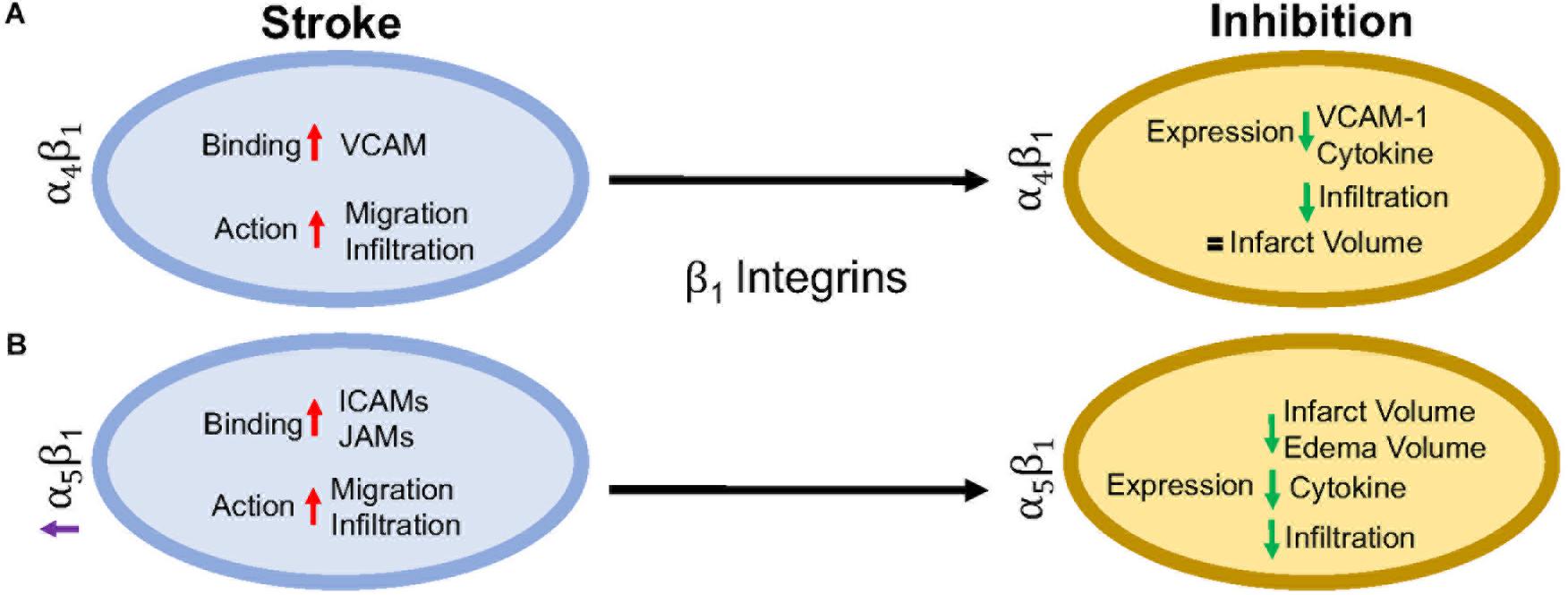
endothelial cell or extracellular matrix (Campanero et al., 1994; Caimi et al., 2001). Inhibition of the β1 integrin, just as with β2 integrin inhibition, does not fully stop leukocyte rolling. However, when both β1 and β2 integrins are inhibited, complete leukocyte arrest occurs (Lobb and Hemler, 1994; Pierini et al., 2000). This suggests that both β1 and β2 integrins are necessary for leukocyte migration, regardless of expression load. Of all the β1 integrins, both α4β1 and α5β1 appear to be the most highly expressed and the most studied in post-stroke inflammation. The other β1 integrin expressed on leukocytes, α9β1, has not been studied in the context of stroke as its expression and role has not yet been fully elucidated in the brain. Figure 3 summarizes the results discussed in this section.
FIGURE 3
Figure 3. Representative image of the β1 integrin response following experimental stroke and inhibitory antibody treatment in preclinical trials. Inhibition of (A) α4β1 and (B) α5β1 integrins post-stroke responses and effects.
α4β1 Integrin
α4β1, also known as CD49d/CD29 VLA-4 (very late antigen-4),
is localized primarily to leukocytes (neutrophils, monocytes,
lymphocytes, macrophages, etc.) and microglia as a leukocyte-specific
receptor. Additionally, α4 will also dimerize with β4, which is found in gut endothelium (Hammond et al., 2014). Activation of α4β1 integrin results from the binding of upregulated chemokines to GPCRs in the same manner as αLβ2 as discussed above (Vajkoczy et al., 2001). This stimulates binding to α4β1’s preferred ligand, VCAM-1, but experiments have shown some preference for paxillin ICAM-1 (Steffen et al., 1994; Ghandour et al., 2007), and fibronectin (Hart and Greaves, 2010) as well. Interestingly, instead of using the β1 submit of the heterodimer for binding, integrin α4β1 uses its α subunit of α4β1 to mediate binding to VCAM-1 (Luo et al., 2007).
Preclinical ischemic stroke studies targeting α4β1
have shown increasingly varied results. Most researchers reported a
decrease in VCAM-1 expression, cytokine production, and infiltrating
leukocytes (Liesz et al., 2011; Langhauser et al., 2014; Llovera et al., 2015),
but this reduction in inflammation did not result in reduced infarct
volumes or functional deficit following analysis of a randomized
preclinical trial involving six different centers (Llovera et al., 2015).
Langhauser et al went one step further and found that no treatment
paradigm (prophylactic or therapeutic) and no model (transient or
permanent) showed efficacy (Langhauser et al., 2014). On the other hand, both Becker, 2002 and Relton et al., 2000 found that inhibition of α4
improved both infarct volumes and functional deficits. When a
preclinical randomized control trial was implemented at multiple
centers, researchers found efficacy only in patients with small infarct
volumes (Llovera et al., 2015). Collectively, these contradictory results may be caused by a couple of scenarios, 1) the varying expression of integrin α4β1 expression following ischemic stroke resulting in continued leukocyte infiltration, or 2) integrin α4β1
is not a primary driver of post-stroke pathophysiology, but other
factors, including other integrins, promote leukocyte migration (Hammond et al., 2014).
α5β1 Integrin
α5β1, also known as CD49e/CD29 and
VLA-5, plays an as yet largely undetermined role in inflammation, with
studies primarily limited to cell culture. What is known is that
leukocytes express different β1 integrins with α5β1 composing around 50% of all β1 –integrins expressed on neutrophils (Pierini et al., 2000) and monocytes (Pacifici et al., 1994). Additionally, α5β1 is necessary for leukocyte adhesion. Only inhibition of both α5β1 and β2 integrins completely blocks adhesion in vitro (Pierini et al., 2000), while inhibition of α5β1 alone prevents transmigration across the BBB (Labus et al., 2018) in vitro.
As an RGD receptor, fibronectin has been shown to be the primary and
preferred [over other potential ligands such as fibrinogen (Suehiro et al., 1997)] ligand for α5β1 on endothelial cells and leukocytes (Schaffner et al., 2013; Bharadwaj et al., 2017). Importantly, in the presence of activated αLβ2, leukocyte α5β1 binding to fibronectin is enhanced (Bohnsack, 1992; Loike et al., 1999; Gronholm et al., 2016). α5β1 integrin expression is induced by cytokines, particularly TNFα (Li et al., 2011) toward the leading edge of the cell in contrast with other integrins at the uropod (Pierini et al., 2000). Furthermore, α5β1 integrin appears to be highly sensitive to calcium (Pierini et al., 2000), an ion that is increased rapidly following reperfusion (Sandoval and Witt, 2008). Upon calcium buffering, α5β1
expression moves from the front of the cell to the uropod and the
leukocyte becomes elongated. The change in expression localization and
morphology is attributed to non-movement as the leukocyte cannot detach α5β1 from the vascular wall (Pierini et al., 2000). Recently, Edwards et al. (2019) found that inhibition of α5β1
integrin by the small peptide ATN-161 prevented CD45+ leukocytes from
infiltrating the brain parenchyma following the tandem/transient common
carotid artery/middle cerebral artery occlusion model. Additionally,
mice were observed to have reduced BBB permeability, functional
deficits, edema, and infarct volume following middle cerebral artery
occlusion (Roberts et al., 2015; Edwards et al., 2019). Thus, targeting α5β1 after ischemic stroke could be a new avenue for reduction of inflammation following ischemic stroke.
Clinical Implications
The preclinical studies discussed here point towards the potential of targeting β2 and β1
integrins in the treatment of post-stroke inflammation. Though their
potential has not fully been elucidated, many clinical trials, not just
limited to stroke, have been approved in the last 10 years targeting
these integrins.
The most common target for post-stroke inflammation are the β2 integrins. Though some efficacy has been reported, no clinical stroke trials to date have targeted the αL
subunit in stroke patients. However, one clinical trial with the
monoclonal antibody, Efalizumab, has shown promise in decreasing
T-lymphocyte rolling in patients with moderate-severe plaque psoriasis (Lebwhohl et al., 2003).
In preclinical studies targeting αMβ2, a hookworm isolated recombinant glycoprotein targeting rNIF (UK279276) (Zhang et al., 2003) and humanized Hu23F2G (Leukarrest) (Yenari et al., 1998),
were both shown to decrease infarct volume and increase functional
recovery following reperfusion. Both therapies had negligible side
effects in Phase 1 studies and thus were continued to a Phase II study,
respectively, before the trials were halted due to no observed efficacy (Becker, 2002; Krams et al., 2003). The failure to target αMβ2 integrin may be due to the observation that human ischemic stroke patients do not experience the increase in αMβ2 expression as seen in rodent stroke models (Caimi et al., 2001). Interestingly, when given in conjunction with United Kingdom279276, patients experienced a slight improvement (Krams et al., 2003), but no follow-up has been conducted. This interesting effect may be worth additional investigation in future clinical trials.
Clinical inhibition of β1 integrins, on the other hand, is small and varied. Of the current clinical trials, one trial has emerged targeting α4β1 in the context of ischemic stroke. The monoclonal antibody targeting the α4 subunit (Natalizumab) has been successful in protecting patients from relapses in multiple sclerosis (Polman et al., 2006) and Crohn’s disease (Sandborn et al., 2015).
However, in a Phase II ischemic stroke study, patients receiving
Natalizumab showed no improvement in infarct growth or neurological
scores over 30 days. Furthermore, two patients (out of 79) died from
serious infections attributed to Natalizumab treatment (Elkins et al., 2017). At this time, there are no further clinical trials planned.
Future Considerations
As discussed in this review, targeting leukocytic
integrins has had limited to no efficacy in clinical trials.
Importantly, these studies have collectively employed only three
different therapeutics and two targets; there are still significant
areas that can be investigated. Though not discussed here, most
preclinical investigations have focused on the ligands themselves rather
than the integrin as the therapeutic target, highlighting the continued
importance of integrins in stroke.
It is also important to note that preclinical studies
carried out in rodents inadequately model the post-stroke
pathophysiology that patients experience. Preclinical stroke research is
also typically limited, focusing on one species, sex, and age that do
not necessarily match the demographic of stroke patients (see Kahle and Bix, 2012
for a review of this topic). Furthermore, as the changes following
stroke and/or reperfusion are inadequately understood, identifying
appropriate therapeutic targets that translate from the lab to clinical
trials, has been particularly challenging.
However, this does not suggest abandoning therapeutic
trials for ischemic stroke. As mentioned above, stroke is a leading
cause of death and disability, separate from cardiovascular disease.
This will not improve without intervention with our aging and obese
population. Fortunately, with the advent of stroke mortality-altering
therapies, i.e., t-PA and endovascular mechanical thrombectomy, our
financial burden has shifted to aftercare. When we review the amount of
trials performed for thrombolytic agents (Multicentre Acute Stroke Trial–Italy (MAST-I) Group, 1995; National Institute of Neurological Disorders and Stroke rt-Pa Stroke Study Group, 1995; The Multicenter Acute Stroke Trial–Europe Study Group, 1996) and endovascular thrombectomy [MR CLEAN (Berkhemer et al., 2015), ESCAPE (Goyal et al., 2015), EXTEND IA (Campbell et al., 2015), SWIFT PRIME (Saver et al., 2015), and REVASCAT (Jovin et al., 2015)]
as potential treatments of ischemic stroke, it is obvious that the
complexities of stroke affect the outcome of the clinical trial. This
includes, but is not limited to, the time a patient takes to arrive at
an ER, time to treatment, location of the stroke, amount of surrounding
collaterals, current medications and co-morbidities (diabetes, cancer,
etc.), and if the patient has experienced multiple strokes.
Based on current advances, as well as previous failures, a
focus on integrins as a therapeutic target for stroke is emerging. A
significant reason for this focus may be the complex, multi-dimensional
role that integrins appear to play in brain pathophysiology. Integrins
are diverse, existing on all cell types with varying roles depending
upon expression and activation. This complexity can represent a
significant challenge to integrin-targeted therapies inasmuch as such
therapies could have diverse, even unintended off-target effects.
However, we believe that this can be overcome by a better understanding
of how integrin function and expression is altered after stroke, with
the potential to exploit stroke-dependent integrin changes to
therapeutic effect. For example, identifying a specific integrin to be
upregulated in select cells in the post-stroke brain or brain-targeting
cells, but not in other organs, could render it a viable therapeutic
target. This emphasizes the need and importance of preclinical stroke
research to discover and unravel the complexities of integrin biology.
We are confident that such studies will result in viable new stroke
therapies.
Conclusion
In this review, we have implicated integrins as an area
of research for limiting inflammation following ischemic stroke. To
date, therapeutic inhibition of αLβ2, αMβ2, and α4β1
has shown promising results in preclinical studies, but translation to
the clinic has been disappointing. Going forward, more targeted
antibodies to all reactive β1 and β2 integrins
after ischemic stroke may prove more beneficial, but more research needs
to be done to completely understand the human inflammatory response and
how that relates to changes in preclinical models.
Originally published27 Aug 2018Stroke. 2018;49:2261-2267
The central nervous system and the immune system are tightly interconnected through complex communicating networks.1
Immune cells are distributed in specific central nervous system
compartments. Microglia are the innate immune cells resident in the
brain parenchyma. Macrophages surround the blood vessels and also line
the leptomeninges and the choroid plexus together with dendritic cells
and lymphocytes, among other immune cells, where they play
immunosurveillance functions. Therefore, the immune system keeps a close
watch on brain function and reacts when brain homeostasis is lost
because of injuries or diseases. Sterile organ damage may turn immune
cells into harmful agents and for this reason they are regarded as
targets for therapeutic intervention in acute stroke.2
Stroke induces strong inflammatory reactions involving the local
production of cytokines, such as TNF-α (tumor necrosis factor-α) by
various brain cells, including human neurons,3
activation of glial and endothelial cells, blood-brain barrier damage,
and infiltration of different types of leukocytes after an orchestrated
time course.4
Given the variety of leukocyte subsets trafficking to the ischemic
brain tissue, this review will focus on neutrophils,
monocyte/macrophages, and T and NK (natural killer) lymphocytes. For
further information, the readers are addressed to previous reviews on
dendritic cells5 and B lymphocytes.6
The different immune cells are considered separately in the next
sections, but leukocyte infiltration surely comprises intercellular
crosstalks by mechanisms that are not entirely known.
Neutrophils
Neutrophils
are among the first cells attracted to the brain after ischemic stroke
where they are detected in the microvessels within the first hour7 and peak at 1 to 3 days.4,7,8
Neutrophils are short-lived innate immune cells containing different
types of granules with antimicrobial pro-oxidant and proteolytic enzymes
that can damage the tissues. Accordingly, neutrophils are regarded as
detrimental following compelling evidence associating these cells with
blood-brain barrier breakdown and brain injury.7,9 Also, higher blood neutrophil counts are associated with larger infarct volumes in acute ischemic stroke patients.10
Nevertheless, the pathogenic role of neutrophils in ischemic stroke is
still not conclusive. For instance, there are conflicting results in the
literature on the potential benefit of neutrophil depletion in
experimental ischemia models.9,11
Furthermore, we lack entire demonstration that neutrophils reach the
ischemic tissue before substantial neuronal death has occurred.11
Nonetheless, neutrophils can exert detrimental effects already from the
vessel wall. Adhesion of neutrophils to the inflamed endothelium after
ischemia/reperfusion is involved in the no-reflow phenomenon,
obstructing blood flow in precapillary arterioles, postcapillary
venules, and the capillary bed.7,12
In addition, neutrophils in the vessel lumen and at perivascular
locations can damage the blood-brain barrier by releasing proteolytic
enzymes and pro-oxidant molecules (Figure).9 Moreover, neutrophils can produce NETs (neutrophil extracellular traps) promoting clot formation.13 NETs can precipitate thrombotic events and impair tPA (tissue-type plasminogen activator)-induced thrombolysis.14
In turn, thrombolysis may exacerbate detrimental effects of neutrophils
because tPA promotes neutrophil transmigration to the reperfused tissue
by proteolytic activation of plasmin and matrix metalloproteinases.15
These effects might contribute to explain why neutrophilia and high
neutrophil-to-lymphocyte ratio are associated with the risk of
hemorrhagic transformation in ischemic stroke patients treated with tPA.16
After permanent middle cerebral artery occlusion (MCAo) in mice, we
observed the formation of intravascular NETs and found NETs in
perivascular locations and in the brain parenchyma.17
Figure.
Schematic representation of leukocyte infiltration to the ischemic
brain tissue. Neutrophils are attracted to the activated endothelium and
reach perivascular spaces after extravasation from intracerebral
venules and leptomeningeal vessels. Activated neutrophils are
prothrombotic and can damage the blood-brain barrier (BBB). The presence
of neutrophils in the brain parenchyma is observed only under certain
circumstances, but the conditions determining that neutrophils remain in
perivascular spaces or reach the parenchyma are still poorly defined.
Attracted by certain chemokines, immature proinflammatory monocytes
infiltrate the ischemic tissue where they mature to macrophages, acquire
signs of alternative polarization, and seem to be involved in tissue
repair. Current experimental evidence suggests that lymphocytes, in
particular T cells and γδ T cells, play detrimental roles in the acute
phase of stroke by promoting thromboinflammation and tissue damage.
Natural killer (NK) cells are attracted to the ischemic tissue, but
their function is not fully clear. RBC indicates red blood cells.There
is also some controversy on whether neutrophils actually reach the
ischemic brain parenchyma at all. An elegant study by Enzmann et al18
noticed the massive accumulation of neutrophils in perivascular spaces
surrounding venules within the ischemic tissue after
ischemia/reperfusion in mice. Most neutrophils remained perivascular,
and only a few were detected in the brain parenchyma.18
This is an important observation because it highlights that
perivascular spaces are a niche for neutrophils where they accumulate
after transient MCAo. We also detected neutrophils in perivascular
locations and leptomeningeal spaces in the mouse after permanent MCAo.17
These results suggest that, besides extravasating from intracerebral
venules, neutrophils extravasate from leptomeningeal vessels and migrate
from the subpial space along the vessels penetrating the cortex.17 However, our study17 and previous studies7
found neutrophils in the ischemic brain parenchyma using models of
permanent MCAo. Ischemic conditions involving severe endothelial damage,
vessel rupture, and microbleeds or hemorrhagic transformation, are
expected to facilitate the presence of neutrophils in the brain
parenchyma. Other conditions, such as high blood glucose, also promote
neutrophil infiltration.19 A recent study20
analyzed the postmortem brain of 16 ischemic stroke patients and
confirmed the presence of neutrophils in the leptomeninges and
perivascular spaces, but neutrophils were rare in the infarcted
parenchyma with the exception of 1 patient deceased 3 days after stroke
with no signs of infection. Interestingly, the time to death of this
series of patients was 1 day in 2 cases, 3 days in the case above
mentioned, and then times ranged from 8 days to 240 days poststroke.20
Neutrophils display a specific time-window of attraction to the damaged
tissues after acute injuries, and they have a short life in tissues.
Therefore, the time to death of ischemic stroke patients is critical to
look for the presence of neutrophils in the brain parenchyma. More
studies of human tissue within the first days poststroke are necessary
to understand under which conditions neutrophils might gain access to
the infarcted brain parenchyma.
Despite
many advances, there are aspects of neutrophil behavior in stroke that
are still difficult to interpret. For instance, neutrophils with
anti-inflammatory and repair phenotypes were found in the ischemic brain
tissue of experimental animals,21 neutrophils of ischemic stroke patients show a reduced oxidative burst and NET formation,22 and microglia surrounding blood vessels phagocyte neutrophils.23
The possibility that neutrophils were passive bystanders under some
circumstances but active players in others depending on specific
features of the ischemic lesion needs further consideration.
Monocyte/Macrophages
After
brain ischemia, microglia acquire a reactive morphology resembling
macrophages. Classically, immunohistochemical studies have described the
presence of reactive microglia/macrophages peaking at ≈4 days
postischemia in rats or mice, but it was not possible to distinguish
whether these cells derived from resident microglia or they infiltrated
from the periphery. Nowadays, flow cytometry, cell type-specific
fluorescent reporter mice, adoptive transfer of fluorescent cells,
generation of chimeras, and recently identified specific microglia
markers, allow differentiating resident reactive microglia from
infiltrating macrophages. Monocyte infiltration is detected within the
first 24 hours postischemia, peak at 4 days, and some of these cells
persist for weeks and acquire features of tissue macrophages. Immature
CCR2+Ly6Chi proinflammatory monocytes are the subset of monocytes first attracted to the ischemic brain tissue.24–26
These cells might be released by the bone marrow, but a study reported
that monocytes reaching the ischemic brain originate in the spleen.25
Infiltrating
macrophages were classically associated with inflammation and brain
damage after ischemic stroke. In mice, monocyte infiltration is largely
dependent on CCR2 (C-C motif chemokine receptor type 2), the receptor of
the chemokine CCL2 (C-C motif chemokine ligand 2), also known as MCP1
(monocyte chemoattractant protein 1). To investigate the role of
monocytes, several studies used CCR2-deficient mice or CCR2 inhibitors,
with the limitation that besides the subset of Ly6Chi monocytes other cells, like some T cells, also express CCR2. CCR2-deficiency reduced the ischemic brain lesions in mice.27 Challenging this view, CCR2 drug inhibitors exacerbated the brain lesion.28 Furthermore, anti-CCR2 blocking antibodies impaired spontaneous long-term functional recovery,29 depletion of monocytes/macrophages worsened the ischemic lesion,30 and infiltrating macrophages prevented hemorrhagic transformation of the ischemic lesion.24
By systemic injection of fluorescent monocytes after brain ischemia, we
observed fluorescent cells in the subpial space, and along the vessels
penetrating the cortex,26
supporting the view that a subset of infiltrating macrophages establish
persistent interactions with the blood vessels (Figure).
The
phenotype of activated macrophages depends on the environmental
stimuli. The M1 and M2 phenotypes are prototypical states of macrophage
polarization achieved in culture after exposure to certain cytokines.
The M1 phenotype is proinflammatory whereas the M2 phenotype promotes
resolution of inflammation and repair. Macrophages infiltrating the
ischemic tissue, including the Ly6Clo population and some of the Ly6Chi monocytes, acquire features of alternatively polarized M2 macrophages during the first week postischemia.26,28–30
Studies of human ischemic infarcts reported that macrophages initially
showed proinflammatory features that with lesion maturation transformed
into anti-inflammatory phenotypes.20
Interestingly, a study noticed that after ischemia in mice, the
expression of M2 markers increased within the first week but then
decreased, whereas proinflammatory markers persisted and predominated at
week 2, suggesting a long-lasting inflammatory status.31
The
factors that contribute to the time-dependent changes in macrophage
phenotypes in the ischemic brain tissue are not entirely identified.
Increased anaerobic glycolysis and activation of the hypoxia-inducible
factor-1 are associated with proinflammatory M1 phenotypes, whereas
energy production in M2 phenotypes rather relies on fatty acid
oxidation.32
In M1 activated macrophages, arginine metabolism occurs through
inducible nitric oxide synthase leading to generation of reactive oxygen
and nitrogen species that damage proteins, lipids, and DNA. In
contrast, M2 macrophages metabolize arginine through arginase-1
generating polyamines involved in cell division and collagen synthesis,
among other functions.32
However, under pathological conditions 1 single phenotypic feature may
not be sufficient to attribute any specific phenotype to the cells.
Likely, cellular metabolic adaptations to ischemia and reperfusion
together with the cytokine environment and phagocytic activity have an
impact on the phenotype and function of macrophages and microglia.
Lymphocytes
T Cells
Severe stroke reduces the numbers of lymphocytes in the circulation and lymphoid organs.33 In contrast, T-cell numbers increase in the ischemic brain within the first 24 hours and can persist for long times.4
During the first hours after ischemia/reperfusion, T cells facilitate
adhesion of platelets and leukocytes to the vascular endothelium34 causing a phenomenon called thromboinflammation35
by which molecular and cellular players in thrombosis and coagulation
promote proinflammatory pathways exacerbating the brain lesion.36
However, the interaction of T cells with platelets may also have
hemostatic effects preventing hemorrhagic transformation after severe
ischemic stroke.37 T cells are found in subpial and cortical vessels and infiltrating the ischemic lesion.38,39 In addition, the choroid plexus is a gateway for T cells migrating to the periphery of cortical infarction.40 Importantly, CD8+ cytotoxic T cells were detected in human ischemic infarcts.20 Also, ischemic stroke patients show increased frequency of CD4+CD28null cells in blood associated with stroke severity and serum levels of proinflammatory cytokines.41 CD4+CD28null
T cells are an interesting subset of T cells because they have enhanced
effector functions, are associated with senescent T cells, and expand
under inflammatory conditions.42 At later phases, CD4+ cells accumulate in the brain of mice peaking at day 14 and persisting at day 30 after ischemia/reperfusion.43
Furthermore, emerging evidence suggests that antigen-mediated T-cell
responses take place in subacute or chronic stages after stroke and may
worsen stroke outcome.43–47
However, in central nervous system trauma, protective autoimmunity
mediated by T-cell responses is involved in promoting recovery.48
Overall, T cells seem to play innate functions and interact with
players in thrombosis and hemostasis in the acute phase of stroke,
whereas at later stages they exert adaptive functions that could affect
stroke outcome in the long term.
γδ T Cells
Subsets
of unconventional innate T cells with invariant T-cell receptor could
play a role in acute ischemic brain damage. Growing evidence supports
that γδ T cells are pathogenic in experimental brain
ischemia/reperfusion by secreting IL (interleukin)-17 and exacerbating
the inflammatory response.49–51 Moreover, IL-17A+ lymphocytes were detected in the postmortem brain of stroke patients.50 Interestingly, γδ T cells are abundant in the gut from where they seem to traffic to the leptomeninges after brain ischemia.52
T helper 17 cells (Th17) and γδ T cells increase in the blood of stroke
patients in association with increased levels of IL-17A, IL-23, IL-6,
and IL-1β.53
In spite of the fact that IL-17 producing cells are a small subset of
cells, they seem to play a prominent role in orchestrating the
inflammatory response in acute stroke and exacerbating the lesion
(Figure).
Regulatory Lymphocytes
Regulatory
lymphocytes exert immunomodulatory and immunosuppressor functions.
Several lines of evidence support beneficial effects of regulatory T
cells (Treg)54 and regulatory B cells55 in experimental brain ischemia. However, other studies found acute detrimental effects of Treg in brain ischemia/reperfusion56 as previously reviewed.57
Although the number of Treg found in the ischemic brain parenchyma
during the first days poststroke is low, Treg strongly accumulate in the
ischemic lesion 15 days poststroke where potentially they could inhibit
autoimmune responses.43
Increased apoptosis of Tregs, loss of Tregs in peripheral blood, and
impaired suppressive function of the remaining Treg population has been
reported in ischemic stroke patients.53,58,59
However, other studies reported upregulation of Tregs in stroke
patients in spite that decreased Treg function was observed,
particularly in female patients.60 Notably, an increased proportion of Treg cells was reported in the spleen of mice 4 days after transient MCAo.61
NK Cells
NK innate lymphocytes show a rapid and transient increase in the ischemic brain tissue.4,8 A study reported no benefits of depleting NK cells in permanent or transient MCAo.44 In contrast, another study suggested pathogenic actions of NK cells by promoting inflammation and neuronal cytotoxicity.62
This study reported infiltration of NK cells in the ischemic brain
tissue of humans and mice where NK cell numbers peaked as soon as 3
hours postischemia and then declined.62
Interestingly, the β2-nACh-R (nicotinic acetylcholine) receptor seems
to be involved in the NK cell decline observed in the ischemic tissue
from 3 hours postischemia.63
Induced-persistence of NK cells in the ischemic tissue achieved by
interfering with this cholinergic receptor did not modify lesion size
but increased systemic IFNγ (interferon γ), protected from bacterial
infection, and enhanced poststroke survival.63
More studies are needed to validate the putative capacity of brain
infiltrating NK cells to prevent poststroke infection, the role of
central acetylcholine in this process, as well as the suggested rapid
pathogenic effect of NK cells worsening the acute ischemic brain lesion.
Therapeutic Intervention
Strategies
designed to prevent negative actions of leukocytes have been taken to
the clinic in acute ischemic stroke patients but with no success to
date.64 Several studies with drugs blocking the action of neutrophils were investigated.11,64
As an example, the ASTIN trial (Acute Stroke Therapy by Inhibition of
Neutrophils) investigated a compound known as UK-279 276, a recombinant
neutrophil inhibitory factor that selectively binds the CD11b integrin
of macrophage-1 antigen (CD11b/CD18).65
The treatment did not improve recovery above placebo. This trial
followed encouraging results of a few preclinical studies with
UK-279 276 in experimental models of brain ischemia/reperfusion.66 However, only a fifth of patients in the ASTIN trial received tPA.65
Monocyte/macrophages
may acutely exacerbate the inflammatory responses, but experimental
studies have identified their involvement in resolution of inflammation,
vascular protection, and recovery of function,24,27–29
possibly linked to the phagocytic and vasculoprotective roles of these
cells. These protective actions of monocytes are in line with the
beneficial effects of administration of autologous bone marrow–derived
mononuclear cells (MNC) after experimental ischemic stroke.67
MNC contain myeloid and lymphoid cells, as well as hematopoietic and
mesenchymal stem cells. MNC administration 24 hours after MCAo improved
functional recovery, reduced lesion size and proinflammatory cytokines,
and enhanced vessel density and neurogenesis,68 and these benefits were long lasting.69
Furthermore, MNC reduced blood-brain barrier permeability and decreased
the severity of hemorrhagic transformation after tPA in an embolic
stroke model.70 However, MNC therapy did not improve outcome in hypertensive rats.71 MNC reach the periphery of brain infarction soon after administration,68
and then the cells seem to differentiate into smooth muscle cells and
endothelial cells, incorporate into vessel walls, and enhance the growth
of leptomeningeal anastomoses, the circle of Willis, and basilar
arteries.69
Phase I trials administering MNC to ischemic stroke patients have shown
safety, but the clinical efficacy of this cell therapy awaits
demonstration.72
Anti-inflammatory treatments, such as minocycline, have not been successful in the clinic.64 Experimental evidences, including a cross-laboratory preclinical study in mouse models of brain ischemia,73
support the therapeutic potential of the IL-1Ra (IL-1β receptor
antagonist). A recent clinical trial with subcutaneous administration of
IL-1Ra showed safety and reduction of plasma IL-6.74
However, the analysis excluded a major clinical benefit of the
treatment, and negative effects potentially attributable to interactions
of IL-1Ra with tPA became apparent.74
Experimental
studies support damaging effects of T lymphocytes in the acute phase of
stroke. Accordingly, fingolimod, a drug approved for
remitting-relapsing multiple sclerosis that sequesters lymphocytes in
the lymph nodes preventing lymphocyte access to the inflamed tissues,
showed beneficial effects in preclinical studies and small clinical
trials in acute ischemic stroke patients, including patients receiving
thrombolysis.64
By acting on S1P1 (sphingosine-1-phosphate receptor 1), fingolimod
induces sustained lymphopenia, but current data do not show higher
incidence of poststroke infection in patients receiving fingolimod.
Fingolimod also acts on endothelial S1P1 receptor increasing vascular
barrier function that might contribute to the observed benefits of this
drug in ischemic stroke. Given that the benefits of fingolimod seem to
be mediated by S1P1, whereas certain side effects are dependent on other
S1P receptors, selective S1P1 agonists were studied in experimental
stroke showing reduced lesion size after ischemia/reperfusion in mice.75
In contrast to the benefits of blocking T-cell trafficking, systemic
administration of regulatory T lymphocytes in rodent models of ischemic
stroke reduced infarct size, ameliorated the neurological functions,76 and reduced hemorrhagic transformation after tPA.77
Leukocyte
recruitment to inflammatory sites is attenuated by blocking α4β1
integrin (VLA-4 [very late antigen-4]) with natalizumab, an antibody in
clinical use for multiple sclerosis treatment. Blockade of VLA-4 with
CD49d antibody was investigated in a multicentric preclinical study in
mice using 2 different models of cerebral ischemia.78
CD49d antibody attenuated leukocyte infiltration and reduced infarct
volume in small cortical lesions but not in large infarctions.
Natalizumab was investigated in ischemic stroke patients in the ACTION
trial (Effect of Natalizumab on Infarct Volume in Acute Ischemic
Stroke).79
Natalizumab did not meet the primary end point of the study, but
secondary and exploratory end points suggested improvement of clinical
outcomes,64,79
encouraging the second ACTION2 trial. This phase-IIb trial was recently
completed and the notes released by the sponsor (Biogen) state that
natalizumab did not improve clinical outcomes compared with placebo.
Final Remarks
Experimental
studies support detrimental effects of certain types of leukocytes in
acute ischemic stroke. However, to date, this knowledge has not been
translated into clinical treatments. No doubt immunomodulatory
interventions in the acute phase of stroke need fine-tuning and
long-term experimental studies to ensure that repair processes in
subacute and chronic phases are not disturbed and the neurological
deficits are attenuated. Cell therapies based on administration of
autologous MNC have shown promising results in preclinical studies by
promoting functional recovery, but clinical efficacy remains to be
demonstrated. Results of various experimental models of brain ischemia
suggest the possibility that the putative pathogenic contribution of
certain leukocytes to the acute ischemic lesion might differ depending
on lesion severity, regions affected, and degree of reperfusion.
Importantly, most of the studies described above were obtained in young
healthy male mice in spite of the fact that aging, sex, and
comorbidities influence the phenotype and function of immune cells.
Identification of the ischemic conditions where leukocytes might have a
meaningful contribution to the brain lesion, the relevant subsets of
leukocytes, and the time-window for intervention, requires more
investigation. Combining immunomodulatory strategies with reperfusion
therapies offer the opportunity to attenuate negative responses of the
immune system that might impair reperfusion at the microvascular bed or
trigger detrimental effects on the brain tissue.
Acknowledgments
I acknowledge relevant studies used to prepare this article that could not be cited because of word count restriction.
Sources of Funding
Supported by the Spanish Ministerio de Economía y Competitividad (SAF2017-87459-R).
Disclosures
None.
Footnotes
Correspondence
to Anna M. Planas, PhD, Institut d’Investigacions Biomèdiques de
Barcelona (IIBB), Consejo Superior de Investigaciones Científicas
(CSIC), Rosselló 161, Planta 6, 08036-Barcelona, Spain. Email anna.planas@iibb.csic.es
 Rebecca Isabella Sienel1,2†,
  Hiroharu Kataoka3†,
  Seong-Woong Kim4,
  Fatma Burcu Seker1,2 and
  Nikolaus Plesnila1,2*


 Danielle N. Edwards
 Gregory J. Bix