Changing stroke rehab and research worldwide now.Time is Brain! trillions and trillions of neurons that DIE each day because there are NO effective hyperacute therapies besides tPA(only 12% effective). I have 523 posts on hyperacute therapy, enough for researchers to spend decades proving them out. These are my personal ideas and blog on stroke rehabilitation and stroke research. Do not attempt any of these without checking with your medical provider. Unless you join me in agitating, when you need these therapies they won't be there.

What this blog is for:

My blog is not to help survivors recover, it is to have the 10 million yearly stroke survivors light fires underneath their doctors, stroke hospitals and stroke researchers to get stroke solved. 100% recovery. The stroke medical world is completely failing at that goal, they don't even have it as a goal. Shortly after getting out of the hospital and getting NO information on the process or protocols of stroke rehabilitation and recovery I started searching on the internet and found that no other survivor received useful information. This is an attempt to cover all stroke rehabilitation information that should be readily available to survivors so they can talk with informed knowledge to their medical staff. It lays out what needs to be done to get stroke survivors closer to 100% recovery. It's quite disgusting that this information is not available from every stroke association and doctors group.

Showing posts with label glutamate. Show all posts

Tuesday, April 8, 2025

Scientists Capture How Glutamate Opens Neuron Signaling Channels

 Your incompetent doctor and hospital have done nothing to stop the glutamate poisoning part of the neuronal cascade of death I bet! And this research won't trigger ways to stop that problem, will it?

  • glutamate poisoning (6 posts to July 2020)
  • And I'm sure they DID NOTHING with this either!

    generic drug candesartan (brand name: ATACAND®) Blood Pressure Drug Helps Alzheimer's June 2018

     This line from there is instructive:

    The scientists found that candesartan prevented glutamate-induced neuronal death. 

    The latest here:

    Scientists Capture How Glutamate Opens Neuron Signaling Channels

    In an effort to understand how brain cells exchange chemical messages, scientists say they have successfully used a highly specialized microscope to capture more precise details of how one of the most common signaling molecules, glutamate, opens a channel and allows a flood of charged particles to enter. The finding, which resulted from a study led by Johns Hopkins Medicine researchers, could advance the development of new drugs that block or open such signaling channels to treat conditions as varied as epilepsy and some intellectual disorders. 

    A report on the experiments, funded by the National Institutes of Health and in collaboration with scientists at UTHealth Houston, was published March 26 in the journal Nature. 

    Neurons are the cellular foundation of the brain, and the ability to experience our environment and learn depends on [chemical] communications between neurons."

    Edward Twomey, Ph.D., assistant professor of biophysics and biophysical chemistry at the Johns Hopkins University School of Medicine

    Scientists have long known that a major molecule responsible for neuron-to-neuron communications is the neurotransmitter glutamate, a molecule abundant in the spaces between neurons. Its landing place on neurons is a channel called an AMPA receptor, which interacts with glutamate, and then acts like a pore that takes in charged particles. The ebb and flow of charged particles creates electrical signals that form communications between neurons. 

    To figure out details of the miniscule movements of AMPA receptors (at the level of single atoms), researchers used a very high-powered microscope to image these channels during specific steps in the communications processes. For the study, the scientists used a cryo-electron microscope (cryo-EM) in a facility at the Johns Hopkins University School of Medicine. 

    Typically, scientists find it easier to study cell samples that are chilled, a state that provides a stable environment. But at normal body temperature, Twomey's team found that the AMPA receptors and glutamate activity increased, providing more opportunities to capture this process in cryoEM images. 

    To that end, the scientists purified AMPA receptors, taken from lab-grown human embryonic cells that are used widely in neuroscience research to produce such proteins. Then, they heated the receptors to body temperature (37 degrees Celsius or 98.6 degrees Fahrenheit) before exposing them to glutamate. Immediately after this, the receptors were flash frozen and analyzed with cryoEM to get a snapshot of the AMPA receptors bound to the major signaling molecule, glutamate. 

    Twomey's previous research has shown that drugs such as perampanel, used to treat epilepsy, act as a door stopper around the AMPA receptor to limit the channel from opening and reducing the abundance of activity known to happen in brain cells of people with epilepsy. 

    Twomey says the findings could be used to develop new drugs that bind to AMPA receptors in different ways that either open or close the signaling channels of brain cells. 

    "With each new finding, we are figuring out each of the building blocks that enable our brains to function," says Twomey. 

    Additional scientists who contributed to the work are Anish Kumar Mondal from Johns Hopkins and Elisa Carrillo and Vasanthi Jayaraman from UTHealth Houston.

    Funding for the research was provided by the National Institutes of Health (R35GM154904, R35GM122528), the Searle Scholars Program and the Diana Helis Henry Medical Research Foundation.

    Source:
    Journal reference:

    Mondal, A. K., et al. (2025). Glutamate gating of AMPA-subtype iGluRs at physiological temperatures. Nature. doi.org/10.1038/s41586-025-08770-0.

    Saturday, June 8, 2024

    Brain’s Oxygen Deprivation Mechanism Hinders Memory Formation

     Ask your competent? doctor how long after your stroke this memory problem continues and how EXACTLY your doctor is treating the problem. NO treatment? You don't have a functioning stroke doctor!

    Brain’s Oxygen Deprivation Mechanism Hinders Memory Formation

    Summary: Oxygen deprivation in the brain triggers a feedback loop involving glutamate and nitric oxide, causing anoxia induced long-term potentiation (aLTP). This process disrupts regular memory-enhancing mechanisms, potentially explaining memory loss post-stroke. The study offers insights into treating memory problems in stroke patients.

    Key Facts:

    1. aLTP occurs during temporary oxygen deprivation in the brain, impairing memory.
    2. Glutamate and nitric oxide form a feedback loop that sustains aLTP.
    3. Disrupting this loop may help restore normal memory function after strokes.

    Source: OIST

    When we learn something new, our brain cells (neurons) communicate with each other through electrical and chemical signals. If the same group of neurons communicate together often, the connections between them get stronger. This process helps our brains learn and remember things and is known as long-term potentiation or LTP.  

    Another type of LTP occurs when the brain is deprived of oxygen temporarily – anoxia-induced long-term potentiationor aLTP. aLTP blocks the former process, thereby impairing learning and memory. Therefore, some scientists think that aLTP might be involved in memory problems seen in conditions like stroke. 

    Researchers at the Okinawa Institute of Science and Technology (OIST) and their collaborators have studied the aLTP process in detail. They found that maintaining aLTP requires the amino acid glutamate, which triggers nitric oxide (NO) production in both neurons and brain blood vessels. This process forms a positive glutamate-NO-glutamate feedback loop.

    Their study, published in iScience, indicates that the continuous presence of aLTP could potentially hinder the brain’s memory strengthening processes and explain the memory loss observed in certain patients after experiencing a stroke.  

    The brain’s response to low oxygen 

    When there is a lack of oxygen in the brain, glutamate, a neurotransmitter, is released from neurons in large amounts. This increased glutamate causes the production of NO. NO produced in neurons and brain blood vessels boosts glutamate release from neurons during aLTP. This glutamate-NO-glutamate loop continues even after the brain gets enough oxygen. 

    “We wanted to know how oxygen depletion affects the brain and how these changes occur,” Dr. Han-Ying Wang, a researcher in the former Cellular and Molecular Synaptic Function Unit at OIST and lead author of the study, stated.

    “It’s been known that nitric oxide is involved in releasing glutamate in the brain when there is a shortage of oxygen, but the mechanism was unclear.”  

    During a stroke, when the brain is deprived of oxygen, amnesia – the loss of recent memories – can be one of the symptoms. Investigating the effects of oxygen deficiency on the brain is important because of the potential medicinal benefits.

    “If we can work out what’s going wrong in those neurons when they have no oxygen, it may point in the direction of how to treat stroke patients,” Dr. Patrick Stoney, a scientist in OIST’s Sensory and Behavioral Neuroscience Unit and former member of the Cellular and Molecular Synaptic Function Unit, explained. 

    Brain tissues from mice were placed in a saline solution, mimicking the natural environment in the living brain. Normally, this solution is oxygenated to meet the high oxygen demands of brain tissue. However, replacing the oxygen with nitrogen allowed the researchers to deprive the cells of oxygen for precise lengths of time.  

    The tissues were then examined under a microscope and electrodes were placed on them to record electrical activity of the individual cells. The cells were stimulated in a way that mimics how they would be stimulated in living mice. 

    Stopping memory and learning activity 

    The scientists found that maintaining aLTP requires NO production in both neurons and in blood vessels in the brain. Collaborating scientists from OIST’s Optical Neuroimaging Unit showed that in addition to neurons and blood vessels, aLTP requires the activity of astrocytes, another type of brain cell. Astrocytes connect and support communication between neurons and blood vessels. 

    “Long-term maintenance of aLTP requires continuous synthesis of nitric oxide. NO synthesis is self-sustaining, supported by the NO-glutamate loop, but blocking molecular steps for NO-synthesis or those that trigger glutamate release eventually disrupt the loop and stop aLTP,” Prof. Tomoyuki Takahashi, leader of the former Cellular and Molecular Synaptic Function Unit at OIST, explained.  

    Notably, the cellular processes that support aLTP are shared by those involved in memory strengthening and learning (LTP). When aLTP is present, it hijacks molecular activities required for LTP and removing aLTP can rescue these memory enhancing mechanisms.

    This suggests that long-lasting aLTP may obstruct memory formation, possibly explaining why some patients have memory loss after a short stroke. 

    Prof. Takahashi emphasized that the formation of a positive feedback loop formed between glutamate and NO when the brain is temporarily deprived of oxygen is an important finding. It explains long-lasting aLTP and may offer a solution for memory loss caused by a lack of oxygen.  

    About this memory and neuroscience research news

    Author: Tomomi Okubo
    Source: OIST
    Contact: Tomomi Okubo – OIST
    Image: The image is credited to Neuroscience News

    Original Research: Open access.
    “Anoxia-induced hippocampal LTP is regeneratively produced by glutamate and nitric oxide from the neuro-glial-endothelial axis” by Han-Ying Wang et al. iScience

    Tuesday, February 9, 2021

    Study of Neuroprotection by a Combination of the Biological Antioxidant (Eucalyptus Extract) and the Antihypertensive Drug Candesartan against Chronic Cerebral Ischemia in Rats

     By using the benign word 'neuroprotection' 100% of stroke survivors won't understand how important this is. But rewording it to 'Stopping the neuronal cascade of death by a Combination of the Biological Antioxidant (Eucalyptus Extract) and the Antihypertensive Drug Candesartan against Chronic Cerebral Ischemia in Rats' would perk up the ears considerably and get to asking their doctors what the fuck they are doing about stopping the neuronal cascade of death in the first week. And a few medical malpractice suits asking for $1000 a dead neuron might concentrate the stroke medical worlds mind. I lost 5.4 billion neurons that first week. 5.4 trillion dollars!

    But your doctor should already be using Candesartan on you.

    generic drug candesartan (brand name: ATACAND®) Blood Pressure Drug Helps Alzheimer's June 2018

     This line from there is instructive:

    The scientists found that candesartan prevented glutamate-induced neuronal death.

    The latest here:

    Study of Neuroprotection by a Combination of the Biological Antioxidant (Eucalyptus Extract) and the Antihypertensive Drug Candesartan against Chronic Cerebral Ischemia in Rats

    1
    Neuroscience Research Center, Faculty of Medical Sciences, Lebanese University, Beirut P.O. Box 6573/14, Lebanon
    2
    Rammal Hassan Rammal Research Laboratory, Physiotoxicity (PhyTox), Faculty of Sciences, Lebanese University, Beirut P.O. Box 6573/14, Lebanon
    3
    Plateforme de recherche et d’analyse en sciences de l’environnement (EDST-PRASE), Beirut P.O. Box 6573/14, Lebanon
    4
    Aix Marseille University, CNRS, Centrale Marseille, iSm2, 13397 Marseille, France
    *
    Authors to whom correspondence should be addressed.
    †
    These authors contributed equally to this work.
    Academic Editor: Raffaele Capasso
    Molecules 2021, 26(4), 839; https://doi.org/10.3390/molecules26040839 (registering DOI)
    Received: 5 December 2020 / Revised: 28 January 2021 / Accepted: 30 January 2021 / Published: 5 February 2021
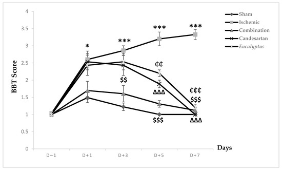
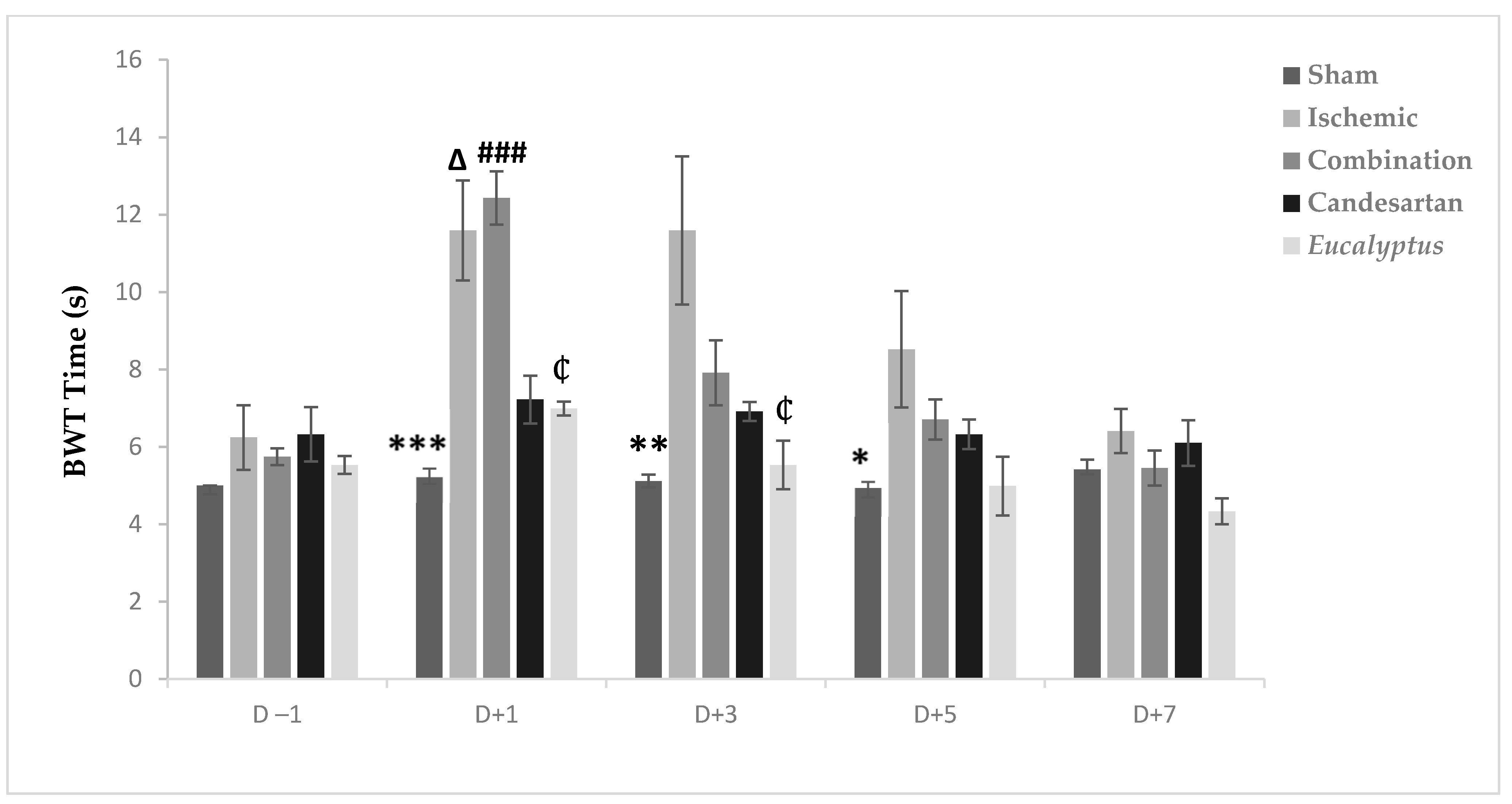
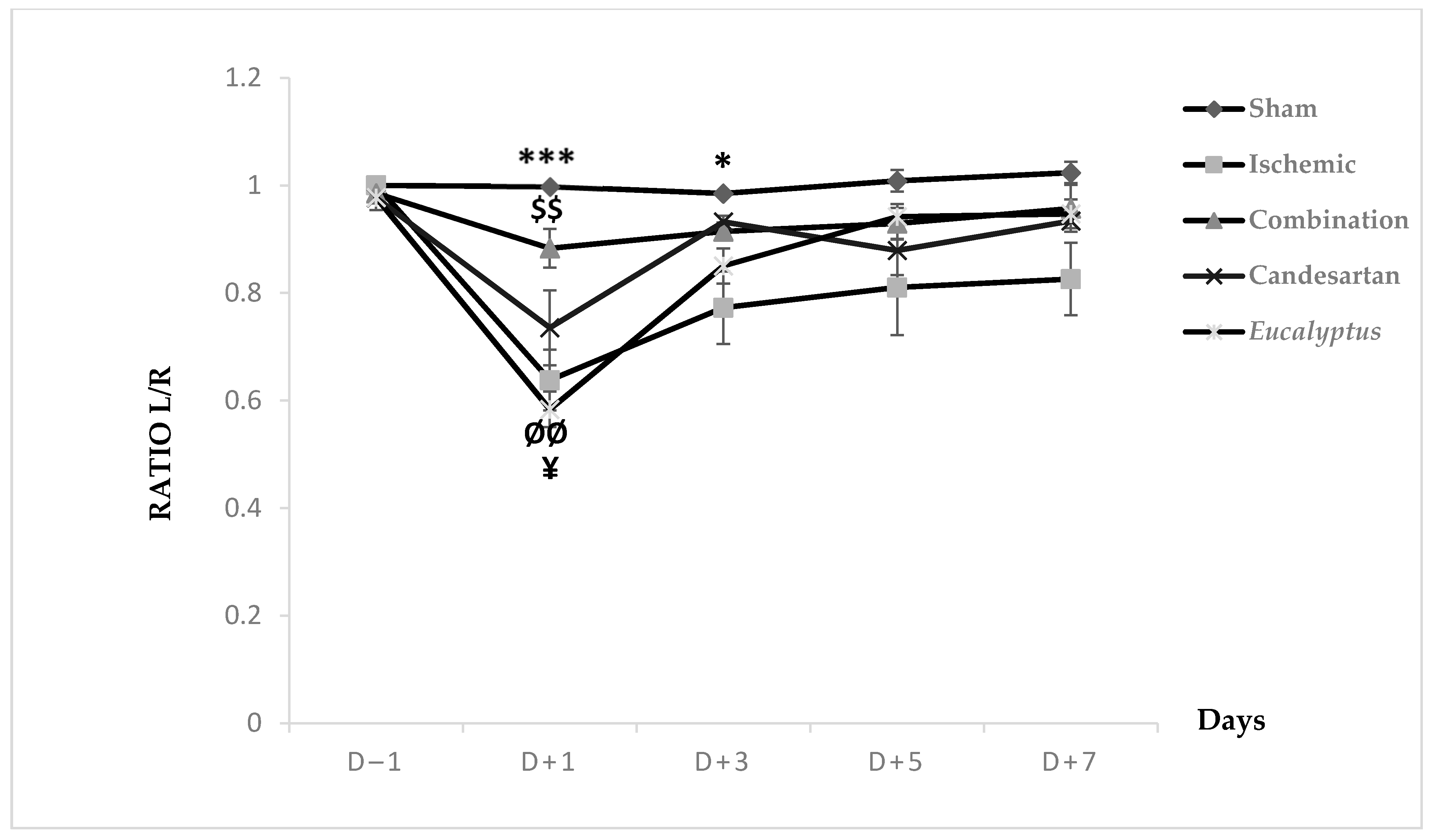
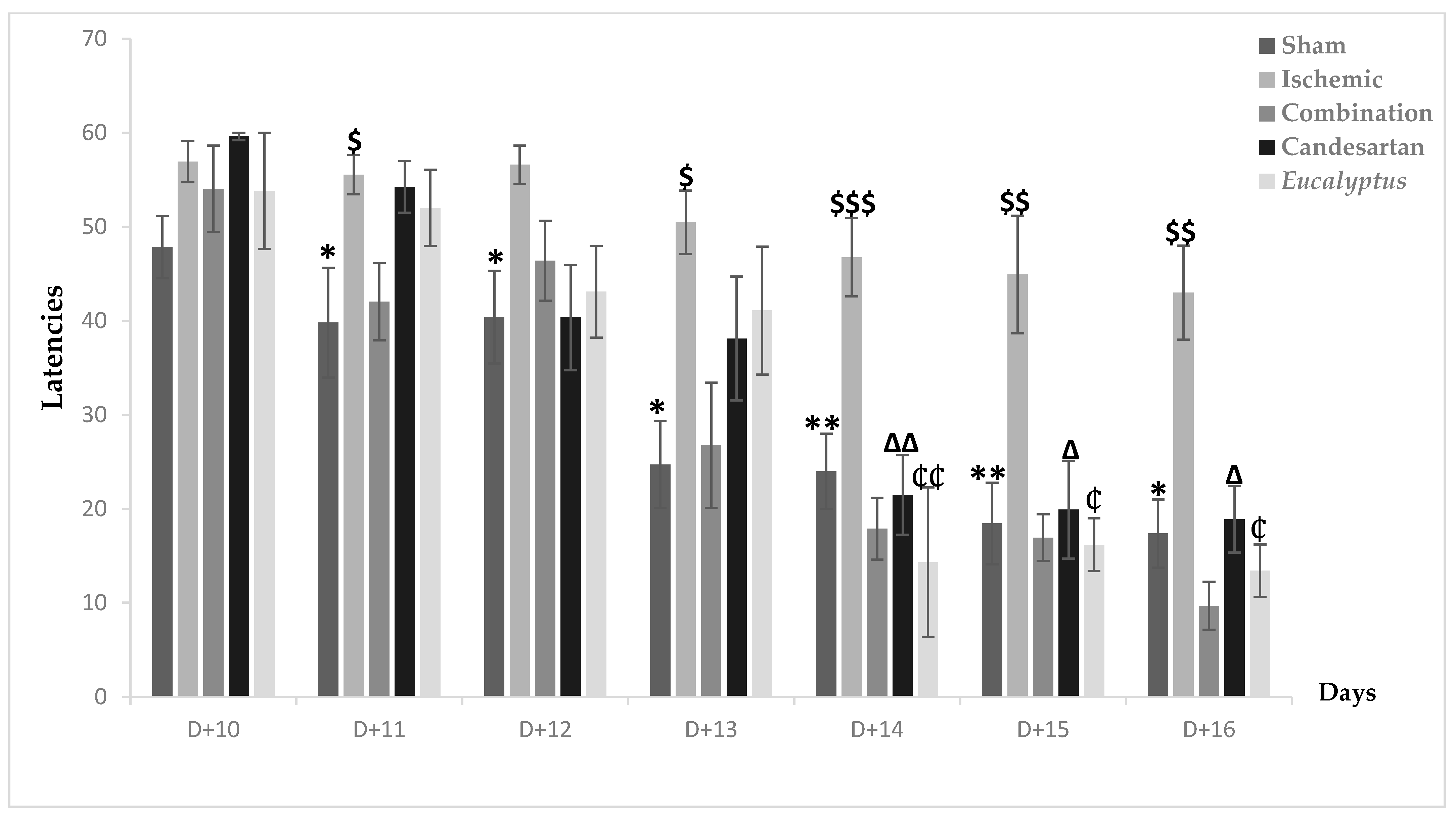
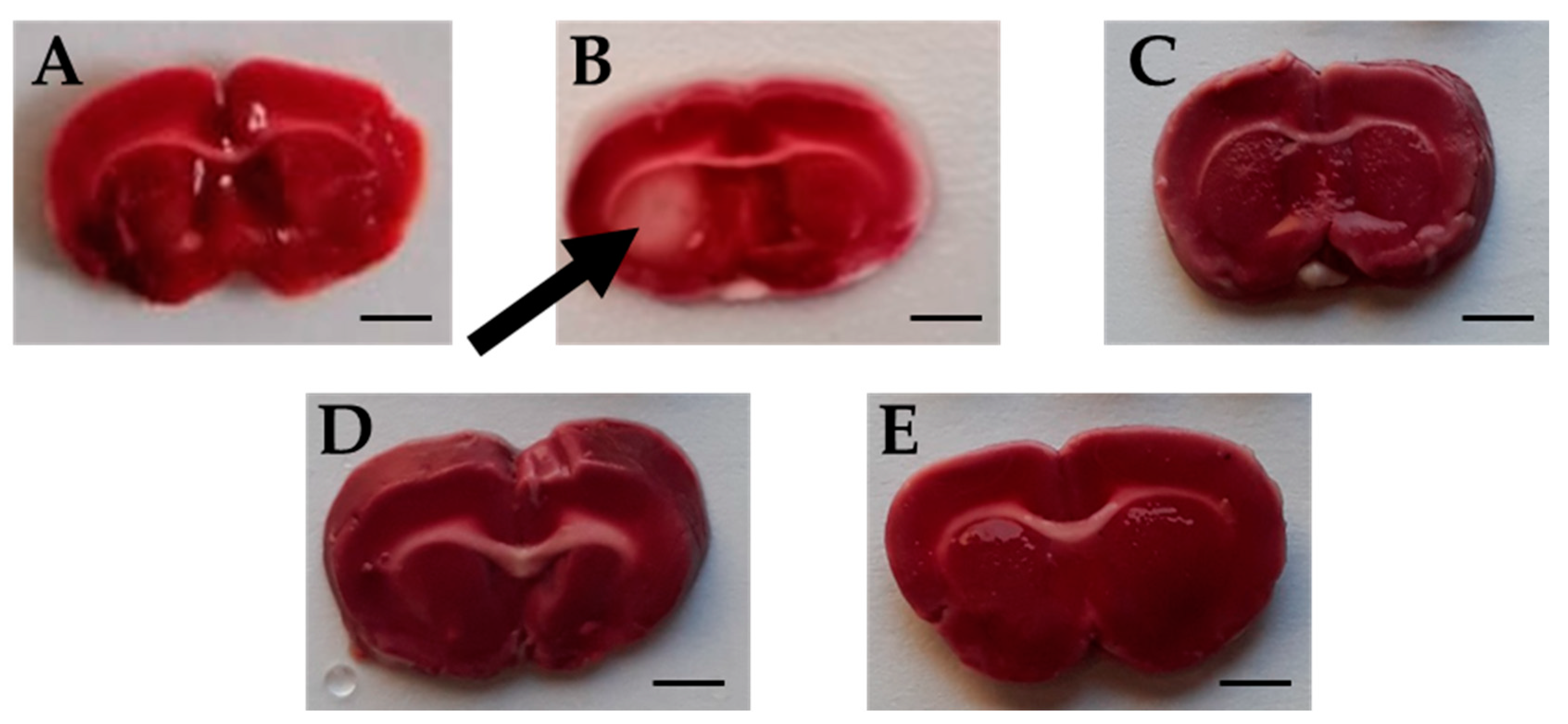
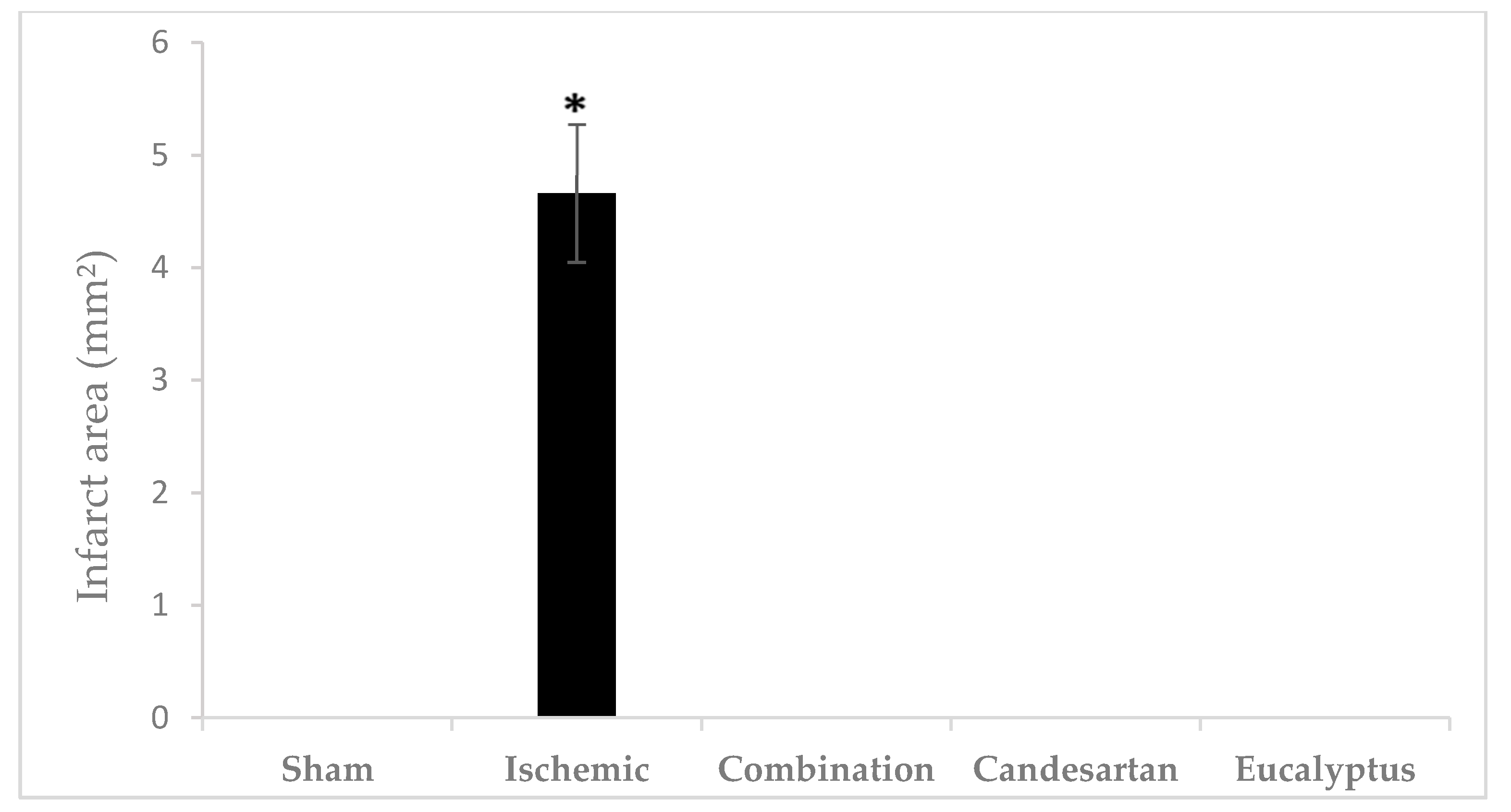
    Chronic cerebral ischemia with a notable long-term cessation of blood supply to the brain tissues leads to sensorimotor defects and short- and long-term memory problems. Neuroprotective agents are used in an attempt to save ischemic neurons from necrosis and apoptosis, such as the antioxidant agent Eucalyptus. Numerous studies have demonstrated the involvement of the renin-angiotensin system in the initiation and progression of cardiovascular and neurodegenerative diseases. Candesartan is a drug that acts as an angiotensin II receptor 1 blocker. We established a rat model exhibiting sensorimotor and cognitive impairments due to chronic cerebral ischemia induced by the ligation of the right common carotid artery. Wistar male rats were randomly divided into five groups: Sham group, Untreated Ligated group, Ischemic group treated with Eucalyptus (500 mg/kg), Ischemic group treated with Candesartan (0.5 mg/kg), and Ischemic group treated with a combination of Eucalyptus and Candesartan. To evaluate the sensorimotor disorders, we performed the beam balance test, the beam walking test, and the modified sticky test. Moreover, the object recognition test and the Morris water maze test were performed to assess the memory disorders of the rats. The infarct rat brain regions were subsequently stained using the triphenyltetrazolium chloride staining technique. The rats in the Sham group had normal sensorimotor and cognitive functions without the appearance of microscopic ischemic brain lesions. In parallel, the untreated Ischemic group showed severe impaired neurological functions with the presence of considerable brain infarctions. The treatment of the Ischemic group with a combination of both Eucalyptus and Candesartan was more efficient in improving the sensorimotor and cognitive deficits (p < 0.001) than the treatment with Eucalyptus or Candesartan alone (p < 0.05), by the comparison to the non-treated Ischemic group. Our study shows that the combination of Eucalyptus and Candesartan could decrease ischemic brain injury and improve neurological outcomes. View Full-Text
    ▼ Show Figures

    Figure 1


    Tuesday, October 1, 2019

    Potential Neuroprotective Treatment of Stroke: Targeting Excitotoxicity, Oxidative Stress, and Inflammation

    We don't need potential we DEMAND actual.  WHEN THE HELL will you provide that? Maybe 50 years from now after you are the 1 in 4 per WHO that has a stroke?

    Potential Neuroprotective Treatment of Stroke: Targeting Excitotoxicity, Oxidative Stress, and Inflammation

    • Department of Neurology, The Second Xiangya Hospital of Central South University, Changsha, China
    Stroke is a major cause of death and adult disability. However, therapeutic options remain limited. Numerous pathways underlie acute responses of brain tissue to stroke. Early events following ischemic damage include reactive oxygen species (ROS)-mediated oxidative stress and glutamate-induced excitotoxicity, both of which contribute to rapid cell death within the infarct core. A subsequent cascade of inflammatory events escalates damage progression. This review explores potential neuroprotective strategies for targeting key steps in the cascade of ischemia–reperfusion (I/R) injury. NADPH oxidase (NOX) inhibitors and several drugs currently approved by the U.S. Food and Drug Administration including glucose-lowering agents, antibiotics, and immunomodulators, have shown promise in the treatment of stroke in both animal experiments and clinical trials. Ischemic conditioning, a phenomenon by which one or more cycles of a short period of sublethal ischemia to an organ or tissue protects against subsequent ischemic events in another organ, may be another potential neuroprotective strategy for the treatment of stroke by targeting key steps in the I/R injury cascade.

    Introduction

    Although stroke is a major cause of death and adult disability, therapeutic options remain limited. The development of new treatments including potential pharmaceutical agents is therefore of great importance. The acute responses of brain tissue to cerebral ischemia are complex. First, oxidative stress, which plays an essential role in the pathogenesis of cerebral ischemia–reperfusion (I/R) injury (Zalba et al., 2007; Carbone et al., 2015), is caused by increased reactive oxygen species (ROS) production and decreased activity levels of scavenger enzymes and protective antioxidants (De Silva and Miller, 2016; Grochowski et al., 2017). Second, glutamate, the most abundant excitatory neurotransmitter, acts as a potent neurotoxin under pathological conditions. Increased extracellular glutamate levels play an essential role in ischemia-mediated cytotoxicity through N-methyl-D-aspartate (NMDA) and α-amino-3-hydroxy-5-methyl-4-isoxazolepropionic acid (AMPA) ionotropic glutamate receptors (Chuang et al., 2011; Vaarmann et al., 2013; Khanna et al., 2015; Yoo et al., 2017). Third, minutes to hours after cerebral ischemia onset, a series of inflammatory events are triggered following the activation of resident cells including microglia. Several signals contribute to the two main activation phenotypes: classically activated (M1) and alternatively activated (M2) (Kim et al., 2015; Bonaventura et al., 2016; Fu and Yan, 2018). Microglia are sensitive to signaling through receptors such as toll-like receptors (TLRs) and peroxisome proliferator-activated receptor-γ (PPAR-γ) (Kim et al., 2015). Primary signals that upregulate inflammatory mediators include damage-associated molecular patterns (DAMPs) (Macrez et al., 2011; Bonaventura et al., 2016). Other signals are hyaluronan and pathogen-associated molecular patterns (PAMPs). Many DAMPs and PAMPS are sensed by TLRs (Macrez et al., 2011). The M1 phenotype promotes the release of inflammatory mediators such as nitric oxide and ROS. This leads to increased cell death and blood–brain barrier dysfunction, triggering the release of chemokines, activating matrix metalloproteinase (MMP)-9, and upregulating adhesion molecules. The M2 phenotype is activated by anti-inflammatory cytokines such as interleukin-4, which may inhibit inflammation and promote tissue repair and wound healing (Macrez et al., 2011; Kim et al., 2015; Bonaventura et al., 2016). The development of novel neuroprotective strategies to target key steps in this cascade may represent promising therapeutic options. Therefore, this review explores potential neuroprotective strategies for halting the cascade of I/R injury. These neuroprotective strategies include NADPH oxidase (NOX) inhibitors and drugs currently approved by the Food and Drug Administration to treat other diseases but show promise as new drugs for the treatment of stroke in animal experiments and clinical trials. Ischemic conditioning may be another neuroprotective strategy for stroke.

    More at link.

    Tuesday, June 5, 2018

    generic drug candesartan (brand name: ATACAND®) Blood Pressure Drug Helps Alzheimer's

    You may need this, but talk to your doctor first. If this drug prevents glutamate-induced neuronal death then is this the solution to this particular cause(glutamate poisoning) of the neuronal cascade of death?

    Would this be useful as pretreatment to prevent your likely chance of getting dementia? Your doctor needs to find out that answer. Otherwise you're screwed.

    Your chances of getting dementia.

    1. A documented 33% dementia chance post-stroke from an Australian study?   May 2012.

    2. Then this study came out and seems to have a range from 17-66%. December 2013.

    3. A 20% chance in this research.   July 2013.

    4. Dementia Risk Doubled in Patients Following Stroke September 2018 

    5. Parkinson’s Disease May Have Link to Stroke March 2017

    generic drug candesartan (brand name: ATACAND®) Blood Pressure Drug Helps Alzheimer's 


    WASHINGTON  — In laboratory neuronal cultures, an FDA-approved drug used to treat high blood pressure reduced cell damage often linked to Alzheimer’s disease, say researchers at Georgetown University Medical Center (GUMC) and the National Institutes of Health.
    Continued below video...
    Print Friendly Version of this pagePrint Get a PDF version of this webpagePDF
    They say their work, published online in the journal Alzheimer's Research and Therapy, provides information supporting the potential effect of the generic drug candesartan (brand name: ATACAND®) — as well as other Angiotensin receptor blockers (ARBs) for the early treatment of Alzheimer’s disease.

    “Our findings make sense in many ways,” says the study’s senior author Juan M. Saavedra, MD, from GUMC’s Department of Pharmacology and Physiology.  “Hypertension reduces blood flow throughout the body and brain and is a risk factor of Alzheimer’s disease. Previous epidemiological studies found that Alzheimer’s progression is delayed in hypertensive patients treated with ARBs.”

    Using neuronal cultures, the researchers explored the action of candesartan on the neurotoxic effects of exposure to excessive glutamate, a demonstrated injury factor in the early stages of Alzheimer’s disease.

    The scientists found that candesartan prevented glutamate-induced neuronal death. They conducted in-depth gene analyses of the laboratory results, demonstrating that candesartan prevented neuronal inflammation and many other pathological processes, including alterations in amyloid metabolism, a hallmark of Alzheimer’s disease.

    The study’s first author, Abdel G. Elkahloun, PhD, from the Comparative Genomics and Cancer Genetics Branch of the National Human Genome Research Institute, then compared gene expression in the neuronal cultures with published gene databases of autopsy samples from Alzheimer’s disease patients. “The correlations were impressive — the expression of 471 genes that were altered by excess glutamate in our cultures were also altered in brain autopsy samples from patients who suffered from Alzheimer’s disease. Candesartan normalized expression of these genes in our cultures,” Elkahloun says.

    “We hypothesize that candesartan, or other members of the ARB group, may not only slow progression of Alzheimer’s but also prevent or delay its development
    ,” Saavedra says.
    The researchers say this work has immediate translational value, supporting testing candesartan, or other ARBs, in controlled clinical studies on patients at early stages of Alzheimer’s disease. 

    MORE INFORMATION:
    Roman Hafko, PhD, formerly of the National Institute of Mental Health, also contributed to this work and is an author of the paper.
    The work was supported by grants from the National Institutes of Health including the National Human Genome Research Institute (MD 20892) and the National Institute of Mental Health (MH 002762-16). The authors report having no personal financial interests related to the study.
    SOURCE:
    Georgetown University Medical Center
    Georgetown University Medical Center is an internationally recognized academic medical center with a three-part mission of research, teaching and patient care (through MedStar Health). GUMC’s mission is carried out with a strong emphasis on public service and a dedication to the Catholic, Jesuit principle of cura personalis -- or "care of the whole person." The Medical Center includes the School of Medicine and the School of Nursing & Health Studies, both nationally ranked; Georgetown Lombardi Comprehensive Cancer Center, designated as a comprehensive cancer center by the National Cancer Institute; and the Biomedical Graduate Research Organization (BGRO), which accounts for the majority of externally funded research at GUMC including a Clinical Translation and Science Award from the National Institutes of Health. 

    Alzheimer's Weekly Store

    Friday, May 25, 2018

    Moderate ultraviolet light exposure boosts the brainpower of mice thanks to increased production of the neurotransmitter glutamate

    Is this too fucking hard for your doctors and stroke hospitals to do followup clinical research for this on humans?
    https://www.the-scientist.com/?articles.view/articleNo/54603/title/Could-a-Dose-of-Sunshine-Make-You-Smarter-/&utm_content=71706132&
    The sun’s ultraviolet (UV) radiation is a major cause of skin cancer, but it offers some health benefits too, such as boosting production of essential vitamin D and improving mood. Today (May 17), a report in Cell adds enhanced learning and memory to UV’s unexpected benefits.
    Researchers have discovered that, in mice, exposure to UV light activates a molecular pathway that increases production of the brain chemical glutamate, heightening the animals’ ability to learn and remember.
    “The subject is of strong interest, because it provides additional support for the recently proposed theory of ultraviolet light’s regulation of the brain and central neuroendocrine system,” dermatologist Andrzej Slominski of the University of Alabama who was not involved in the research writes in an email to The Scientist.
    “It’s an interesting and timely paper investigating the skin-brain connection,” notes skin scientist Martin Steinhoff of University College Dublin’s Center for Biomedical Engineering who also did not participate in the research. “The authors make an interesting observation linking moderate UV exposure to . . . [production of] the molecule urocanic acid. They hypothesize that this molecule enters the brain, activates glutaminergic neurons through glutamate release, and that memory and learning are increased.”
    While the work is “fascinating, very meticulous, and extremely detailed,” says dermatologist David Fisher of Massachusetts General Hospital and Harvard Medical School, “it does not imply that UV is actually good for you. . . . Across the board, for humanity, UV really is dangerous.”
    Wei Xiong of the University of Science and Technology of China who led the research did not set out to investigate the effects of UV light on the brain or the skin-brain connection. He stumbled upon his initial finding “almost accidentally,” he explains in an email to The Scientist. Xiong and his colleagues were using a mass spectrometry technique they had recently developed for analyzing the molecular contents of single neurons, when their results revealed the unexpected presence of urocanic acid—a little-known molecule produced in the skin in response to UV light.
    “It was a surprise because we checked through all the literature and found no reports of the existence of this small molecule in the central nervous system,” writes Xiong.
    With little information to go on, Xiong and his colleagues decided to see whether UV light could also boost levels of urocanic acid in the brain. They exposed shaved mice to a low-dose of UVB—responsible for sunburn in humans—for 2 hours, then performed mass spectrometry on the animals’ individual brain cells. Sure enough, levels of urocanic acid increased in neurons of the animals exposed to the light, but not in those of control animals.
    Urocanic acid can absorb UV rays and, as a result, may be able to protect skin against the sun’s harmful effects. But in the liver and other peripheral tissues, the acid is also known to be an intermediate molecule generated in the metabolic pathway that converts histidine to glutamate. Given glutamate’s role in the brain as an excitatory neurotransmitter, Xiong and his colleagues were interested to test whether the observed UV-dependent increase in urocanic acid in neurons might be coupled with increased glutamate production. It was.
    Next, the team showed that UV light enhanced electrical transmission between glutaminergic neurons in brain slices taken from animals exposed to UV, but not in those from control animals. This UV-induced effect was prevented when the researchers inhibited activity of the enzyme urocanase, which converts urocanic acid to glutamate, indicating that the acid was indeed the mediator of the UV-induced boost in glutaminergic activity.
    Lastly, the team showed that mice exposed to UV performed better in motor learning and recognition memory tasks than their unexposed counterparts. And, as before, treating the animals with a urocanase inhibitor prevented the UV-induced improvements in learning and memory. Administering urocanic acid directly to animals not exposed to ultraviolet light also spurred similar learning and memory improvements to those achieved with UV exposure.
    Whether the results obtained in mice, which are nocturnal and rarely see the sun, will hold true in humans is yet to be determined. But, Fisher says, if the results do hold, the finding that urocanic acid alone can enhance learning and memory might suggest “a way to utilize this information to benefit people without exposing them to the damaging effects of UV.”
    H. Zhu et al., “Moderate UV exposure enhances learning and memory by promoting a novel glutamate biosynthetic pathway in the brain,” Cell, doi: 10.1016/j.cell.2018.04.014, 2018.

    Friday, March 16, 2018

    A new study reveals the drug QNZ-46 may help to protect patients at risk of stroke

    Whom is going to followup and see if this tackles the glutamate poisoning cause in the neuronal cascade of death? Or will fucking nothing be done since we have NO stroke leadership or strategy?Does this mean myelin in the white matter is damaged and we need our doctors to get a protocol for myelin repair done?
    http://neurosciencenews.com/qnz-46-stroke-8640/?
    Source: Plymouth University.
    New research shows how the novel drug QNZ-46 can help to lessen the effects of excess release of glutamate in the brain – the main cause of brain injury in stroke.
    Published in Nature Communications, the study shows how identifying the source of damaging glutamate in stroke leads to discovery of brain protection with QNZ-46, a novel form of preventative treatment with clinical potential.
    Existing studies show that restricted blood supply promotes the excess release of glutamate. The glutamate binds to receptors, over-stimulating them and leading to the break-down of myelin – the protective sheath around the nerve fibre (axon).
    Previous studies had focused on the brain’s grey matter – the area where all of the synapses operate. Now the new study focuses on white matter – the part of the brain the connects all of the grey matter together – and demonstrates that the glutamate release from axons themselves contributes to damaging myelin.
    The study, led by Professor Robert Fern at the Plymouth University Peninsula Schools of Medicine and Dentistry (PUPSMD), is the first direct comparison of vesicular fusion within different cellular components in white matter, and it reveals extensive fusion in axons – a mechanism previously thought to be absent from white matter.

    Tuesday, January 23, 2018

    Blood Glutamate Levels Are Closely Related to Acute Lung Injury and Prognosis after Stroke

    So you've identified a problem but offered no solution. You stroke survivors are going to have to solve this on their own.
    https://www.frontiersin.org/articles/10.3389/fneur.2017.00755/full?
    imageWei Bai1†, imageWei Li2†, imageYa-Lei Ning1, imagePing Li1, imageYan Zhao1, imageNan Yang1, imageYu-Lin Jiang1, imageZe-Ping Liang3, imageDong-Po Jiang3, imageYing Wang2, imageMeng Zhang2* and imageYuan-Guo Zhou1*
    • 1Molecular Biology Center, State Key Laboratory of Trauma, Burn, and Combined Injury, Research Institute of Surgery and Daping Hospital, Third Military Medical University, Chongqing, China
    • 2Department of Neurology, Research Institute of Surgery and Daping Hospital, Third Military Medical University, Chongqing, China
    • 3Department of ICU, Research Institute of Surgery and Daping Hospital, Third Military Medical University, Chongqing, China
    Background: Acute lung injury (ALI) is a serious complication of stroke that occurs with a high incidence. Our preclinical results indicated that ALI might be related to blood glutamate levels after brain injury. The purpose of this study was to assess dynamic changes in blood glutamate levels in patients with stroke and to determine the correlation between blood glutamate levels, ALI, and long-term prognosis after stroke.
    Methods: Venous blood samples were collected from controls and patients with stroke at admission and on the third and seventh day after the onset of stroke. Patients were followed for 3 months. The correlations among blood glutamate levels, severities of stroke and ALI, and long-term outcomes were analyzed, and the predictive values of blood glutamate levels and severity scores for ALI were assessed.
    Results: In this study, a total of 384 patients with stroke were enrolled, with a median age of 59 years. Patients showed significantly increased blood glutamate levels within 7 days of stroke onset (p < 0.05), and patients with more severe injuries showed higher blood glutamate levels. Moreover, blood glutamate levels were closely related to the occurrence (adjusted odds ratio, 3.022, p = 0.003) and severity (p < 0.001) of ALI and the long-term prognosis after stroke (p < 0.05), and they were a more accurate predictor of ALI than the more commonly used severity scores (p < 0.01).
    Conclusion: These results indicated that an increased blood glutamate level was closely related to the development of ALI and a poor prognosis after stroke.
    Clinical Trial Registration: http://www.chictr.org.cn, identifier ChiCTR-RPC-15006770.

    Wednesday, January 17, 2018

    A master regulatory network restoring brain glutamate homeostasis is coordinately activated in stroke

    Somewhere in these 29 pages something might be useful. Which our great stroke association would analyze and distribute worldwide. Because right now your doctor and stroke hospital are not updating their stroke recovery protocols at all, they don't have any. 

    Do you prefer your doctor, hospital and board of director's incompetence NOT KNOWING? OR NOT DOING?

    A master regulatory network restoring brain glutamate homeostasis is coordinately activated in stroke

      Mariko Kobayashi1,3, Corey Anderson2, Corinne Benakis2, Michael J. Moore1, Aldo Mele1, John J. Fak1, Christopher Y. Park1, Ping Zhou2, Josef Anrather2, Costantino Iadecola2, Robert B. Darnell1,3. 1Laboratory of Molecular Neuro-Oncology and Howard Hughes Medical Institute, The Rockefeller University, 1230 York Avenue, New York, NY 10065, USA. 2Fell Family Brain and Mind Research Institute, Weill Cornell Medicine, 407 East 61st Street, New York, NY, 10065, USA. 3Correspondence: mkobayashi@rockefeller.edu, darnelr@rockefeller.edu

    ABSTRACT
    Altered miRNA expression in various disease states have been identified, but their global targets contributing to the collective regulatory power to promote or attenuate pathology remains poorly defined. Here we applied a combination of hi-throughput RNA profiling techniques, including AGO CLIP, miRNAseq, RNAseq and ribosomal profiling, to develop an unbiased and comprehensive view of miRNA:mRNA functional interactions following ischemia/reperfusion (IR) injury in the mouse brain. Upon acute I/R insult miR-29 family members were most prominently lost, with corresponding de-regulation of their global target sites. This leads to a dynamic, cascading mode of miR-29 target transcript activation, orchestrated by an initial translational activation and subsequent increase in target mRNA levels. Unexpectedly, activated genes include factors essential for glutamate signaling and reuptake, indicating a fundamental role for this regulatory network in modulating criticalendogenous neuroprotective programs to restore brain homeostasis. We integrated this data with human brain AGO CLIP profiles to infer target site variants that determine miRNA binding and to explore the role of non-coding site polymorphisms in stroke. Together these results establish a new strategy for understanding RNA regulatory networks in complex neurological disease.

    Tuesday, October 31, 2017

    Nitric Oxide Signaling in Neurodegeneration and Cell Death

     This is fascinating because nitric oxide is so useful in blood pressure management. 85 posts on nitric oxide so your doctor can inform you when you should be getting it post stroke.
    http://www.sciencedirect.com/science/article/pii/S1054358917300819


    Abstract

    In this tribute to Solomon H. Snyder (Sol) we discuss the mechanisms by which nitric oxide (NO) kills neurons. We provide a historical perspective regarding the discovery that glutamate excitotoxicity is mediated by NO. It also contains a discussion of the discovery that neuronal nitric oxide synthase (nNOS) catalytic activity accounts for NADPH diaphorase activity and its localization in the central nervous system. NADPH diaphorase/nNOS neurons are unique in that they are resistant to toxic effects of excess glutamate and that they are resistant to neurodegeneration in a variety of neurodegenerative diseases. NADPH diaphorase/nNOS neurons are resistant to neurotoxicity and neurodegeneration through the overexpression of manganese superoxide dismutase. The review also delves into the mechanisms by which NO kills neurons including NO's activation of the glyceraldehyde-3-phosphate dehydrogenase-dependent cell pathway. In addition, there is a review of parthanatos in which NO combines with the superoxide anion (
    ) to form peroxynitrite (ONOO−) that damages DNA and activates poly (ADP-ribose) (PAR) polymerase (PARP). This ultimately leads to activation of the PARP-dependent apoptosis-inducing factor-associated nuclease, the final executioner in NO-dependent cell death. Finally, there is a discussion of potential targets that are under development that target the mechanisms by which NO kills neurons.

    Keywords

    NADPH diaphorase
    Nitric oxide
    Neuronal nitric oxide synthase
    Parthanatos
    Poly (ADP-ribose) polymerase

    Choose an option to locate/access this article:

    Check if you have access through your login credentials or your institution.