Use the labels in the right column to find what you want. Or you can go thru them one by one, there are only 34,148 posts. Searching is done in the search box in upper left corner. I blog on anything to do with stroke. DO NOT DO ANYTHING SUGGESTED HERE AS I AM NOT MEDICALLY TRAINED, YOUR DOCTOR IS, LISTEN TO THEM. BUT I BET THEY DON'T KNOW HOW TO GET YOU 100% RECOVERED. I DON'T EITHER BUT HAVE PLENTY OF QUESTIONS FOR YOUR DOCTOR TO ANSWER.
Changing stroke rehab and research worldwide now.Time is Brain!trillions and trillions of neuronsthatDIEeach day because there areNOeffective hyperacute therapies besides tPA(only 12% effective). I have 523 posts on hyperacute therapy, enough for researchers to spend decades proving them out. These are my personal ideas and blog on stroke rehabilitation and stroke research. Do not attempt any of these without checking with your medical provider. Unless you join me in agitating, when you need these therapies they won't be there.
What this blog is for:
My blog is not to help survivors recover, it is to have the 10 million yearly stroke survivors light fires underneath their doctors, stroke hospitals and stroke researchers to get stroke solved. 100% recovery. The stroke medical world is completely failing at that goal, they don't even have it as a goal. Shortly after getting out of the hospital and getting NO information on the process or protocols of stroke rehabilitation and recovery I started searching on the internet and found that no other survivor received useful information. This is an attempt to cover all stroke rehabilitation information that should be readily available to survivors so they can talk with informed knowledge to their medical staff. It lays out what needs to be done to get stroke survivors closer to 100% recovery. It's quite disgusting that this information is not available from every stroke association and doctors group.
Showing posts with label glymphatic system. Show all posts
Showing posts with label glymphatic system. Show all posts
Strengthening the body's natural
biological clock may improve recovery after a stroke by enhancing the
brain's ability to clear waste and reduce inflammation, according to a
new study published in the Journal of Clinical Investigation.
Researchers
from the University of Rochester Medical Center found that
interventions aimed at reinforcing circadian rhythms — including timed
light exposure, melatonin, a body clock-targeting drug, and
time-restricted feeding — improved recovery outcomes in mouse models of
stroke.
The
study also showed improvements in the glymphatic system, the brain's
waste-clearance network that helps remove harmful substances and
inflammatory signals while delivering nutrients through cerebrospinal
fluid.
The rest is behind a paywall at the Sunday Guardian.
Ask your competent? doctor for this test AND THE PROTOCOLS THAT PROMOTE THIS DRAINAGE!
I'm sorry, your doctor isn't competent enough to do that simple task? And the board of directors is so incompetent they have no standards for staff performance?
Your brain has its own cleaning crew. Called the The takeaway This large study suggests that glymphatic function is a measurable marker of brain aging. Lifestyle factors like blood pressure control, strength training for women, and lung health for men may help slow the process. The research makes a strong case for personalized, proactive approaches to keeping your brain healthy as you age. system, this network of channels flushes out waste while you sleep, including proteins linked to Alzheimer's and other brain diseases.
For years, researchers suspected this system played a role in how our brains age, but measuring it in living people was nearly impossible. Now, a major new study has changed that and pinpointed specific lifestyle factors that could help keep your brain younger.
How researchers tracked the brain's waste-clearing system
The glymphatic system works mostly while you sleep, using fluid to clear toxins from brain tissue. While animal studies have shown how important it is, checking how well it works in humans has been tricky until recently.
In a recent study, researchers analyzed glymphatic function1 in 40,488 people from the UK Biobank using a brain imaging tool called the DTI-ALPS index. Their goal was to figure out whether this measurement could reliably track brain aging and identify factors we can actually change. They tested their model against two other datasets to make sure it held up.
The DTI-ALPS index lined up with several markers of aging: actual age, telomere length (a sign of how old your cells are), brain structure, and thinking skills.
Put simply, people with better glymphatic function tended to have brains that looked biologically younger.
When researchers built a model using this index to predict brain age, it worked well.
This suggests that glymphatic function isn't just linked to brain health; it may be a useful way to measure how fast your brain is aging.
Blood pressure below 120 & sex-specific factors made a difference
Perhaps the most useful finding: keeping systolic blood pressure below 120 mmHg was strongly tied to slower brain aging. That's lower than the typical "normal" cutoff of 120/80, suggesting tighter blood pressure control may matter more than we thought when it comes to the brain.
The study also found differences between men and women. In women, musculoskeletal health (think bones and muscles) was a key factor in brain aging. In men, lung function mattered more. These findings suggest that the best lifestyle approach may depend on your sex.
What you can do to support your brain's cleaning system
Based on this research, here are some practical strategies to consider:
Keep blood pressure in check: Aim for systolic BP below 120 mmHg through diet (less sodium, more potassium-rich foods), regular cardiovascular exercise, stress management, and medication if your doctor recommends it.
For women, focus on muscle and bone health: Strength training and weight-bearing exercise may support brain aging outcomes. Building muscle two to three times per week is a solid starting point.
For men, prioritize lung health: Aerobic exercise, breathing practices, and not smoking can help keep your lungs working well. Activities like running, cycling, or swimming are especially helpful.
Get quality sleep: The glymphatic system is most active during deep sleep. Aim for seven to nine hours of restful sleep, and address issues like sleep apnea that can get in the way of waste clearance.
The takeaway
This large study suggests that glymphatic function is a measurable marker of brain aging. Lifestyle factors like blood pressure control, strength training for women, and lung health for men may help slow the process. The research makes a strong case for personalized, proactive approaches to keeping your brain healthy as you age.
Summary: Researchers have discovered that problems with the brain’s waste-clearing system—the glymphatic system—may significantly raise the risk of developing dementia. In one of the largest studies to date, MRI data from 40,000 adults revealed that impaired cerebrospinal fluid (CSF) flow predicts dementia risk years before symptoms appear.
The findings show that cardiovascular issues like high blood pressure can damage this system, worsening toxin buildup in the brain. Improving sleep quality and managing blood pressure could help keep this system functioning and lower dementia risk.
Key Facts:
Waste Clearance Link: Impaired glymphatic system function predicted dementia risk in 40,000 adults.
Cardiovascular Impact: High blood pressure and other vascular risk factors disrupted brain waste flow.
Preventive Potential: Good sleep and blood pressure control may protect glymphatic function and reduce dementia risk.
Source: University of Cambridge
Problems with the brain’s waste clearance system could underlie many cases of dementia and help explain why poor sleep patterns and cardiovascular risk factors such as high blood pressure increase the risk of dementia.
A study led by researchers at the University of Cambridge found that impaired movement of cerebrospinal fluid (CSF) – the clear liquid that cushions and cleans the brain – predicted risk of dementia later in life among 40,000 adults recruited to the UK Biobank.
Their findings are published today in Alzheimer’s & Dementia: The Journal of the Alzheimer’s Association.
In the healthy brain, the so-called glymphatic system serves to clear out toxins and waste materials, keeping the brain healthy. Only discovered as recently as 2012, this system functions by flushing CSF through the brain along tiny channels around blood vessels known as perivascular spaces. It collects waste then drains out of the brain, helping keep it clean and healthy.
The glymphatic system is thought to be important in protecting against many of the common forms of dementia, which are often characterised by the build-up of toxic substances in the brain – for example, Alzheimer’s disease sees amyloid ‘plaques’ and tau ‘tangles accumulate in brain tissue.
One of the most common forms of dementia is vascular dementia, caused by reduced blood flow to the brain. The most common cause of this type of dementia is cerebral small vessel disease, which affects the small blood vessels in the brain.
But the impact of cerebral small vessel disease is even greater because it also interacts with other dementias making them worse; for example, a study of nuns in the US found that among those nuns whose brains showed signs of Alzheimer’s disease post mortem, only around a half exhibited symptoms of dementia – but this increased to around nine in 10 if they also had cerebral small vessel disease.
Professor Hugh Markus and colleagues at the University of Cambridge wanted to see whether cerebral small vessel disease and other cardiovascular risk factors damage the glymphatic system – and whether this in turn increases the risk of dementia.
Until recently, it has only been possible to study glymphatic function in mice, but recent advances in MRI scanning have made it possible to study it indirectly in humans. Even so, it was only possible to do this practically in relatively small numbers, but Yutong Chen, while a medical student at the University of Cambridge, developed machine learning algorithms capable of assessing glymphatic functions from MRI scans at scale.
The team applied the algorithm to MRI scans taken from around 40,000 adults in UK Biobank. They found three biomarkers – biological signatures – associated with impaired glymphatic function assessed at baseline, predicted the risk of dementia occurring over the subsequent decade.
One of these was DTI-ALPS, a measure of the diffusion of water molecules along the perivascular spaces. Another was the size of the choroid plexus, where the CSF is produced. The third measure reflected the flow velocity of CSF into the brain.
Yutong Chen, from the Department of Clinical Neurosciences at Cambridge, said: “Although we have to be cautious about indirect markers, our work provides good evidence in a very large cohort that disruption of the glymphatic system plays a role in dementia. This is exciting because it allows to ask: how can we improve this?”
Further analysis showed that several cardiovascular risk factors impaired glymphatic function – and hence increased dementia risk, and that this was partly via causing cerebral small vessel disease, which is visible in the MRI scans.
First author Hui Hong, now a radiologist at the Second Affiliated Hospital of Zhejiang University, Hangzhou, China, said: “We already have evidence that small vessel disease in the brain accelerates diseases like Alzheimer’s, and now we have a likely explanation why. Disruption to the glymphatic system is likely to impair our ability to clear the brain of the amyloid and tau that causes Alzheimer’s disease.”
The research suggests possible approaches for reducing dementia risk. One is to look at strategies for improving glymphatic function. Sleep plays an important role in glymphatic function, and so disrupted sleep patterns are likely to impair its ability to clear toxins. Alternatively, there may be existing medicines that could be repurposed, or new ones that could be developed, to improve glymphatic function.
Another possible approach is to treat vascular risk factors such as high blood pressure. This is supported by recent studies: the SPRINT MIND trial, for example, showed that intensive blood pressure control (maintaining a systolic blood pressure of less than 120 mm Hg) led to a 20% reduction in cognitive decline or dementia compared to participants in the standard treatment group.
Professor Markus, who leads the Stroke Research Group at the University of Cambridge and is a Fellow of Clare Hall, Cambridge, said: “We already know the importance of cardiovascular risk factors when it comes to dementia, and our findings further emphasise this link.
“At least a quarter of all dementia risk is accounted for by common risk factors like blood pressure and smoking. If these impair glymphatic function, then we can intervene. Treating high blood pressure or encouraging people to stop smoking would be an achievable way to helping the glymphatic system work better.”
Professor Bryan Williams, Chief Scientific and Medical Officer at the British Heart Foundation, said:“This study offers us a fascinating glimpse into how problems with the brain’s waste clearance system could be quietly increasing the chances of developing dementia later in life.
“By improving our understanding of the glymphatic system, this study opens exciting new avenues for research to treat and prevent dementia. It also emphasises the importance of managing known cardiovascular risk factors, such as high blood pressure, for reducing dementia risk.”
Funding: The research was funded by the British Heart Foundation, with additional support from the National Institute for Health and Care Research Cambridge Biomedical Research Centre.
Key Questions Answered:
Q: What is the glymphatic system and why is it important?
A: The glymphatic system is the brain’s cleaning network that flushes out toxins and waste through cerebrospinal fluid (CSF). Disruption of this system can lead to toxin buildup, potentially contributing to dementia.
Q: How do cardiovascular factors affect dementia risk?
A: High blood pressure and small vessel disease impair glymphatic flow, reducing the brain’s ability to remove waste such as amyloid and tau proteins associated with Alzheimer’s disease.
Q: Can improving sleep or lowering blood pressure reduce dementia risk?
A: Yes. The study suggests that deep, regular sleep and treating vascular risk factors may enhance waste clearance in the brain, offering practical ways to protect against cognitive decline.
About this dementia and neurology research news
Author: Craig Brierley Source: University of Cambridge Contact: Craig Brierley – University of Cambridge Image: The image is credited to Neuroscience News
The sleep and stroke
neurologist at Barrow Neurological Institute detailed how improving
glymphatic function may prevent strokes and enhance recovery from both
hemorrhagic and ischemic events. [WATCH TIME: 5 minutes]
New Biomarker Links Brain Waste Clearance to Vascular Dementia
·
Summary: A
new study has identified a biomarker, DTI-ALPS, which connects
glymphatic system dysfunction to vascular dementia. By analyzing over
3,750 participants, researchers found that lower DTI-ALPS scores
correlated with worse executive function, highlighting the glymphatic
system’s role in clearing brain waste.
The study also uncovered a
potential pathway linking impaired waste clearance to cognitive decline,
mediated by free water accumulation in white matter. These findings
provide a robust tool for clinical trials and potential interventions,
including lifestyle changes and medications, to enhance glymphatic
function and treat vascular dementia.
3 Key Facts:
DTI-ALPS Role: DTI-ALPS scores measure glymphatic system health, with lower scores linked to cognitive impairment.
Pathway Identified: Impaired glymphatic function may lead to free water accumulation, white matter damage, and cognitive decline.
Diverse Validation: The biomarker was independently validated across four cohorts with diverse demographics.
Source: USC
A
new study from the Keck School of Medicine of USC has tested a
biomarker linked to vascular dementia across four separate groups and
proposed an explanation for how cognitive impairment arises.
The findings were just published in Alzheimer’s & Dementia®: The Journal of the Alzheimer’s Association.
Vascular
dementia is the second most common form of dementia following
Alzheimer’s disease. It has similar symptoms, including problems with
memory, decision-making and language, and represents a significant
public health problem as the global population continues to age.
> The
researchers found that lower DTI-ALPS scores, which indicated damage to
the glymphatic system, were associated with worse executive function.
Credit: Neuroscience News
The condition is
usually caused by cerebral small vessel disease (cSVD), which damages
the brain’s small blood vessels—but researchers don’t yet know the exact
mechanism linking cSVD to dementia.
One theory involves problems with the glymphatic system, which helps clear waste from the brain.
A
team of researchers from the Mark and Mary Stevens Neuroimaging and
Informatics Institute (Stevens INI), part of the Keck School of
Medicine, as well as researchers from the Biomarkers for Vascular
Contributions to Cognitive Impairment and Dementia (MarkVCID)
consortium, have found new evidence in support of that theory.
With
funding from the National Institutes of Health, the group analyzed
brain scans and cognitive tests from a total of 3750 people.
The
researchers then used technique known Diffusion Tensor Image Analysis
along the Perivascular Space – or DTI-ALPS – to determine how well the
glymphatic system was functioning in each person based on their brain
scans.
The researchers compared the DTI-ALPS results with the
cognitive test results and found that people with lower DTI-ALPS scores
also performed lower on cognitive tests.
The study confirmed that a
low DTI-ALPS score is a biomarker for cSVD and suggests that glymphatic
damage may be driving cognitive decline.
“The most significant
finding is that we found a clear link between DTI-ALPS and cognitive
function in all four cohorts, with ages ranging from middle-age through
older adulthood,” said Danny J. J. Wang, PhD, the study’s senior author
and a professor of neurology and radiology and director of imaging
technology innovation at the Keck School of Medicine’s Stevens INI.
Wang
and his team also analyzed the progression of symptoms across study
participants, finding a possible pathway to explain how glymphatic
problems lead to cognitive impairment.
Their results provide a
target for clinical researchers seeking to develop treatments for
vascular dementia, Wang said, and may also prove useful for treating
symptoms of Alzheimer’s disease.
Validating the biomarker
The
DTI-ALPS biomarker relies on magnetic resonance imaging (MRI) to
measure water movement along perivascular spaces, fluid-filled regions
around the brain’s blood vessels that are a key part of the glymphatic
system.
If researchers detect changes in DTI-ALPS score, that can
indicate damage and suggest that the waste clearance system is not
functioning as it should.
In the present study, Wang and his team
analyzed MRI scans to collect a measure of DTI-ALPS for each
participant. They compared those measurements to each person’s level of
executive function, a composite score of cognition that includes memory,
attention, planning, emotion regulation and other abilities that tend
to suffer as dementia progresses.
The researchers found that
lower DTI-ALPS scores, which indicated damage to the glymphatic system,
were associated with worse executive function.
That link was
verified independently in four separate participant groups—from the
MarkVCID consortium; the University of California, Davis; the University
of California, San Francisco; and the Framingham Heart Study—with a
total of 3750 participants.
Independently validating the DTI-ALPS
biomarker in each of the four cohorts provides strong evidence for the
glymphatic system’s role in cSVD and vascular dementia, Wang said.
The
racial and ethnic diversity of the participant groups, as well as the
range of ages included (averaging between 56 and 76 years of age across
cohorts) also suggests that the findings are robust and can be
generalized to a broader patient population.
Treatments for vascular dementia
Once
the team linked problems with glymphatic function to declines in
executive function, they moved on to the next question — why?
To
find out more, the researchers conducted a mediation analysis, which
studies the process or mechanism connecting two or more variables. In
this case, they found that another biomarker—”free water” or excess
water in the brain’s white matter—helped explain the link between
glymphatic problems and cognitive decline.
In this potential
pathway, “first waste clearance is impaired, which causes accumulation
of free water in the brain’s white matter. That leads to tissue damage
and eventually to cognitive impairment,” said the paper’s first author,
Xiaodan Liu, MD, PhD, a former postdoctoral researcher at the USC
Stevens INI, now an assistant researcher in radiology at the University
of California, San Francisco.
More research, including
longitudinal work, is needed to confirm whether each step in that
pathway is causal. But the team’s findings indicate that the DTI-ALPS
score biomarker for vascular dementia is robust and ready to be used in
clinical trials, Wang said.
Those studies could explore enhancing
glymphatic function as a way to treat vascular dementia. Lifestyle
changes such as exercising more and improving sleep quality are one way
to do that, Wang said, and future studies may also reveal medications
that can help.
The findings could also provide clues for how to
treat Alzheimer’s disease, which has been linked to low DTI-ALPS scores
in other studies.
About this research
In
addition to Wang, the study’s other authors are Xiaodan Liu, Xingfeng
Shao and Kay Jann from the Laboratory of fMRI Technology, Mark and Mary
Stevens Neuroimaging and Informatics Institute, Keck School of Medicine
of USC, University of Southern California; Steven Cen and John M.
Ringman from the Department of Neurology, Keck School of Medicine of
USC, University of Southern California; Pauline Maillard and Charles S.
DeCarli from the University of California, Davis; Giuseppe Barisanofrom Stanford University; Arvind Caprihanfrom
the Mind Research Network, Albuquerque, New Mexico; Hanzhang Lu from
Johns Hopkins University School of Medicine; Konstantinos Arfanakis from
the Illinois Institute of Technology and Rush University Medical
Center, Chicago, Illinois; Brian T. Gold from the University of
Kentucky; Sudha Seshadri, Claudia L. Satizabal and Mohamad
Habes from the University of Texas Health Science Center at San Antonio,
San Antonio, Texas; Alexa S. Beiser from Boston University; Joel H.
Kramer and Lara Stablesfrom the University of California,
San Francisco; Herpreet Singh, Kristin Schwab and Steven M. Greenberg
from Massachusetts General Hospital, Boston, Massachusetts; and Karl G.
Helmer from Harvard Medical School, Massachusetts General Hospital and
the Massachusetts Institute of Technology.
Funding: This
work was supported by the National Institute of Neurological Disorders
and Stroke and the National Institute on Aging, part of the National
Institutes of Health [U24NS100591, UH3NS100599, UH3NS100605,
UH3NS100588, UH3NS100608, UH3NS100606, UH3NS100598 and UH3NS100614]
About this neurology research news
Author: Laura LeBlanc Source: USC Contact: Laura LeBlanc – USC Image: The image is credited to Neuroscience News
Didn't your competent? doctor start creating protocols on this years ago? NO? So you don't have a functioning stroke doctor, do you? Your doctor was incompetent for DOING NOTHING before and will stay incompetent by doing nothing now to get this to human testing! There are NO excuses for such behavior!
Summary: New research in mice reveals that aging
slows the brain’s ability to clear out harmful waste, contributing to
neurological disorders like Alzheimer’s and Parkinson’s. Scientists have
found that restoring function in the brain’s waste-clearing system,
known as the glymphatic system, can reverse these age-related effects.
Using
a clinically approved drug, researchers increased the efficiency of
waste removal, offering a potential treatment strategy for age-related
brain diseases.
Key facts:
Aging slows the brain’s waste-clearing process, increasing the risk of neurological disorders.
The glymphatic system, responsible for brain detoxification, can be restored to youthful efficiency.
A known drug successfully revived waste-clearing in aged mice, showing promise for future therapies.
Source: University of Rochester
Alzheimer’s,
Parkinson’s, and other neurological disorders can be seen as “dirty
brain” diseases, where the brain struggles to clear out harmful waste.
Aging is a key risk factor because, as we grow older, our brain’s
ability to remove toxic buildup slows down.
However, new
research in mice demonstrates that it’s possible to reverse age-related
effects and restore the brain’s waste-clearing process.
The
new research combines advanced imaging and particle tracking techniques
to describe for the first time in detail the route via the cervical
lymph vessels in the neck through which half of dirty CSF exits the
brain. Credit: Neuroscience News
“This
research shows that restoring cervical lymph vessel function can
substantially rescue the slower removal of waste from the brain
associated with age,” said Douglas Kelley, PhD, a professor of
Mechanical Engineering in the University of Rochester Hajim School of
Engineering and Applied Sciences.
“Moreover, this was accomplished with a drug already being used clinically, offering a potential treatment strategy.”
Kelley is one of the lead authors of the study, which appears in the journal Nature Aging, along with Maiken Nedergaard, MD, DMSc, co-director the University’s Center for Translational Neuromedicine.
First
described by Nedergaard and her colleagues in 2012, the glymphatic
system is the brain’s unique waste removal process that uses
cerebrospinal fluid (CSF) to wash away excess proteins generated by
energy hungry neurons and other cells in the brain during normal
activity.
This discovery pointed the way for potential new
approaches to treat diseases commonly associated with the accumulation
of protein waste in the brain, such Alzheimer’s (beta amyloid and tau)
and Parkinson’s (alpha-synuclein).
In healthy and young brains,
the glymphatic system does a good job of flushing away these toxic
proteins, however, as we age, this system slows, setting the stage for
these diseases.
A network of tiny pumps draws waste from the brain
Once
laden with protein waste, CSF in the skull needs to make its way to the
lymphatic system and ultimately the kidneys, where it is processed
along with the body’s other waste.
The
new research combines advanced imaging and particle tracking techniques
to describe for the first time in detail the route via the cervical
lymph vessels in the neck through which half of dirty CSF exits the
brain.
In addition to measuring the flow of CSF, the researchers were able
observe and record the pulsing of lymph vessels in the neck that helps
draw CSF out of the brain.
“Unlike the cardiovascular system
which has one big pump, the heart, fluid in the lymphatic system is
instead transported by a network of tiny pumps,” said Kelley.
These
microscopic pumps, called lymphangions, have valves to prevent backflow
and are strung together, one after another, to form lymph vessels.
The
researchers found that as the mice aged, the frequency of contractions
decreased, and the valves failed. As a result, the speed of dirty CSF
flowing out of the brains of older mice was 63 percent slower compared
to younger animals.
Known drug restarts flow of brain cleaning fluids
The
team then set out to see if they could revive the lymphangions and
identified a drug called prostaglandin F2α, a hormone-like compound
commonly used medically to induce labor and known to aid smooth muscle
contraction.
The lymphangions
are lined with smooth muscle cells, and when the researchers applied the
drug to the cervical lymph vessels in older mice, the frequency of
contractions and the flow of dirty CSF from the brain both increased,
returning to a level of efficiency found in younger mice.
“These vessels are conveniently located near the surface of the skin,
we know they are important, and we now know how to accelerate
function,” said Kelley.
“One can see how this approach, perhaps
combined with other interventions, could be the basis for future
therapies for these diseases.”
Additional contributors to the
study include first authors Ting Du, Aditya Raghunandan, and Humberto
Mestre, and Virginia Plá, Guojun Liu, Antonio Ladrón-de-Guevara, Evan
Newbold, Paul Tobin, Daniel Gahn-Martinez, Saurav Pattanayak, Qinwen
Huang, and Weiguo Peng with the University of Rochester.
Funding: The
research was supported with funding from National Institute of
Neurological Disorders and Stroke, the Lundbeck Foundation, the Novo
Nordisk Foundation, the Human Frontier Science Program, the Miriam and
Sheldon G. Adelson Medical Research Foundation, the Simons Foundation,
the EU Joint Programme – Neurodegenerative Disease Research, the US Army
Research Office, the National Center for Complementary and Integrative
Health, and the BRAIN Initiative.
About this neurology and aging research news
Author: Mark Michaud Source: University of Rochester Contact: Mark Michaud – University of Rochester Image: The image is credited to Neuroscience News
Make sure your doctor and hospital are closely following this so when results come out, you get notified of the interventions needed. The idea is to prevent your likely chances of getting dementia.
Your risk of dementia, has your doctor told you of this?
Summary: A new study aims to assess the
usefulness of a new technology that could speed up and enhance the
clearing of metabolic waste via the glymphatic system as a person
sleeps.
Source: University of North Carolina
The U.S. Department of Defense is funding the first human
trial of a device to speed up and enhance the natural system of brain
cleansing that occurs when we sleep.
The trial will be conducted among 90 people at three trial sites –
University of North Carolina, University of Washington School of
Medicine, and a collaboration between Oregon Health & Science
University and the Brain Electrophysiology Laboratory (BEL). Results are
expected in the fall of 2022.
Summary: A new study aims to assess the
usefulness of a new technology that could speed up and enhance the
clearing of metabolic waste via the glymphatic system as a person
sleeps.
Source: University of North Carolina
The U.S. Department of Defense is funding the first human
trial of a device to speed up and enhance the natural system of brain
cleansing that occurs when we sleep.
The trial will be conducted among 90 people at three trial sites –
University of North Carolina, University of Washington School of
Medicine, and a collaboration between Oregon Health & Science
University and the Brain Electrophysiology Laboratory (BEL). Results are
expected in the fall of 2022.
Recent discoveries point to the
importance of quality sleep for clearance of brain metabolic waste
through the newly-discovered brain glymphatic system.
If sleep is disrupted, so are these crucial processes, leading to
cognitive impairment – things like faulty motor coordination, attention
deficits, slower processing speed, decreased decision-making
capabilities, and hampered short-term memory, in addition to increasing
risk of neurodegenerative disease later in life.
These issues can have life-or-death consequences for service members
in the U.S. military, which is why the Department of Defense is funding
innovative research initiatives, including this three-year,
$4.3-million, project with the ultimate goal of helping service members
overcome acute sleep deprivation and chronic sleep restriction.
The scientists leading this effort are from UNC-Chapel Hill, the
University of Washington School of Medicine, the Brain Electrophysiology
Lab Oregon Health & Science University, and Montana State
University.
“Our approach is to continue to
validate novel imaging approaches of the human glymphatic system while
assessing novel technology to improve glymphatic clearance and cognitive
function,” said co-principal investigator Dawn Kernagis, PhD, assistant
professor of neurosurgery at UNC School of Medicine.
“If this works, it would have major implications for service members
and potentially anyone with diagnosed sleep dysfunction. It could also
have implications for people with other neurological conditions, such as
traumatic brain injury, Alzheimer’s disease, and other dementias.”
This project, titled “Augmented Neurophysiology of Sleep and
Performance Readiness,” is part of the Medical Technology Enterprise
Consortium, a collaboration between industry and academia to facilitate
research and development activities, in cooperation with the U.S. Army
Medical Research and Development Command and other Department of Defense
agencies in the biomedical sciences to protect, treat, and optimize the
health and performance of military personnel.
This effort was years in the making, starting with the discovery of
the glymphatic system by co-principal investigator Jeffrey Iliff, PhD
and Maiken Nedergaard, MD. In 2013, Science Magazine called it one of the Top 10 discoveries of the year.
Iliff, a professor of psychiatry and
behavioral sciences and neurology at the University of Washington
School of Medicine, said the biology is straightforward. Cerebral spinal
fluid surrounds the brain, and as we sleep, fluid washes through brain
cells and supports clearance of difference wastes. Iliff’s TED Talk on this process has been viewed more than 5 million times.
Scientists think this glymphatic function is at the heart of the
restorative power of sleep. And so, scientists think disrupting sleep
disrupts the glymphatic system, impairing cognition and potentially
impacting long-term brain health.
Functional
prototype to test in-home sleep treatment. Electronics and battery are
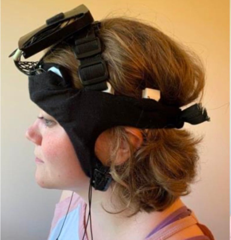
perched on top of the head. Next generation of the device will have
electronics/battery integrated in the headband. Credit: BEL Company
“Improving glymphatic function, whether pharmacologically or by means
of a device, could improve the cognitive effects of acute sleep
deprivation and chronic sleep restriction,” said Iliff.. “Our lab’s
research over the past eight years on brain-waste clearance in animals
has helped define glymphatic biology. Now we hope to see if we can use
what we’ve learned to help people overcome poor or interrupted sleep and
the brain dysfunction that follows.”
The proposed technology for
improving glymphatic function is called Augmented Neural Oscillation
Driver or ‘AugNOD’, an easily-to-use, wireless, combined
electroencephalography (EEG)/transcranial electrical stimulation
headband that can be applied before sleep to monitor and improve slow
wave sleep and glymphatic clearance. Project co-principal investigator
Don Tucker, PhD, professor emeritus at the University of Oregon and BEL
CEO, created the technology.
The research team has also developed and validated an imaging and
analysis system, including the use of magnetic resonance imaging to
measure glymphatic exchange of fluids when individuals are given an IV
containing contrasting agents. The new study will validate novel imaging
approaches developed at the University of Washington School of Medicine
that do not require contrast agents.
Through complete modeling conducted at the University of Montana, the
team will also be able to capture long-range fluid transport and
diffusion in the brain, as well as changes in brain blood vessel
pressure; both are closely linked to glymphatic pathway function.
“Beyond evaluating potential technology for improving glymphatic
function, the resulting imaging and modeling approaches that will be
validated in this study could be incredibly impactful for the
neuroscience and neurological research fields given the limited options
for studying a potentially critical system for short and long-term brain
health,” Kernagis said.
You'll have to ask your doctor if your brain glymphatic system was impaired from your stroke and what protocols they have to restore that system. You do expect your doctor to know EXACTLY what damage your stroke caused AND THE PROTOCOLS that will fix them. Or are you OK with your doctor's incompetence in not getting you 100% recovered? Getting you recovered is your doctor's responsibility,don't let him/her dump it on you by using the 'get out of jail free' statement: 'All strokes are different,all stroke recoveries are different.' Laugh maniacally in their face and ask them;'You expect me to believe that crapola excuse?'
Summary: A new study aims to assess the
usefulness of a new technology that could speed up and enhance the
clearing of metabolic waste via the glymphatic system as a person
sleeps.
Source: University of North Carolina
The
U.S. Department of Defense is funding the first human trial of a device
to speed up and enhance the natural system of brain cleansing that
occurs when we sleep.
The trial will be conducted among
90 people at three trial sites – University of North Carolina,
University of Washington School of Medicine, and a collaboration between
Oregon Health & Science University and the Brain Electrophysiology
Laboratory (BEL). Results are expected in the fall of 2022.
Recent
discoveries point to the importance of quality sleep for clearance of
brain metabolic waste through the newly-discovered brain glymphatic
system.
If sleep is disrupted, so are these crucial processes,
leading to cognitive impairment – things like faulty motor coordination,
attention deficits, slower processing speed, decreased decision-making
capabilities, and hampered short-term memory, in addition to increasing
risk of neurodegenerative disease later in life.
These issues can
have life-or-death consequences for service members in the U.S.
military, which is why the Department of Defense is funding innovative
research initiatives, including this three-year, $4.3-million, project
with the ultimate goal of helping service members overcome acute sleep
deprivation and chronic sleep restriction.
The scientists leading
this effort are from UNC-Chapel Hill, the University of Washington
School of Medicine, the Brain Electrophysiology Lab Oregon Health &
Science University, and Montana State University.
“Our approach is
to continue to validate novel imaging approaches of the human
glymphatic system while assessing novel technology to improve glymphatic
clearance and cognitive function,” said co-principal investigator Dawn
Kernagis, PhD, assistant professor of neurosurgery at UNC School of
Medicine.
“If this works, it would have major implications for
service members and potentially anyone with diagnosed sleep dysfunction.
It could also have implications for people with other neurological
conditions, such as traumatic brain injury, Alzheimer’s disease, and
other dementias.”
This project,
titled “Augmented Neurophysiology of Sleep and Performance Readiness,”
is part of the Medical Technology Enterprise Consortium, a collaboration
between industry and academia to facilitate research and development
activities, in cooperation with the U.S. Army Medical Research and
Development Command and other Department of Defense agencies in the
biomedical sciences to protect, treat, and optimize the health and
performance of military personnel.
This effort was years in the making, starting with the discovery of
the glymphatic system by co-principal investigator Jeffrey Iliff, PhD
and Maiken Nedergaard, MD. In 2013, Science Magazine called it one of the Top 10 discoveries of the year.
Iliff,
a professor of psychiatry and behavioral sciences and neurology at the
University of Washington School of Medicine, said the biology is
straightforward. Cerebral spinal fluid surrounds the brain, and as we
sleep, fluid washes through brain cells and supports clearance of
difference wastes. Iliff’s TED Talk on this process has been viewed more than 5 million times.
Scientists
think this glymphatic function is at the heart of the restorative power
of sleep. And so, scientists think disrupting sleep disrupts the
glymphatic system, impairing cognition and potentially impacting
long-term brain health.
Functional
prototype to test in-home sleep treatment. Electronics and battery are
perched on top of the head. Next generation of the device will have
electronics/battery integrated in the headband. Credit: BEL Company
“Improving glymphatic function, whether pharmacologically or by means
of a device, could improve the cognitive effects of acute sleep
deprivation and chronic sleep restriction,” said Iliff.. “Our lab’s
research over the past eight years on brain-waste clearance in animals
has helped define glymphatic biology. Now we hope to see if we can use
what we’ve learned to help people overcome poor or interrupted sleep and
the brain dysfunction that follows.”
1Department of Neurosurgery, Renji Hospital, School of Medicine, Shanghai Jiao Tong University, Shanghai, China
2Central Laboratory, Renji Hospital, Shanghai Jiao Tong University School of Medicine, Shanghai, China
The glymphatic system (GS) is a novel defined brain-wide
perivascular transit network between cerebrospinal fluid (CSF) and
interstitial solutes that facilitates the clearance of brain metabolic
wastes. The complicated network of the GS consists of the periarterial
CSF influx pathway, astrocytes-mediated convective transport of fluid
and solutes supported by AQP4 water channels, and perivenous efflux
pathway. Recent researches indicate that the GS dysfunction is
associated with various neurological disorders, including traumatic
brain injury, hydrocephalus, epilepsy, migraine, and Alzheimer’s disease
(AD). Meanwhile, the GS also plays a pivotal role in the
pathophysiological process of stroke, including brain edema, blood–brain
barrier (BBB) disruption, immune cell infiltration, neuroinflammation,
and neuronal apoptosis. In this review, we illustrated the key
anatomical structures of the GS, the relationship between the GS and the
meningeal lymphatic system, the interaction between the GS and the BBB,
and the crosstalk between astrocytes and other GS cellular components.
In addition, we contributed to the current knowledge about the role of
the GS in the pathology of stroke and the role of AQP4 in stroke. We
further discussed the potential use of the GS in early risk assessment,
diagnostics, prognostics, and therapeutics of stroke.
Introduction
Clearing the metabolic wastes and maintaining the fluid
homeostasis are important for brain function. In most organs, the
lymphatic network is responsible for the wastes clearance and fluid
drainage (Ikomi et al., 2012).
However, a hallmark of the brain is the absence of typical lymphatic
structures. Due to the presence of blood–brain barrier (BBB), the
movement of solutes and ions in the brain is strictly restricted.
Cerebrospinal fluid (CSF) has been considered to be important for the
exchange of water-soluble metabolites; however, its mechanisms remain
largely unknown. Iliff et al. (2012)
reported the existence of the glymphatic system (GS) in the central
nervous system (CNS), which is an alternative clearance system located
in the perivascular space and aquaporin-4 (AQP4) dependent (Iliff et al., 2012).
Emerging evidence from human studies and rodent models suggests that
the GS is crucial for maintaining brain health, and dysfunction of GS is
closely associated with various neurological disorders, including
aging, neurodegeneration, and acute brain injury (de Leon et al., 2017; Ringstad et al., 2017).
In parallel, the meningeal lymphatic vessels were discovered and
demonstrated to participate in solutes transport and in immune
surveillance (Aspelund et al., 2015; Louveau et al., 2015, 2016; Antila and Karaman, 2017).
Stroke, a major cause of death and disability, affects over 800,000 individuals annually (Coutts, 2017).
It has been well-recognized that the GS plays a crucial role in the
pathophysiology of stroke, including brain edema, blood–brain barrier
(BBB) disruption, immune cell infiltration, neuroinflammation, and
neuronal apoptosis (Ji et al., 2021).
Targeting the GS, therefore, has provided potential for the early risk
assessment, diagnosis, prognosis, and therapeutic of stroke. In this
review, we summarize the latest research progress in the GS, including
the anatomy and function, the interaction with the meningeal lymphatic
systems and the BBB, and the communication between astrocytes and other
GS cellular components. We emphasize the role of the GS in
pathophysiology of different stroke subtypes, especially the role of
AQP4 in the pathophysiology of stroke. In the end, we summarize the
concerns and give some perspectives for future research.
This will only become useful if those future studies occur and create protocols to prevent this edema. But since we have NO STROKE LEADERSHIP OR STRATEGY, nothing will occur. Incompetence reigns supreme in stroke and will continue until survivors are in charge.
Cerebral edema following ischemic stroke is predictive of the severity of the eventual stroke related damage, however the effective treatment is limited. The glymphatic system is a recently identified waste clearance pathway in the brain, found in the paravascular space and mainly composed of astrocytes and their aquaporin-4 (AQP4) water channels. In this review, we primarily focus on the role of the glymphatic system in the formation of cerebral edema after ischemic stroke. There is still no definite conclusion whether the influx of cerebrospinal fluid (CSF) in the glymphatic system is increased or not after ischemic stroke. However, the reduced interstitial fluid (ISF) clearance after ischemic stroke is definite. Additionally, AQP4 as the most important part of glymphatic system plays a complex bimodal in cerebral edema after ischemic stroke. Most of the research has found that AQP4 deletion in animals reduced cerebral edema after acute ischemic stroke compared with wild type animal models. The mislocalization of astrocytic AQP4 was also presented after ischemic stroke. As the cerebral edema after ischemic stroke is difficult to treat, we discuss several potential treatment targets related to glymphatic system. More studies are needed to explore the role of glymphatic system in the formation of cerebral edema after ischemic stroke and develop probable treatment strategies.
Or this latest one only tested in mice and probably decades away from common usage? Assuming of course that your doctor and stroke hospital have enough competence/responsibility to get this research going in humans. Can you wait that long? I'll be doing the wine thing.
Even if this works you will then need a SPECIFIC SLEEP PROTOCOL so your glymphatic system can clear the waste from your brain while you sleep.
Nanodevices are the newest weapon in medicine’s growing arsenal to fight
Alzheimer’s. They capture dangerous peptides before they can assemble
to form Alzheimer’s plaques in the brain. Find out how.
Alzheimer's disease is the sixth leading cause of death in the United
States, affecting one in 10 people over the age of 65. Scientists are
engineering nanodevices to disrupt processes in the brain that lead to
the disease.
People who are affected by Alzheimer's disease have a specific type of
plaque, made of self-assembled molecules called β-amyloid (Aβ) peptides,
that build up in the brain over time. This buildup is thought to
contribute to loss of neural connectivity and cell death. Researchers
are studying ways to prevent the peptides from forming these dangerous
plaques in order to halt development of Alzheimer's disease in the
brain.
How it works
In a multidisciplinary study, scientists at the U.S. Department of
Energy's (DOE) Argonne National Laboratory, along with collaborators
from the Korean Institute of Science and Technology (KIST) and the Korea
Advanced Institute of Science and Technology (KAIST), have developed an
approach to prevent plaque formation by engineering a nano-sized device
that captures the dangerous peptides before they can self-assemble.
The β-amyloid peptides arise from the breakdown of an amyloid precursor
protein, a normal component of brain cells," said Rosemarie Wilton, a
molecular biologist in Argonne's Biosciences division. "In a healthy
brain, these discarded peptides are eliminated."
In brains prone to the development of Alzheimer's, however, the brain
does not eliminate the peptides, leaving them to conglomerate into the
destructive plaques.
"The idea is that, eventually, a slurry of our nanodevices could collect
the peptides as they fall away from the cells -- before they get a
chance to aggregate," added Elena Rozhkova, a scientist at Argonne's
Center for Nanoscale Materials (CNM), a DOE Office of Science User
Facility.
Decorating the surface
The researchers covered the surface of the new nanodevice with fragments
of an antibody -- a type of protein -- that recognizes and binds to the
Aβ peptides. The surface of the nanodevice is spherical and porous, and
its craters maximize the available surface area for the antibodies to
cover. More surface area means more capacity for capturing the sticky
peptides.
To find the optimal coating, the scientists first searched previous
literature to identify antibodies that have high affinity to Aβ
peptides. It was important to choose an antibody that attracts the
peptides but doesn't bind to other molecules in the brain. Then the
team, led by Wilton, produced the antibodies in bacteria and tested
their performance.
A full antibody molecule can be up to a few dozen nanometers long, which
is big in the realm of nanotechnology. However, only a fraction of this
antibody is involved in attracting the peptides. To maximize the
effectiveness and capacity of the nanodevices, Wilton's group produced
tiny fragments of the antibodies to decorate the nanodevice's surface.
Engineering and testing the nanodevice
The scientists at CNM constructed the base of the porous, spherical
nanodevices out of silica, a material that has long been used in
biomedical applications due to its flexibility in synthesis and its
nontoxicity in the body. Coated with the antibody fragments, the
nanodevices capture and trap the Aβ peptides with high selectivity and
strength.
"Many attempts to prevent Alzheimer's have focused on inhibiting enzymes
from cutting β-amyloid peptides off of the cell's surface," said
Rozhkova, who led the project at CNM. "Our elimination approach is more
direct. We've taken building blocks from nanotechnology and biology to
engineer a high-capacity 'cage' that traps the peptides and clears them
from the brain."
At CNM, the scientists tested the effectiveness of the devices by
comparing how the peptides behaved in the absence and presence of the
nanodevices. Using in vitro transmission electron microscopy (TEM), they
observed a notable decline in peptide aggregation in the presence of
the nanodevices. They further analyzed the interactions using confocal
laser scanning microscopy and microscale thermophoresis measurement, two
additional techniques for characterizing interactions at the nanoscale.
The scientists also performed small-angle X-ray scattering to study the
processes that make the nanodevices porous during synthesis. The
researchers performed the X-ray characterization, led by Byeongdu Lee, a
group leader in Argonne's X-ray Science division, at beamline 12-ID-B
of the lab's Advanced Photon Source (APS), a DOE Office of Science User
Facility.
These studies supported the case that the nanodevices sequester the
peptides from the pathway to aggregation by more than 90 percent
compared to the control silica particles without the antibody fragments.
However, the devices still needed to demonstrate their effectiveness
and safety within cells and brains.
Joonseok Lee -- who originally proposed this experiment at Argonne as a
Director's Postdoctoral Appointee and pioneered the design for the
nanodevice -- continued the study of the therapeutic potential of this
device at KIST and KAIST.
"The Director's Postdoctoral Position is a rare opportunity offered at
Argonne that allows for unique research projects and cross-field
collaborations that might not otherwise be possible," said Rozhkova. "We
have incredible minds at the lab who want to explore topics that don't
fall under a predefined area of research, and this program encourages
this creativity and innovation."
Models Demonstrated Safety, Efficacy
The in vivo experiments -- experiments that took place in living cells
-- performed by Lee and his collaborators showed that the nanodevices
are nontoxic to cells. They also tested the effectiveness of the devices
in the brains of mice with Alzheimer's, demonstrating around 30 percent
suppression of plaque formation in brains containing the nanodevices
compared to control brains. The research on mice was conducted at KIST
and KAIST in South Korea with appropriate government approvals.
This study combined the strengths of antibody engineering and
nanotechnology, the power of two DOE User Facilities at Argonne and
innovative collaboration resulting from the laboratory's postdoctoral
program to explore a technological approach to preventing Alzheimer's.
Using a similar approach, scientists may also be able to pair the silica
nanoparticles with different antibodies that target molecules related
to other neurodegenerative diseases, such as Huntington's disease and
Parkinson's disease, which also involve abnormal protein aggregation.
The porous nanoparticles may be further upgraded for use in imaging
applications including fluorescent imaging and magnetic resonance
imaging. SOURCE:
To me, this means the goal for tPA administration is 3 minutes after stroke onset. 'Think your stroke team can do that?' Since you will never meet that goal you are going to have to go down the difficult route of solving the 5 causes of the neuronal cascade of deathin the first week. 100% recovery is still expected.
Salty fluid regularly flushes through the brain to clear away toxins
and waste, but after a stroke, this liquid floods the organ, drowning
its cells.
Swelling in the brain, known as cerebral edema, occurs after stroke
as water flows into brain cells and the space surrounding them. For
years, scientists thought this excess fluid came from blood, but new
evidence suggests that the water springs from another source entirely:
the sodium-rich cerebrospinal fluid that permeates the brain. These
results come from both live mouse models and human tissue.
Strokes
occur when a blockage plugs a blood vessel in the brain, or a vessel
completely ruptures. Without an adequate energy supply, brain cells can
no longer police which particles pass through their membranes. Within
minutes, the neurons swell
like overfilled beach balls and begin to short-circuit, accrue damage
and die. Hours later, the tightly woven tissue lining blood vessels in
the brain, the blood-brain barrier, also begins to malfunction, and the
entire organ takes on water.
"For over 60 years, people thought
this accumulation of fluid was coming from the blood" leaking through
the compromised blood-brain barrier, said study lead author Dr. Humberto
Mestre, a clinician and current doctoral student at the University of
Rochester Medical Center (URMC) Center for Translational Neuromedicine.
But cerebral edema sets in long before
the blood-brain barrier breaks down, leading Mestre and his colleagues
to wonder whether the water actually comes from somewhere else.
"No
one had looked at these alternate fluid sources," Mestre said.
Cerebrospinal fluid, which makes up about 10% of the fluid found in the
mammalian cranial cavity, stood out as a promising candidate, he added.
In
the brain, cerebrospinal fluid flows through the glymphatic system, a
network of tubing that winds along paths carved out by the organ's veins
and arteries, according to a 2015 report in the journal Neurochemical Research.
The fluid flows just outside the blood vessels, held in place by a
"doughnut-shaped tunnel" of cells. (Picture a length of wire,
representing an artery, resting inside a rubber hose, which acts like
the outer tunnel filled with fluid.) As muscles along the arteries
contract, the nearby cerebrospinal fluid gets pushed along its route and
picks up metabolic wastes on the way. Besides taking out the trash, the
glymphatic system may also help distribute fats, sugars and other important compounds within the brain.
Although
crucial in a healthy brain, in the aftermath of a stroke, the
glymphatic system goes haywire and drives the onset of edema, Mestre and
his co-authors found. "The cerebrospinal fluid is actually the primary
driver of swelling right after the stroke happens," Mestre said.
Staying the flood
The
role of cerebrospinal fluid in stroke eluded scientists for decades, in
part, because no technology existed to observe a stroke unfolding in
real time, Mestre said.
He and his co-authors combined several
techniques to observe the change in fluid flow in mice experiencing
stroke. The team peered into the animals' brains using both MRI and a
two-photon microscope, which uses light and fluorescent chemicals to
image living tissues. "We can basically image what the cerebrospinal
fluid is doing while the stroke is happening," Mestre said. By infusing
the fluid with radioactive particles, the researchers could also
determine how the flow rate changed over time.
Using these
methods, the team determined that edema takes hold of the mouse brain
"as early as 3 minutes" after stroke, long before the blood-brain
barrier began to leak, Mestre said. As brain cells short-circuit, they
spew chemical messengers known as neurotransmitters and potassium into
the space beyond their membranes. Nearby cells react to the influx of
chemicals and, in turn, short-circuit. As these electrical storms sweep
through the brain, muscles
within the blood vessels contract and create a pocket of space between
themselves and the surrounding glymphatic system. Salty cerebrospinal
fluid gets sucked into the resulting vacuum, pulling water molecules
along with it.
"Wherever sodium
is accumulating, water is going to follow it," Mestre said. The team
could watch this game of follow-the-leader unfold in select areas of the
brain but could not track water flow in the whole organ at once. Using a
computer model to simulate the entire glymphatic network, however, they
were able to predict how constricting blood vessels would drive the
flow of water through a whole mouse brain after stroke.
To connect
the dots between mice and humans, the authors examined the brain tissue
of patients who had died from ischemic stroke, wherein a blood clot
blocks a blood vessel in the brain. The mouse and human brains
accumulated fluid in the same regions, namely areas through which the
glymphatic system runs and picks up wastes. Given the strong correlation
between animals and people, "these findings could provide a conceptual
basis for development of alternative treatment strategies," the authors
noted.
The team tested one of these strategies in mice by
blocking a water channel on astrocytes, cells in the brain that help
direct water through the glymphatic system. Mice that lacked the channel
were slower to develop edema after stroke, suggesting that a similar
treatment could show promise in human patients. In addition to blocking
water flow, future treatments could potentially prevent edema by slowing
the spread of stroke-induced electrical activity in the brain, the
authors added. These electrical storms continue to barrage the brain for days after stroke, inciting edema each time they happen.
The
harmful waves of electrical activity seen in ischemic stroke also
appear in concert with "virtually every [central nervous system]
injury," Mestre said. The new study hints that the glymphatic system may
play roles in conditions where there's bleeding in and around the brain, traumatic brain injury and even migraine,
although such connections remain "purely speculative." Someday, the
glymphatic system could offer doctors a whole new strategy for treating
acute brain injuries, Mestre said.






 Tao Lv
 Bing Zhao1,
  Xiaohua Zhang