Changing stroke rehab and research worldwide now.Time is Brain! trillions and trillions of neurons that DIE each day because there are NO effective hyperacute therapies besides tPA(only 12% effective). I have 523 posts on hyperacute therapy, enough for researchers to spend decades proving them out. These are my personal ideas and blog on stroke rehabilitation and stroke research. Do not attempt any of these without checking with your medical provider. Unless you join me in agitating, when you need these therapies they won't be there.

What this blog is for:

My blog is not to help survivors recover, it is to have the 10 million yearly stroke survivors light fires underneath their doctors, stroke hospitals and stroke researchers to get stroke solved. 100% recovery. The stroke medical world is completely failing at that goal, they don't even have it as a goal. Shortly after getting out of the hospital and getting NO information on the process or protocols of stroke rehabilitation and recovery I started searching on the internet and found that no other survivor received useful information. This is an attempt to cover all stroke rehabilitation information that should be readily available to survivors so they can talk with informed knowledge to their medical staff. It lays out what needs to be done to get stroke survivors closer to 100% recovery. It's quite disgusting that this information is not available from every stroke association and doctors group.

Showing posts with label knee. Show all posts

Monday, October 10, 2022

A unilateral robotic knee exoskeleton to assess the role of natural gait assistance in hemiparetic patients

You didn't give us any information on how much use of this is needed to 100% recover, so you didn't do your job properly. But I blame your mentors and senior researchers for that failure in not providing correct instructions on the point of stroke research. 100% RECOVERY, NOTHING LESS!

A unilateral robotic knee exoskeleton to assess the role of natural gait assistance in hemiparetic patients

Abstract

Background

Hemiparetic gait is characterized by strong asymmetries that can severely affect the quality of life of stroke survivors. This type of asymmetry is due to motor deficits in the paretic leg and the resulting compensations in the nonparetic limb. In this study, we aimed to evaluate the effect of actively promoting gait symmetry in hemiparetic patients by assessing the behavior of both paretic and nonparetic lower limbs. This paper introduces the design and validation of the REFLEX prototype, a unilateral active knee–ankle–foot orthosis designed and developed to naturally assist the paretic limbs of hemiparetic patients during gait.

Methods

REFLEX uses an adaptive frequency oscillator to estimate the continuous gait phase of the nonparetic limb. Based on this estimation, the device synchronically assists the paretic leg following two different control strategies: (1) replicating the movement of the nonparetic leg or (2) inducing a healthy gait pattern for the paretic leg. Technical validation of the system was implemented on three healthy subjects, while the effect of the generated assistance was assessed in three stroke patients. The effects of this assistance were evaluated in terms of interlimb symmetry with respect to spatiotemporal gait parameters such as step length or time, as well as the similarity between the joint’s motion in both legs.

Results

Preliminary results proved the feasibility of the REFLEX prototype to assist gait by reinforcing symmetry. They also pointed out that the assistance of the paretic leg resulted in a decrease in the compensatory strategies developed by the nonparetic limb to achieve a functional gait. Notably, better results were attained when the assistance was provided according to a standard healthy pattern, which initially might suppose a lower symmetry but enabled a healthier evolution of the motion of the nonparetic limb.

Conclusions

This work presents the preliminary validation of the REFLEX prototype, a unilateral knee exoskeleton for gait assistance in hemiparetic patients. The experimental results indicate that assisting the paretic leg of a hemiparetic patient based on the movement of their nonparetic leg is a valuable strategy for reducing the compensatory mechanisms developed by the nonparetic limb.

Monday, December 7, 2020

Knee sleeve

 In order not to trigger my bursitis I have been methodically increasing my left leg stride, both forward and behind. That increases my knee snapping. Which one of my physical therapists should have told me about and what to do about it. Hell, there have been 10 million survivors a year for a long time. At least a few of them have had this problem and solved it. It should have been put in that public database of all stroke protocols and research.  So I bought a knee sleeve, not the pull on kind since I thought that might be a problem pulling on one handed depending on how tight it was. So I got a velcro one, which I have to use my right knee to hold it in place while I fasten it down. 

There is a hot or cold pack that can be inserted over the patella.
 

It works pretty well, snapping is considerably reduced.

Sunday, November 29, 2020

Towards a robotic knee exoskeleton control based on human motion intention through EEG and sEMG signals

I looked but didn't find anything that suggested that my problem of knee snapping was addressed by any of these. I'm assuming that this is because my pre-motor cortex is mostly dead. My doctor explained absolutely nothing about why my deficits were occurring, he was completely useless.

 Towards a robotic knee exoskeleton control based on human motion intention through EEG and sEMG signals

 A.C.Villa-Parra a,b,
D.Delisle-Rodríguez a,c, 
A. López-Delis c, 
T. Bastos-Filho a,*,
R. Sagaró d, 
A. Frizera-Neto a
a Post-Graduate Program in Electrical Engineering, Universidade Federal do Espírito Santo, Vitória,Brazil
b GIIB, Universidad Politécnica Salesiana, Cuenca, Ecuador
c Center of Medical Biophysics, Universidad de Oriente, Santiago,Cuba
d  Mechanical and Design Engineering Department, Universidad de Oriente, Santiago,Cuba

Abstract

The integration of lower limb exoskeletons with robotic walkers allows obtaining a system to improve mobility and security duringgait rehabilitation. In this work, the evaluation of human motion intention (HMI) based on electroencephalogram (EEG) and surface electromyography (sEMG) signals are analyzed for a knee exoskeleton control as a preliminary study for gait neuro-rehabilitation with a hybrid robotic system. This system consists of the knee exoskeleton H2 and the UFES’s Smart Walker, which are used to restore the neuromotor control function of subjects with neural injuries. An experimental protocol was developed to identify patterns to control the exoskeleton in accordance with the HMI-based on EEG/sEMG. The EEG and sEMG signals are recorded during the following activities: stand-up/sit-down and knee flexion/extension. HMI is analyzed through  both event-related desynchronization/synchronization (ERD/ERS) and slow cortical potential, as well as the myoelectric  pattern classification related to lower limb. The feature extraction from sEMG signals is based on vector combinations in time and frequency domain which are used for a pattern classification stage trough an artificial neural network with Levenberg Marquadt training algorithm and support vector machine. Preliminary results shown that a combination of EEG/sEMG signals can be used to define a control strategy for the robotic system.©2015The Authors.Published by Elsevier

Friday, July 31, 2020

Passive-elastic knee-ankle exoskeleton reduces the metabolic cost of walking

Useless for us, tested in healthy subjects.  And since we have NO STROKE LEADERSHIP,  we have no one to ask to get this tested in stroke subjects. 

Passive-elastic knee-ankle exoskeleton reduces the metabolic cost of walking


Abstract

Background

Previous studies have shown that passive-elastic exoskeletons with springs in parallel with the ankle can reduce the metabolic cost of walking. We developed and tested the use of an unpowered passive-elastic exoskeleton for walking that stores elastic energy in a spring from knee extension at the end of the leg swing phase, and then releases this energy to assist ankle plantarflexion at the end of the stance phase prior to toe-off. The exoskeleton uses a system of ratchets and pawls to store and return elastic energy through compression and release of metal springs that act in parallel with the knee and ankle, respectively. We hypothesized that, due to the assistance provided by the exoskeleton, net metabolic power would be reduced compared to walking without using an exoskeleton.

Methods

We compared the net metabolic power required to walk when the exoskeleton only acts at the knee to resist extension at the end of the leg swing phase, to that required to walk when the stored elastic energy from knee extension is released to assist ankle plantarflexion at the end of the stance phase prior to toe-off. Eight (4 M, 4F) subjects walked at 1.25 m/s on a force-measuring treadmill with and without using the exoskeleton while we measured their metabolic rates, ground reaction forces, and center of pressure.

Results

We found that when subjects used the exoskeleton with energy stored from knee extension and released for ankle plantarflexion, average net metabolic power was 11% lower than when subjects walked while wearing the exoskeleton with the springs disengaged (p = 0.007), but was 23% higher compared to walking without the exoskeleton (p < 0.0001).

Conclusion

The use of a novel passive-elastic exoskeleton that stores and returns energy in parallel with the knee and ankle, respectively, has the potential to improve the metabolic cost of walking. Future studies are needed to optimize the design and elucidate the underlying biomechanical and physiological effects of using an exoskeleton that acts in parallel with the knee and ankle. Moreover, addressing and improving the exoskeletal design by reducing and closely aligning the mass of the exoskeleton could further improve the metabolic cost of walking.

Introduction

Reducing the metabolic cost of walking through use of assistive devices such as exoskeletons would allow humans to walk further with less effort and fatigue and could allow those with physical disabilities to be able to walk. The idea of creating mechanical devices to assist human movement and reduce metabolic cost has been around since the year 1890 [1]. During the twentieth century, many scientists have focused their efforts on creating mechanical devices that reduce the metabolic cost of human movement [2], and during the last decade, use of an unpowered passive-elastic exoskeleton has reduced the metabolic cost of walking compared to walking without an exoskeleton by improving efficiency (quotient of mechanical and metabolic power) [3, 4].
Distinct biomechanical tasks needed to walk, such as supporting body weight and redirecting/accelerating the center of mass, require leg muscle force and work, and thus incur a metabolic cost. The single limb support phase of walking has been modelled as an inverted pendulum [5,6,7]. In this model, the body’s mass is represented by a point mass and the stance leg by a rigid massless strut [6, 7]. During the single support phase, mechanical energy is conserved through the phasic exchange of kinetic and gravitational energy. However, the muscles of the leg must produce force to support body weight during single support and thus require metabolic energy [8]. The muscles of the leg must also generate mechanical work to transition body mass from step to step during the double support phase and this incurs a greater metabolic cost than body weight support [8,9,10]. Redirecting the center of mass during the step-to-step transition requires approximately 45% of the overall net metabolic power; whereas supporting body weight requires approximately 28% of the overall net metabolic power needed for steady-speed level-ground walking [8]. To facilitate walking, the muscles surrounding the ankle, knee, and hip joints dissipate and generate mechanical work; these changes in negative and positive energy could be exploited by a passive-elastic exoskeleton to reduce metabolic cost.
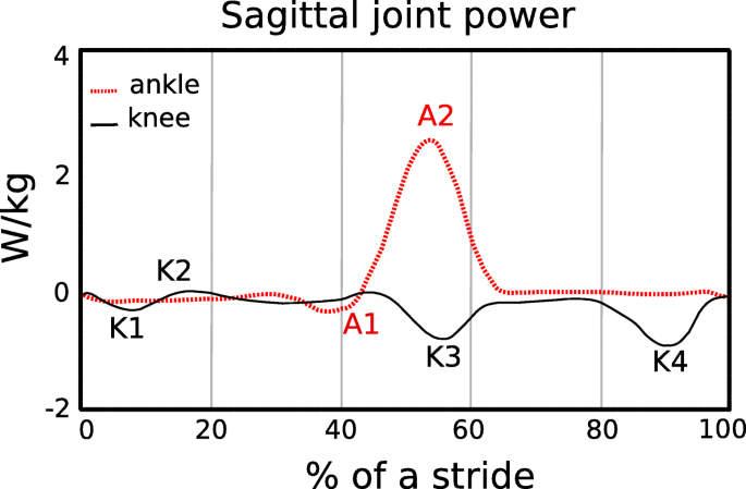
The muscles surrounding the ankle joint are primarily responsible for absorbing/producing power to facilitate the redirection of the center of mass during the step-to-step transition [11]. Over a stride, the muscles surrounding the knee joint dissipate or absorb/store net negative mechanical power and work, whereas the muscles surrounding the ankle and hip joints generate net positive mechanical power and work [12]. Negative and positive peaks in joint power indicate when mechanical energy is absorbed and generated, respectively, during a stride (Fig. 1). Negative peak power indicates eccentric contraction of the ankle plantar-flexor muscles from heel-strike through tibial progression, knee extensor muscles during heel-strike, rectus femoris during late stance, and biceps femoris during late stance (Fig. 1). Positive power regions primarily correspond to concentric contraction of the ankle plantar-flexor muscles during late stance, knee extensor muscles during early stance and hip flexor muscles during early swing phase. All these muscle contractions incur a metabolic cost. Thus, an exoskeleton that stores energy corresponding with the eccentric contraction of the knee extensor muscles and returns this energy during the concentric contraction of the ankle plantar-flexor muscles could decrease the metabolic cost of walking.

Fig. 1
figure1
Average sagittal plane ankle (A), and knee (K) joint mechanical power during level ground walking at 1.10 m/s over a stride for one leg, starting at heel strike. Data are from a previous study [13]. Negative peak power regions for the ankle and knee joints are denoted as A1 and K1, K3, and K4, respectively. Mechanical power and thus energy, is dissipated/absorbed during negative ankle (A1) and knee (K4) joint minimums [11]. At ~ 35–40% of the stride, the ankle plantar-flexors contract eccentrically to control ankle joint dorsiflexion. During terminal swing (K4), the hamstrings contract eccentrically to slow the speed of the swinging leg and avoid knee hyperextension just prior to the subsequent heel-strike (~ 90% of the stride). Positive mechanical power regions are labelled as A2 and K2 and correspond to the concentric contraction of the ankle plantar-flexors during late stance and the knee extensors during early stance, respectively
In order to reduce the metabolic cost of walking, use of an exoskeleton should not alter kinematic gait parameters such as stride length and step width, or kinetic parameters such as ground reaction forces. Previous studies have shown that when people walk with stride lengths and stride frequencies different from preferred, the metabolic cost of walking increases [14,15,16]. Walking speed is the product of stride length and stride frequency. At a fixed walking speed, the relationship between stride frequency and metabolic cost is represented by a U–shaped curve with the minimum metabolic cost corresponding to the preferred stride frequency [17]. Similarly, previous studies show that when humans walk with wider or narrower step widths compared to preferred, their metabolic cost increases [18,19,20]. Step width indicates the lateral distance between the midlines of the feet [21]. At a fixed walking speed, metabolic cost increases with the square of step width [19]. Thus, use of an exoskeleton that results in changes to stride length, stride frequency and step width compared to preferred could increase the metabolic cost of walking.
The development of wearable devices such as exoskeletons has been motivated by the challenge to reduce the metabolic cost of walking. In 1890, Nicholas Yagn conceptualized and received a patent for the first exoskeleton for assisting walking, running, and jumping using pneumatically powered gas bags [1]. Since then, many investigators have developed electrically powered or battery-powered lower limb exoskeletons for medical applications, neurorehabilitation therapy, augmentation, and military use [2, 22,23,24,25]. Most of the recent powered exoskeletons use actuators to provide assistance at the ankle joint during powered plantarflexion at the end of the stance phase of walking [25,26,27,28,29,30]. Specifically, use of powered exoskeletons has reduced the muscle activity and lower limb joint work needed by the user during level-ground walking compared to wearing the exoskeleton with the power turned off. Together with the timing of the assistance, the weight of these devices (12 kg to 38 kg [22]) may be one of the reasons why use of a powered exoskeleton does not decrease metabolic cost compared to normal walking without any wearable system [24].
With new methodological innovations, current research shows that exoskeletons can improve the metabolic cost of walking. Malcolm et al. [30] used optimal actuation timing predicted by a mathematical model combined with a tethered electrically-powered exoskeleton that assists ankle plantarflexion, and found that use of the exoskeleton reduced the metabolic cost of walking at 1.38 m/s by 6.0 ± 2.0% (mean ± SD) compared to walking without the exoskeleton. Mooney et al. [25] developed a battery-powered exoskeleton that utilizes a mathematical model to control the magnitude of positive mechanical power provided by the exoskeleton during ankle powered plantarflexion, and reduced metabolic cost by 8 ± 3% compared to walking without an exoskeleton at 1.5 m/s. Thus, through the optimal timing and magnitude of applied power, use of a powered exoskeleton can reduce the metabolic cost of walking.
The way that an exoskeleton is attached to a person can affect the assistance provided to the person and thus the metabolic cost of walking. Panizzolo et al. [31] have investigated the use of an exosuit equipped with compliant textiles that provide assistance instead of rigid structures, such as those used in other powered exoskeletons. This approach aimed to improve the interface between the exoskeleton and the body [32] and reduce the mass on distal body segments to have less of an effect on metabolic cost [33, 34]. In particular, the exosuit was designed to provide assistance during both ankle joint plantarflexion at the end of the stance phase and hip joint flexion during the early swing phase. Using this exosuit, net metabolic power in the powered condition was 14.2 ± 6.1% lower than in the unpowered condition, but net metabolic power was not reduced with respect to normal walking at 1.5 m/s.
Use of passive-elastic exoskeletons has reduced the metabolic cost of walking by enhancing the mechanism of elastic energy storage and return at the ankle joint, with springs in parallel to the Achilles tendon [3, 25]. Recent studies demonstrate that storing and returning elastic energy during the phases of ankle joint negative and positive mechanical power can significantly reduce metabolic cost [3, 35]. Collins et al. [3] has shown that use of a passive-elastic exoskeleton in parallel with the ankle joint that stores and returns energy during the negative and positive phases of ankle joint power (Fig. 1) reduced metabolic cost by 7.2 ± 2.6% (mean ± SD) compared to walking without an exoskeleton at 1.25 m/s. Many others have investigated how use of a passive-elastic exoskeleton, which does not require an external power supply and is not equipped with sensors or actuators, affects walking [3, 36,37,38,39]. Panizzolo et al. [3] demonstrated that it is possible to reduce the metabolic cost of walking by more than 3% with a passive device that assists the hip joint compared to normal walking. Rome et al. [38] found that use of rubber bands in parallel with the hip joint can reduce the metabolic costs of carrying loads during walking, whereas Dean et al. [39] has shown that the use of a two-joint passive-elastic exoskeleton that works in parallel with the hip and knee joints can reduce the activity of lower limb muscles compared to normal walking [39].
We aimed to determine if a passive-elastic exoskeleton in parallel with the knee and ankle joints could reduce the metabolic cost of walking. We built an exoskeleton that stores energy from knee extension during the late leg swing phase, which corresponds to negative peak knee power (Fig. 1, K4) since it represents the greatest magnitude of energy absorption/storage during the stride [12]. Then, we designed the exoskeleton to release the energy stored from knee extension to assist ankle powered plantarflexion, which corresponds to positive peak ankle power during late stance (Fig. 1, A2). We designed our experiments to test three hypotheses. First, we hypothesized that the use of a passive-elastic exoskeleton that resists knee extension during the late leg swing phase would reduce metabolic power during level-ground walking compared to walking without an exoskeleton. Second, we hypothesized that the use of a passive-elastic exoskeleton that stores energy from knee extension during the late leg swing phase and returns energy for ankle powered plantarflexion during late stance would reduce metabolic power during level-ground walking compared to walking without an exoskeleton. Third, we hypothesized that the use of a passive-elastic exoskeleton would not change stride length, ground contact time, peak ground reaction forces, and step width during level ground walking compared to walking without an exoskeleton.

Material and methods

Participants

Eight healthy subjects [4 M and 4 F, mean ± SD age: 25 ± 3 years, mass: 73 ± 15 kg, height: 174 ± 10 cm, standing leg length: 83 ± 5 cm] participated in the study. We measured their leg lengths from the greater trochanter to the medial malleolus and averaged the right and left leg lengths. All subjects gave informed written consent before participating according to the University of Colorado Boulder Institutional Review Board.
We measured metabolic rates, ground reaction forces, and center of pressure while subjects walked on a dual-belt force measuring treadmill with the exoskeleton springs disengaged (no springs), engaged in parallel with the knee only, engaged in parallel with the knee and ankle, engaged in parallel with the knee only but with a longer engagement rope length, and engaged in parallel with the knee and ankle but with a longer engagement rope length, and without the exoskeleton.

Description of the exoskeleton

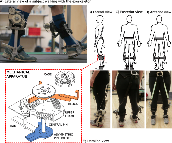
We custom-made the passive-elastic exoskeleton, which consists of a lightweight aluminum frame secured to the lower leg with a modified knee brace (Ottobock HealthCare LP, Austin, US). The exoskeleton is equipped with a mechanical apparatus comprised of six parts (Fig. 2, panel e). The primary frame has a central pin that is fixed on the external side of a modified knee brace, and all the other parts are attached to this frame. An asymmetric pin holder includes two small pins and rotates around the central pin, and an upper frame is fixed to the primary frame and holds the system of springs and a pawl. A block, with three arms and a ratchet wheel rotates about the central pin and compresses the system of springs on the upper frame. A case, which contains a spiral spring is attached distally on the longest arm of the block. In addition, the exoskeleton includes two inextensible ropes (r1, r2) that are fixed proximally on the anterior and posterior side of a belt worn by the subject positioned just above the iliac spines and glutei (Fig. 2), and distally to the exoskeletal mechanical apparatus on the pawl (r1) and the asymmetric pin holder (r2). Specifically, r2 is comprised of two parts (Fig. 2b). The superior part consists of a 4 cm wide piece of nylon webbing that lies posteriorly over the middle of the gluteus, whereas the lower part is a nylon rope that is attached to the mechanical apparatus. 4 cm wide pieces of nylon webbing were used to increase the surface area on the skin and prevent potential discomfort related to the pressure exerted by the rope on the skin. A third inextensible rope (r3) that originates from the exoskeletal case, is attached to the ankle frame surrounding the subject’s heel (Fig. 2a). The total mass of the exoskeleton is ~ 1.4 kg per leg.

Fig. 2
figure2
a Lateral view of a subject walking on the treadmill while using the exoskeleton just as the rope (r3) provides plantarflexion assistance to the trailing leg at the end of the stance phase. b Lateral, c Posterior, and d Anterior views of the exoskeleton attached to the body and corresponding pictures of a subject wearing the exoskeleton with ropes attached from the anterior belt to the apparatus (r1), posterior belt to the apparatus (r2) and apparatus to the ankle frame (r3). E) An exploded view of the mechanical apparatus. We designed the upper frame so that it could engage up to three linear springs. We used two springs during the experimental sessions in order to attain an angular spring stiffness of 17.45 Nm/rad. The mechanical apparatus is comprised of a frame that anchors the upper frame to the braces on the shank. That frame provides a central pin for the rotation of the asymmetric pin holder and block. The case rotates around a pin (not shown), fixed on the longer arm of the block
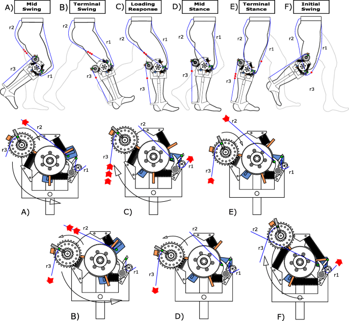
We 3D printed plastic ratchets and a pawl that were attached to the lateral portion of the brace and allowed energy from the movement of the knee to be stored through compression of the metal springs. The exoskeleton is attached to each leg, but we describe the effects of the exoskeleton for the right leg (Fig. 3). At the end of the swing phase, after hip extension is maximal, r2 is tensioned and rotates the asymmetric pin holder counter-clockwise (Fig. 3a), which pushes the block against the system of springs and compresses them (Fig. 3b). At the same time, the 3D-printed pawl engages the ratchet wheel to keep the spring compressed and prevent the clockwise rotation of the block (Fig. 3b, c, d). As r2 slackens, the asymmetric pin holder returns to its original position and the knee can freely flex from the loading response through mid-stance without any interaction with the engaged mechanical apparatus. Prior to the beginning of the experimental trial, we set the length of r2 to ensure it only tensioned in late swing, at maximum hip flexion. As the shank moves forward, due to its inertia, the tension in rope r2 provides a force that compresses the springs and allows the exoskeleton to decelerate the shank prior to heel strike. Also, the biological ankle’s motion is not affected by the exoskeleton due to a spiral spring inside the case. This spring allows the case to rotate from mid-swing through mid-stance (Fig. 3a, b, c, d), while r3 remains slack and does not interfere with the motion of the ankle. r3 only undergoes tension during late stance, when the hip extends, and the case rotation is locked. During this phase, just prior to powered plantarflexion, r1 is extended along the anterior side of the upper leg, disengages the pawl, and releases the compressed springs (Fig. 3e). The pawl disengagement allows the system of springs to release the stored energy; the block rotates clockwise lifting the case attached to its longer arm, which pulls up on r3 and assists the ankle joint during the push–off phase (Fig. 3f). The posterior case, attached to the longer arm of the block, rotates freely during a stride except for at the end of the stance phase, when the central ratchet is released. Ankle joint plantarflexion is therefore assisted by the elastic energy return of the exoskeleton. The exoskeleton does not constrain the biological range of motion of the ankle joint, and it is positioned at the same height and just lateral to the ankle, which connects the brace and heel frame of the exoskeleton. The ankle frame does not include a bearing but is designed to have low friction and the lever arm of the ankle frame is approximately 0.125 m.

Fig. 3
figure3
Engagement and disengagement of the exoskeleton mechanical apparatus during different phases of a walking stride. The upper portion of the figure shows the action of the exoskeleton during a stride and the lower portion provides a detailed view of the knee mechanism. Red arrows indicate the direction that ropes r1, r2 and r3 are moving during a specific phase (in panel E, red arrows are also used to indicate the extension of the linear springs). a During Mid-Swing, r2 begins to stretch due to knee extension and pulls the central pin. This knee extension movement continues until (b) Terminal Swing, and results in the rotation of the block which, in turn, compresses the springs. r3 also undergoes tension and causes the counterclockwise rotation of the case during Terminal Swing (considering the lateral side of the right leg). c During the Loading Response, both r2 and r3 become slack as the hip extends, which causes clockwise rotation of the central pin and case, respectively. d During Mid-Stance, r3 is stretched again as the hip rotates over the ankle and the case is locked so that there is no additional counterclockwise rotation. e During Terminal Stance, r1 is stretched due to hip extension, which pulls the pawl out of the ratchet, and allows the linear springs to extend and release their stored elastic energy. In this way, the block rotates clockwise and pulls on r3, which provides assistance during ankle powered plantarflexion. f At the subsequent Initial Swing only r1 is still stretched, while all of the other components of the device are disengaged


Thursday, May 7, 2020

A new lower limb portable exoskeleton for gait assistance in neurological patients: a proof of concept study

Even looking at the website I could get nothing if this does dorsiflexion or preventing ankle rolling, but it does say it is contraindicated for severe spasticity. Seems to only be available in Madrid. Road trip,(Whoops, COVID-19). I would be concerned that this is actually preventing real recovery like AFOs do, but it does allow your hospital to get you out the door faster and declare you are recovered.

Website here: Marsi Active Knee 


A new lower limb portable exoskeleton for gait assistance in neurological patients: a proof of concept study


Abstract

Background

Few portable exoskeletons following the assist-as-needed concept have been developed for patients with neurological disorders. Thus, the main objectives of this proof-of-concept study were 1) to explore the safety and feasibility of an exoskeleton for gait rehabilitation in stroke and multiple sclerosis patients, 2) to test different algorithms for gait assistance and measure the resulting gait changes and 3) to evaluate the user’s perception of the device.

Methods

A cross-sectional study was conducted. Five patients were recruited (4 patients with stroke and 1 with multiple sclerosis). A robotic, one-degree-of-freedom, portable lower limb exoskeleton known as the Marsi Active Knee (MAK) was designed. Three control modes (the Zero Force Control mode, Mode 1 and Mode 3) were implemented. Spatiotemporal gait parameters were measured by the 10-m walking test (10MWT), the Gait Assessment and Intervention Tool (G.A.I.T.) and Tinetti Performance Oriented Mobility Assessment (gait subscale) before and after the trials. A modified QUEST 2.0 questionnaire was administered to determine each participant’s opinion about the exoskeleton. The data acquired by the MAK sensors were normalized to a gait cycle, and adverse effects were recorded.

Results

The MAK exoskeleton was used successfully without any adverse effects. Better outcomes were obtained in the 10MWT and G.A.I.T. when Mode 3 was applied compared with not wearing the device at all. In 2 participants, Mode 3 worsened the results. Additionally, Mode 3 seemed to improve the 10MWT and G.A.I.T. outcomes to a greater extent than Mode 1. The overall score for the user perception of the device was 2.8 ± 0.4 95% CI.

Conclusions

The MAK exoskeleton seems to afford positive preliminary results regarding safety, feasibility, and user acceptance. The efficacy of the MAK should be studied in future studies, and more advanced improvements in safety must be implemented.


Wednesday, September 13, 2017

Wearable sensors to predict improvement following an exercise intervention in patients with knee osteoarthritis

These sensors should be able to be used to diagnose EXACT problems in stroke knees and then prescribe EXACT stroke protocols to correct those problems. This is so fucking simple. Objectively diagnose the problem, create a stroke protocol that fixes the problems, distribute that information worldwide.  And stroke survivors recover.

Wearable sensors to predict improvement following an exercise intervention in patients with knee osteoarthritis


Journal of NeuroEngineering and Rehabilitation201714:94
Received: 7 February 2017
Accepted: 7 September 2017
Published: 12 September 2017

Abstract

Background

Muscle strengthening exercises consistently demonstrate improvements in the pain and function of adults with knee osteoarthritis, but individual response rates can vary greatly. Identifying individuals who are more likely to respond is important in developing more efficient rehabilitation programs for knee osteoarthritis. Therefore, the purpose of this study was to determine if pre-intervention multi-sensor accelerometer data (e.g., back, thigh, shank, foot accelerometers) and patient reported outcome measures (e.g., pain, symptoms, function, quality of life) can retrospectively predict post-intervention response to a 6-week hip strengthening exercise intervention in a knee OA cohort.

Methods

Thirty-nine adults with knee osteoarthritis completed a 6-week hip strengthening exercise intervention and were sub-grouped as Non-Responders, Low-Responders, or High-Responders following the intervention based on their change in patient reported outcome measures. Pre-intervention multi-sensor accelerometer data recorded at the back, thigh, shank, and foot and Knee Injury and Osteoarthritis Outcome Score subscale data were used as potential predictors of response in a discriminant analysis of principal components.

Results

The thigh was the single best placement for classifying responder sub-groups (74.4%). Overall, the best combination of sensors was the back, thigh, and shank (81.7%), but a simplified two sensor solution using the back and thigh was not significantly different (80.0%; p = 0.27).

Conclusions

While three sensors were best able to identify responders, a simplified two sensor array at the back and thigh may be the most ideal configuration to provide clinicians with an efficient and relatively unobtrusive way to use to optimize treatment.