Use the labels in the right column to find what you want. Or you can go thru them one by one, there are only 33,819 posts. Searching is done in the search box in upper left corner. I blog on anything to do with stroke. DO NOT DO ANYTHING SUGGESTED HERE AS I AM NOT MEDICALLY TRAINED, YOUR DOCTOR IS, LISTEN TO THEM. BUT I BET THEY DON'T KNOW HOW TO GET YOU 100% RECOVERED. I DON'T EITHER BUT HAVE PLENTY OF QUESTIONS FOR YOUR DOCTOR TO ANSWER.
Changing stroke rehab and research worldwide now.Time is Brain!trillions and trillions of neuronsthatDIEeach day because there areNOeffective hyperacute therapies besides tPA(only 12% effective). I have 523 posts on hyperacute therapy, enough for researchers to spend decades proving them out. These are my personal ideas and blog on stroke rehabilitation and stroke research. Do not attempt any of these without checking with your medical provider. Unless you join me in agitating, when you need these therapies they won't be there.
What this blog is for:
My blog is not to help survivors recover, it is to have the 10 million yearly stroke survivors light fires underneath their doctors, stroke hospitals and stroke researchers to get stroke solved. 100% recovery. The stroke medical world is completely failing at that goal, they don't even have it as a goal. Shortly after getting out of the hospital and getting NO information on the process or protocols of stroke rehabilitation and recovery I started searching on the internet and found that no other survivor received useful information. This is an attempt to cover all stroke rehabilitation information that should be readily available to survivors so they can talk with informed knowledge to their medical staff. It lays out what needs to be done to get stroke survivors closer to 100% recovery. It's quite disgusting that this information is not available from every stroke association and doctors group.
Showing posts with label don't have a functioning stroke doctor. Show all posts
Showing posts with label don't have a functioning stroke doctor. Show all posts
There
is a need for novel treatments that lower the risk of major adverse
cardiovascular events and secondary inflammatory brain injury after an
intracerebral hemorrhage (ICH).
METHODS:
We
performed a double-blind, placebo-controlled, pilot randomized clinical
trial at 11 centers across Canada to determine the feasibility of
testing colchicine after an acute ICH. We recruited adults presenting
within 48 hours of ICH onset with vascular neuroimaging evidence or risk
factors for atherosclerosis. Participants were randomized to oral
colchicine 0.5 mg daily or placebo and followed to a common study
termination date. The primary feasibility outcome was the recruitment
rate (participants/center per year). Secondary feasibility outcomes
included retention of participants at 6 months and medication adherence
at 12 months. This trial is registered (ClinicalTrials.gov ID:
REGISTRATION: URL: https://www.clinicaltrials.gov; Unique identifier: NCT05159219).
RESULTS:
Between
August 2022 and March 2024, 52 participants were allocated to
colchicine 0.5 mg daily and 48 participants to placebo daily.
Participants were, on average, 68 years old, and 60% were male. The mean
time from ICH onset to randomization was 35 hours. The average
recruitment rate was 8.9 participants/site per year. Retention at 6
months was 92% (colchicine 91% versus placebo 93%). Following
randomization, 14 participants (27%) in the colchicine group and 13
participants (27%) in the placebo group permanently discontinued the
study drug. Excluding participants who died or permanently discontinued
the study intervention, 12-month medication adherence was 97%, with
similar rates between the colchicine and placebo groups (100% versus
93%). We detected no difference in predefined exploratory efficacy or
safety end points between the 2 groups over the median follow-up time of
364 days.
CONCLUSIONS:
It
is feasible to test low-dose colchicine after an acute ICH. Future
randomized clinical trials should account for the high rates of early
permanent study drug discontinuation in this patient population.
Thromboinflammation
is believed to worsen neurological outcomes and contribute to ischemic
vascular events in patients with small vessel disease, including
intracerebral hemorrhage (ICH). The management of patients with ICH is
challenging and requires balancing the benefit of ischemic vascular
preventive treatments against any possible increase in bleeding risk.1 ICH survivors have a constant risk of rebleeding, with the annual rate of ICH recurrence ranging from 1.3% to 7.4%.2
Because of their preexisting vascular risk factors and comorbidities,
ICH survivors are at considerably higher risk of major adverse
cardiovascular events (MACE),3–5 which is further aggravated by the cessation of antithrombotic medications for significant periods after ICH.6
These findings highlight the need for novel treatments that lower the
high vascular risk over the long term in ICH survivors, particularly in
the early post-ICH period when patients are not receiving antithrombotic
medications.
Randomized-controlled clinical
trials (RCTs) have established that colchicine reduces the risk of MACE
(risk ratio, 0.73 [95% CI, 0.65–0.90]) and ischemic stroke (risk ratio,
0.73 [95% CI, 0.58–0.90]) in patients with a history of coronary artery
disease, with no increase in ICH risk.7
Results from an observational study further suggest that patients with
diabetes receiving colchicine treatment have lower risks of stroke, with
the risk reduction for both ischemic and hemorrhagic stroke being
proportionate to the duration of colchicine use.8
In an experimental murine collagenase-induced ICH model, we provided
proof of concept for the safety of oral colchicine after ICH, detecting
no increase in hematoma volume and less perihematomal brain inflammation
at a scaled dose equivalent to the 0.5 mg daily that was used in trials
testing colchicine in patients with coronary artery disease, when
compared with placebo.9
We
initiated the CoVasc-ICH trial (Colchicine for the Prevention of
Vascular Events After an Acute Intracerebral Hemorrhage) to determine
the feasibility of testing colchicine for reducing the risk of MACE and
secondary inflammatory brain injury following an acute spontaneous ICH.
How will your competent? doctor GUARANTEE THIS IS OCCURRING?
Your competent? doctor can explain all the functions and benefits of microglia in all this research! Oh NO, you don't have a functioning stroke doctor, do you?
Summary: Researchers
revealed that aging leads to a massive, previously unrecognized
migration of peripheral blood stem cell-derived immune cells into the
human brain. The team dismantled the long-held dogma that the brain’s
immune environment is an isolated system populated solely by resident
microglia established before birth.
Key Facts
Challenging Neuroimmunology Dogma:
Replaces the longstanding model that brain microglia self-renew
exclusively inside the brain without peripheral input, proving that the
human brain receives continuous immune cell reinforcements from bone
marrow during aging.
DNA Mutation Lineage Tracing:
The team mapped shared somatic mutations in aging blood stem cells and
brain tissue, establishing definitive lineage proof that brain microglia
share a common progenitor with circulating blood cells.
Species-Specific Human Aging Phenomenon:
The migration and functional transformation of peripheral blood cells
into microglial phenotypes is observed in humans but does not occur in
standard laboratory animal models like mice or non-human primates.
Clonal Hematopoiesis Connection:
Builds on prior findings that specific mutant blood stem cell clones
(clonal hematopoiesis) reduce Alzheimer’s risk, showing that peripheral
cells actively infiltrate the central nervous system to alter disease
trajectory.
Platform for Brain Immunotherapies:
Demonstrates that engineered peripheral immune cells can naturally home
to the brain, enabling therapeutic delivery mechanisms designed to
clear amyloid-beta and tau aggregates prior to symptom onset.
Source: Stanford
The
brain’s immune system has long been thought to exist independently from
the rest of the body, complete with its own specialized immune cells
and a blood-brain barrier that limits what can travel into the brain.
Now,
Stanford researchers have found that aging brings with it a large
influx of immune cells into the brain, a discovery that not only upends
current thinking but could also open new avenues for treating
neurological disease.
Peripheral immune cells enter the aging human brain and differentiate into functional microglia. Credit: Neuroscience News
The
researchers, whose out-of-the-box work was supported in part by
the Knight Initiative for Brain Resilience at the Wu Tsai Neurosciences
Institute, described their results July 30, 2026 in the journal Nature.
“We
usually think of the brain as a closed system,” said Julia Belk, a
postdoctoral scholar in pathology at Stanford Medicine and first author
on the new study. “What we found is that actually a lot of immune cells
enter the human brain during aging.”
New to neuroscience
Belk
first developed an interest in the brain as a graduate student in the
Department of Computer Science at Stanford Humanities and Sciences. At
the time, she was also a trainee with Sarafan ChEM-H’s Chemistry/Biology
Interface Predoctoral Training Program, which she described as
formative in helping her develop an approach merging basic science,
computer science, and medicine.
That
program turned out to be a stepping stone to a collaboration
with Siddhartha Jaiswal, a senior author on the new study, an associate
professor of pathology at Stanford Medicine, and a member of
the Institute for Stem Cell Biology and Regenerative Medicine. After
analyzing genetic data on thousands of people, some tracked over the
course of several decades, the team showed that people with specific
clones of immune cells arising from mutated blood stem cells were much
less likely than others to get Alzheimer’s – hinting that those cells
might interact with the brain.
The
team went on to show that some of these mutant cells could somehow make
their way into the brain. Although the mutations involved – known
as clonal hematopoiesis of indeterminate potential – only affect a
minority of people, the finding got the researchers wondering whether
this influx of immune cells was occurring in the broader aging
population.
“Unlike most immune cells,
which are continuously replenished by blood stem cells from the bone
marrow, immune cells in the brain were presumed to renew themselves
throughout the lifespan without contribution from outside the brain,”
said Jaiswal. “Our first study showed that this might not always be the
case.”
That idea ran counter to what
many others believed – that the brain’s specialized immune cells, called
microglia, are all present in the brain from birth, and that no other
immune cells migrate in. Now, Belk and colleagues wondered whether such
migration was not only possible, but perhaps even a common occurrence in
aging.
A cell’s journey
The
idea that immune cells in the blood might have something to do with
Alzheimer’s was unusual – and controversial – so in 2022 Jaiswal and
colleagues sought support from the Knight Initiative for Brain
Resilience, which backs research aimed at reshaping the study of brain
resilience and neurodegenerative disease.
Supported
in part by that Knight Initiative Innovation Award, Belk, Jaiswal, and
co-senior author Howard Chang, the Virginia and D. K. Ludwig Professor
of Cancer Research and a professor of genetics at Stanford Medicine, set
out to see why peripheral immune cells boost Alzheimer’s resilience –
but first, they needed to establish whether microglia really do get
reinforcements from immune cells in the blood.
To
answer the question, they turned to human brain samples. They worked
with samples from the Stanford Rapid Autopsy Center, led by co-author
Jody Hooper, a professor of pathology at Stanford Medicine, and the
University of Washington’s Alzheimer’s Disease Sequencing Project.
Those
efforts gather both blood and post-mortem brain tissue samples from
people with and without Alzheimer’s, giving the researchers a rare
opportunity to compare blood samples directly to post-mortem brain
tissue.
Still, the team needed a way
to determine where immune cells in the brain came from. Since immune
cells are constantly dividing, this means tracing cells’ lineage back to
learn whether their ancestors came from the original population of
microglia – present in the brain from birth – or from blood stem cells
in the bone marrow later in life.
The
team’s key insight was to study DNA from immune cells in both blood and
brain samples, then use shared mutations to trace their lineages – not
unlike a consumer ancestry testing service. Mutations build up randomly
in our blood stem cells as we age, and the immune cell progeny of those
stem cells inherit those mutations.
This
means that two immune cell populations with the same mutations almost
certainly share a common origin, Belk explained. “If we see the same
mutations in the blood and in the brain’s microglia, then we can be very
confident that immune cells in the brain are descendants of those
immune cells in the blood,” she said.
Building
on that idea and drawing on tools from their 2023 study, Belk and
colleagues compared immune cells from blood and brain samples and showed
there was a match: immune cells had indeed moved from the body into the
brain as early as middle age. Additional studies showed that these
peripheral immune cells transformed into specialized microglia in the
brain. That’s something that doesn’t appear to happen in other species
like mice or non-human primates.
Hints of a new approach
In
addition to upending previous expectations, the discovery could also be
an exciting new avenue for the development of brain-focused
immunotherapies, Belk said. “Now that we know that these immune cells
actually can get into the brain, we can think about all kinds of new
engineering strategies to have those peripheral immune cells do useful
things.”
For example, researchers
could imagine engineering immune cells that can chew up amyloid and tau
aggregates associated with neurodegenerative disease, then deliver these
to people as a preventative measure before the aggregates start to
build up.
Since many of the microglia
in aging humans seem to be derived from blood stem cells, the discovery
also opens up a new line of investigation into how factors that
influence blood or bone marrow cells could impact the brain. “Our
findings suggest that the life history of blood stem cells could
influence the risk of brain diseases by altering the microglia,” Jaiswal
said.
Beyond that, Belk highlighted
the excitement of identifying a new aspect of human neuroscience. “I
think this is exciting because this is also a uniquely human feature of
aging that we had no idea about.”
Key Questions Answered:
Q: How did researchers prove that microglial cells in post-mortem brains originated from blood stem cells?
A:
As human blood stem cells age, they naturally accumulate unique,
harmless somatic mutations in their DNA. By sequencing DNA from both
peripheral blood and brain tissue, the researchers identified identical
somatic mutations in both blood cells and brain microglia, providing
genetic proof of a shared lineage.
Q: Why was this peripheral immune infiltration not discovered previously in laboratory animal research?
A:
This phenomenon appears to be a uniquely human feature of aging.
Detailed comparative analyses showed that peripheral blood cells do not
infiltrate the brain and convert into microglia in standard model
organisms such as mice or non-human primates.
Q: What are the therapeutic implications of peripheral immune cells entering the aging brain?
A:
Because peripheral blood cells naturally migrate into the aging brain,
scientists can potentially engineer a patient’s peripheral immune cells
to cross the blood-brain barrier and perform specific protective
functions, such as clearing toxic amyloid-beta and tau protein
aggregates before neurodegeneration progresses.
Editorial Notes:
This article was edited by a Neuroscience News editor.
Journal paper reviewed in full.
Additional context added by our staff.
About this aging and neuroscience research news
Author: Nathan Collins Source: Stanford University Contact: Nathan Collins – Stanford University Image: The image is credited to Neuroscience News
Original Research: Open access. “Somatic mutations reveal the ontogeny of microglia in human aging”
by Julia A. Belk, Yaowen Zhang, Emily E. Reilly, Quanming Shi, Daniel
Dan Liu, Nicole Womack-Gambrel, Maarten van der Linde, Lisa Ma,
Debasmita Paul, Alejandro Medina Enciso, Raja Kalluru, Jacob Weiss, Rui
Li, Anna E. Eastman, Chunfang Zhu, Arnav Chakravarthy, Syed Bukhari,
Dipabarna Bhattacharya, Suyash Raj, Daniel Richard, Simone Brioschi,
Matthew R. Chrostek, Daniel C. Nachun, Christopher M. Arends,
Jayakrishnan Gopakumar, Isak W. Tengesdal, Ademar Bynum, Shaneice
Mitchell, Katalin Sandor, Wenxi Zhang, Badri N. Vardarajan, Inma Cobos,
Donald E. Born, Robert B. West, Anne Brunet, Marco Colonna, Krishna L.
Bharani, Hannes Vogel, Thomas J. Montine, Caitlin S. Latimer, Irving L.
Weissman, Magdalena Matusiak, Jody E. Hooper, C. Dirk Keene, Howard Y.
Chang & Siddhartha Jaiswal. Nature DOI:10.1038/s41586-026-10939-0
Dementia risk was reduced in association with daytime light exposure above 1000 lux, while further reduction in risk was seen in association with longer exposure to brighter light.
HealthDay News — High levels of daytime light exposure are associated with a lower risk for dementia, according to a study based on U.S. data published online June 24 in General Psychiatry. Nana Zheng, Ph.D., from The Affiliated Brain Hospital, Guangzhou Medical University in China, and colleagues examined the associations of daytime and nighttime light exposure with dementia risk in a study involving 87,577 dementia-free participants. Seven-day free-living wrist-worn accelerometry was used to measure daytime and nighttime light exposures.
Overall, 741 participants developed dementia during a median follow-up of 8.1 years. The researchers found that dementia risk was reduced in association with daytime light exposure above 1,000 lux (hazard ratio, 0.84). Further reduction in risk was seen in association with longer exposure to brighter light (e.g., ≥0.70 hour at ≥5,000 lux: hazard ratio, 0.83). Up to 33 percent of the association was mediated by circadian rest-activity rhythms and brain structures. Stronger protective associations were seen in those with high levels of nighttime light exposure, an evening chronotype, or APOE ɛ4 carrier status, with up to a 41 percent risk reduction. Six established dementia predictors were outperformed by <0.70 hours per day of bright daytime light (≥5,000 lux). No significant association was seen for nighttime light with dementia risk. “These findings suggest a targeted approach to mitigate dementia risk by increasing daytime light levels for these populations,” the authors write.
Have your competent? doctor summarize this INTO AN EXACT PROTOCOL! You don't want to do it wrong and your doctor, IF COMPETENT AT ALL, should easily be able to do that. But you don't have a functioning stroke doctor, do you? If your doctor doesn't know about the Merlin app for recognizing bird songs; that's even worse!
A writer’s recent experiences with watching owls helped her recover from a traumatic brain injury—and she’s not alone. New studies are showing how birding can affect brain structure and foster a sense of well-being.
I was 26, thriving in a corporate communications career, flying high. Then, like a bird’s nest in a windstorm, my life collapsed. Suddenly and without warning, I suffered a traumatic brain injury.
Not even a creative writer like me could have scripted a wild plot twist like this. And, little did I know, birds would become a big part of my comeback story.
I rolled my eyes when my neurosurgeon prescribed 90 minutes of walking daily. But walking around my suburban neighborhood in Phoenix, Arizona, became my new reality. So, I walked … and I walked … and I walked.
Behind the scenes, anxiety, stress, and sadness hovered like a cloud. It was hard to control my emotions.
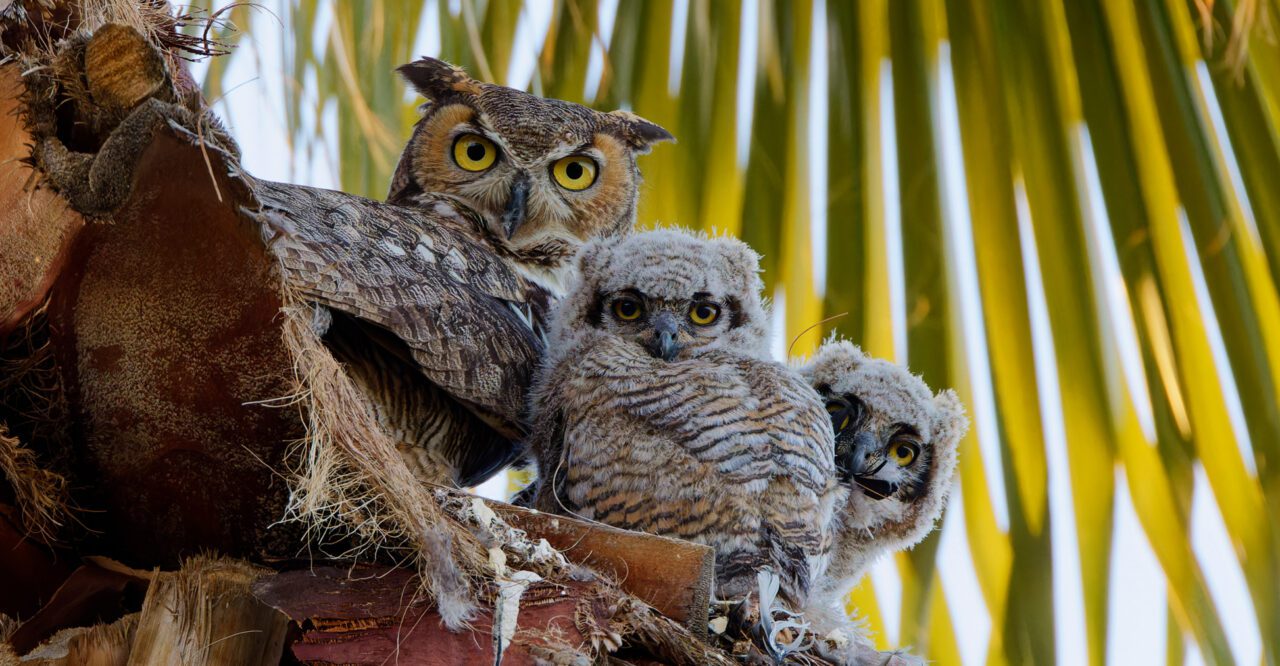
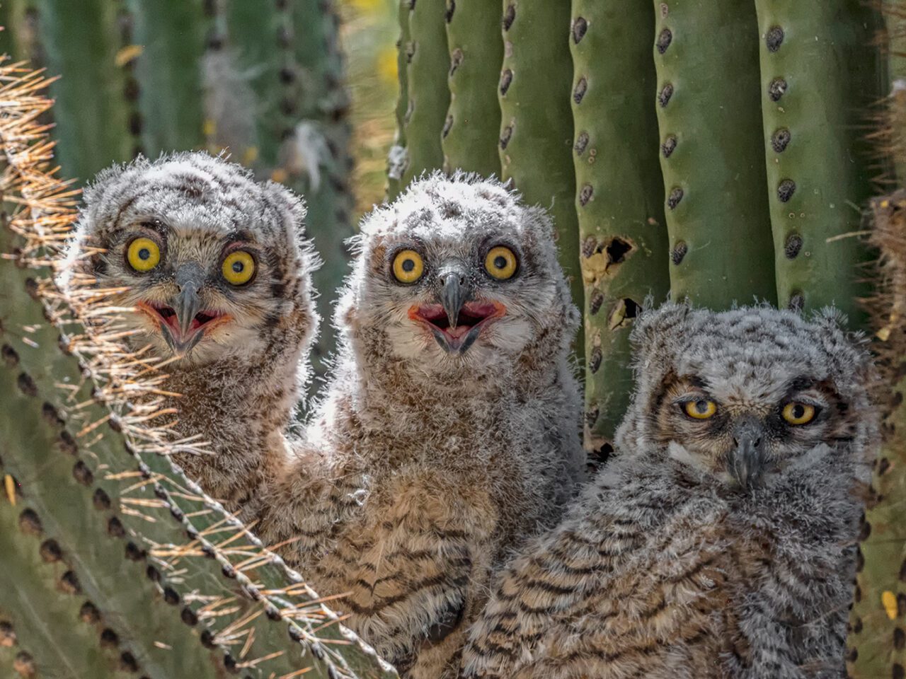
Then, one day on a walk, I spotted a pair of Great Horned Owls and three owlets in a palm tree. Seeing those downy, wide-eyed owlets nestled together stopped me in my tracks. And in that moment, I stopped worrying about my own life and started wondering about theirs.
Learning about the owl family added another purpose to my prescribed walking. I started visiting them every sunrise and sunset—curious, obsessed, and honestly, it just felt better when I was out there. It quickly became part of my daily routine.
Why do I feel so good? Why did I become so attached to birds? Could this be a form of therapy—owl therapy?
It turns out that birds and birding are emerging as a powerful, nature-based prescription for people’s mental well-being, capable of restoring a sense of purpose and inspiring a sense of connection. A new study suggests that building skills in birding may also yield structural benefits in a person’s brain. Research published in February 2026 in the Journal of Neuroscience showed that experienced birders had denser brain matter in the frontoparietal cortex—a region associated with cognition and working memory—than beginners. Birding, wrote the study authors in their research, “leaves a coherent imprint on the brain.”
“Birding strengthens cognitive function by transforming momentary personal experiences into lasting knowledge,” explains Dr. Erik Wing, lead author on the study and a neuroscientist at the Rotman Research Institute in Toronto, Canada.
His studies and work by other researchers in human psychology and social science are showing that birds do more than spark an interest in people’s lives. In fact, birds may help support a person’s cognitive function and mental health across their lifespan.
In his studies at the Rotman Research Institute, one of the world’s preeminent centers for understanding aging and human brain function, Wing explores how deep, accumulated knowledge—such as bird knowledge—changes the brain.
By accumulating sightings and experiences, birders develop complex mental networks as they connect species, behaviors, sounds, and habitats to identify birds. This form of knowledge compounds over time.
“You’re not only learning a new concept—you’re learning something that applies broadly to the entire network,” says Wing. “Everything you learn informs everything else.”
And the benefits to the brain multiply as birders level up their skills. For the study published in the Journal of Neuroscience, Wing and his coauthors conducted MRI brain scans on experienced and beginner birders. The experienced birders—defined primarily by their performance on bird ID tests rather than age alone—had a “tuned cortical network” in their brains, according to the study.
In other words, interconnected learning associated with birding may lead to structural and functional changes through neuroplasticity—the brain’s capacity to rewire and adapt with experience. Wing says that there are multiple routes through which birding can sharpen perception and calm the mind. Identifying subtle patterns—movement, shape, and sound—requires adaptive thinking and integrating our senses.
“Birding combines multiple factors we know support cognitive and mental health—like nature exposure, social interaction, and novelty—into one activity,” Wing says.
And though it demands high-level thinking, birding is also a form of mental rest.
“Birding helps interrupt cycles of stress and rumination by directing attention outward,” Wing says, referencing a 2022 study by German scientists published in the journal Scientific Reports that documented how listening to birdsong reduces anxiety in people. Birding, he says, offers “a moving, engaging focus in the natural world.”
In my case, daily walks for owl therapy were just what I needed to calm my anxious mind. Birding distracted me by giving me space to slow down, stay curious, and change my mental state. I began paying closer attention to what was happening outdoors in my neighborhood, with a growing awareness that deepened my connection to the owls and sparked curiosity about other birds nearby. What once felt like background noise now revealed a world I hadn’t noticed before.
According to Wing, this kind of rapid awakening is often most pronounced in the early stages of birding.
“You progress really fast because you know so little when you start,” he explains, noting that it’s not necessary to put in hundreds of hours up front to feel a sense of reward. Setting simple goals—like seeing 10 different birds in a week—can be both motivating and gratifying.
The benefits of birding experience may also endure across a person’s lifespan. The research published in the Journal of Neuroscience suggests that brain changes associated with birding persist in older experts, who continue to show differences in brain activity and organization compared to their age-matched counterparts. According to Wing, birding engages a variety of brain processes related to memory, attention, and flexible thinking—core components of cognition that are often associated with resilience in aging.
Scientists are also finding that, beyond possibly strengthening cognitive function, birding helps people experience joy, find meaning, and make connections.
Dr. Jenn Lodi-Smith, a professor of psychology at Canisius University in Buffalo, New York, is leading the Spark Bird Project, which collects people’s stories and survey data about how birding sparks transformative experiences that grow into lifelong passions. Since launching the project in 2022, Lodi-Smith has collected more than 500 stories from people about the moment they got hooked on birding. She says the Spark Bird stories are emotionally rich and overwhelmingly positive.
“People describe feelings of joy, curiosity, and pride,” she says. Yet some also reflect on grief or trauma, she says, suggesting that birding can help people process difficult emotions. Stories of recovery, she adds, may highlight the role of birding in emotional healing.
Some days I walked to the owl nest with sadness about my lost career and anxiety about my future. Witnessing the owlets’ wobbly dependence and shaky first flights taught me that recovery is slow and nonlinear—and that accepting support is okay. Their vulnerability and determination helped me let go of rigid expectations, slow down, and find purpose in recovering at my own pace. Gradually, I began to think more positively and see new ways I could be part of the world I shared with the birds—a world bigger than my own struggles.
In Lodi-Smith’s more than 20-year academic career studying identity development—including the psychological construct of Self-Concept Clarity, which describes how clearly defined, consistent, and stable a person’s self-beliefs are—she has found that human mental health isn’t just the absence of stress or illness. It’s the presence of meaning.
“Do people feel a sense of purpose? Do they have a sense of meaning in life?” she says. “One idea we study is generativity—the desire to give back and contribute to the broader world. Birds make us care about birds, but they also make us care about the world we share.”
Lodi-Smith says her research has also explored how the communities that people form can nurture a sense of belonging and identity.
“The birding community offers a space where people can express wonder, awe, and curiosity—emotions that aren’t always encouraged in everyday social roles,” she explains. “Whether a world-renowned ornithologist or a novice, birders share moments of excitement and connection that are deeply meaningful.”
And because birds can be found anywhere that people live, they are a great tool for boosting people’s mental health, says Dr. Katie Holland, a research scientist and social scientist at Virginia Tech University. Holland was a member of the science committee for the State of the Birds 2025 report, and the lead author on a section in that report about birds and mental health that cited studies on how birds ease symptoms of stress, anxiety, and depression in people. According to Holland, those mental health benefits are widely available across every possible demographic group of Americans.
Birding, she says, “can be adapted to diverse landscapes and participants—from those with minimal time or physical ability, to those seeking more elaborate experiences. This inclusivity means the mental health benefits of birding can reach many.”
Holland says the unique flexibility of birding makes it a potentially powerful wellness tool that can be scaled up across communities, at a time of rising need. According to data from the World Health Organization, more than 50% of the world’s population is expected to experience a mental health issue at some point in their lifetime.
“Colleagues of mine at Virginia Tech have also been exploring ways to make birding more accessible for diverse participants and those with disabilities,” she says. “This is important work, and can help make the well-being benefits of this activity available to many more people.”
I am living proof that birds and birding can be a multidimensional mental health tool—one that strengthens cognitive function, restores a sense of purpose, and inspires connection.
And I am continuing to recover. Six years after my traumatic brain injury, I feel more at ease, balanced, and emotionally stable as the clouds of anxiety, stress, and sadness begin to lift—clear signs of my improving mental health. With this progress, I am now working as a freelance writer, photographer, and speaker.
Hearing the owl parents hooting, watching the male deliver food, smiling at the owlets’ first awkward flights and early hunting attempts, and noticing the patterns on their wings as they preened were a few of my most treasured moments from owl therapy. It’s been a powerful force for good in my personal mental health journey—a force rooted in the simple acts of noticing, presence, reconnecting, rebuilding, recalibrating, and reclaiming power. And now when I go birding, I can feel it give me space when I feel stuck, confidence when I feel shattered, and curiosity when I feel uncertain.
It turns out the wildest plot twist in my story wasn’t the injury. It was the ease of recovery, sparked by owls.
I wonder what Great Horned Owls would charge per hour for human therapy? Whatever it is, I’d pay it.
About the Author
Emily Nichols is a bilingual English and Spanish freelance writer, as well as a photographer and speaker. She is a local captain in Phoenix, Arizona, for Birdability, a nonprofit organization that makes birding more accessible and inclusive for people with disabilities and other health concerns. Nichols holds a master’s degree in mass communications from Arizona State University’s Walter Cronkite School of Journalism.