Sulfide-pre-conditioning induces hypoxia tolerance in mice
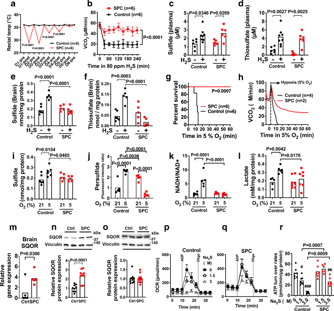
Breathing H2S depresses metabolism and decreases the body temperature of rodents31,32. However, as we previously showed, intermittent breathing of H2S for 5 days makes male wild-type mice tolerant to the hypo-metabolic effects of inhaled H2S (Fig. 1a)33.
Because chronic sulfide exposure might induce enzymes that increase the
capacity to metabolize sulfide, we examined the impact of intermittent H2S breathing for 5 days (sulfide pre-conditioning, SPC) on sulfide metabolism. Mice were studied 24 h after the last H2S exposure (on the 6th day), when the metabolism and body temperature of the mice had completely recovered (Supplementary Fig. 1a). When control (not sulfide pre-conditioned) mice acutely breathed H2S, there was a decrease in whole-body metabolism, as measured by a decreased rate of CO2 production (VCO2) (Fig. 1b). In contrast, in mice that were pre-conditioned with H2S, subsequent acute H2S exposure had no effect on VCO2. Breathing H2S increased plasma levels of sulfide and thiosulfate to a similar extent in control and sulfide pre-conditioned mice (Fig. 1c, d). However, breathing H2S
increased the levels of sulfide and thiosulfate only in the brains of
control mice, but not in the brains of sulfide pre-conditioned mice
(Fig. 1e, f). Thus, sulfide pre-conditioning may induce tolerance to the inhibitory effects of H2S on metabolism by upregulating sulfide catabolism in the brain.
In addition to increased tolerance to breathing H2S, sulfide pre-conditioned mice exhibited marked tolerance to severe hypoxia (5% O2) (Fig. 1g, h and Supplementary Fig. 1b).
To examine the impact of sulfide pre-conditioning on biochemical
changes in the brain during hypoxia, mice were anesthetized with
isoflurane and ventilated with 21% or 5% O2 for 3 min at 37 °C, and brains were harvested and snap-frozen in liquid nitrogen. Breathing 5% O2 decreased brain tissue PO2 below the critical level of 6–8 mmHg within 3 min (Supplementary Fig. 1c). Control mice (not pre-conditioned with H2S) that breathed 5% O2 for 3 min had an acute increase in brain sulfide levels, to an extent similar to that observed after breathing H2S at a dose (80 ppm), which inhibits whole-body metabolism (Fig. 1b, e, i). Breathing 5% O2 also increased brain persulfide levels in control mice (Fig. 1j). Increased levels of sulfide in the brain of control mice were accompanied by an increase in the NADH/NAD+ ratio and lactate levels, indicating impaired oxidative phosphorylation (Fig. 1k, l). In contrast, sulfide pre-conditioned mice breathing 5% O2
for 3 min were protected from sulfide accumulation and decreased
oxidative phosphorylation in the brain. In addition, brain persulfide
levels were decreased in sulfide-pre-conditioned mice breathing 5% O2, suggesting enhanced persulfide consumption during hypoxia (Fig. 1j).
Taken together, the results suggest that sulfide pre-conditioning
prevents accumulation of sulfide and inhibition of oxidative
phosphorylation in the brain of mice after acutely breathing either H2S at 80 ppm or 5% O2.
To
investigate the mechanisms responsible for the inhibition of sulfide
accumulation during hypoxia in mice pre-conditioned with H2S,
we measured the levels of enzymes that synthesize or catabolize
sulfide. Acquired tolerance to acute hypoxia in sulfide pre-conditioned
mice was associated with increased levels of SQOR in the brain, but not
in the heart presumably due to higher baseline SQOR levels in the heart
than in the brain (Fig. 1m–o and Supplementary Fig. 1d).
Increments of brain SQOR levels by sulfide pre-conditioning temporarily
coincided with the acquisition of hypoxia tolerance; sulfide
pre-conditioning for 5 days, but not 2 days, induced hypoxia tolerance
and increased brain SQOR levels (Supplementary Fig. 1e, f).
Levels of other enzymes that metabolize sulfide were not affected by
sulfide pre-conditioning in either brain or the heart (Supplementary
Fig. 2a–c).
Because sulfide pre-conditioning increased SQOR levels in the brain, we
posited that sulfide pre-conditioning upregulates the ability of brain
mitochondria to catabolize sulfide and thereby prevents sulfide-induced
inhibition of oxidative phosphorylation. Sulfide pre-conditioning did
not affect baseline ATP turnover in isolated brain mitochondria as
determined by measuring oxygen consumption rates (OCR). In control mice,
incubation with Na2S (a sulfide donor, that mimics hypoxia)
dose-dependently decreased ATP turnover in isolated mitochondria
obtained from the brain, but not liver that has high basal levels of
SQOR (Fig. 1p and Supplementary Fig. 3a–c). In contrast, the ability of Na2S to depress ATP turnover in the brain mitochondria of sulfide pre-conditioned mice was attenuated (Fig. 1q, r).
Although previous studies suggested that chronic exposure to H2S increases mitochondrial biogenesis34,
sulfide pre-conditioning did not affect the levels of mitochondrial DNA
in the brain or heart of treated mice (Supplementary Fig. 4a). Chronic exposure to hypoxia is known to increase red blood cell mass and the oxygen affinity of hemoglobin35.
However, five days of sulfide pre-conditioning did not affect either
hemoglobin levels or the oxygen dissociation curve of murine red blood
cells. Hydrogen sulfide was previously shown to upregulate
hypoxia-inducible factor-1α (HIF-1α) and one of its canonical targets
vascular endothelial growth factor (VEGF) in vascular endothelial cells
and to activate HIF-1α in C. elegans36,37.
Although sulfide pre-conditioning increased mRNA levels of glucose
transporter-1 (GLUT-1), mRNA levels of VEGF, hemoxygenase-1 (HO-1), and
erythropoietin (EPO), and protein levels of VEGF, GLUT-1, and lactate
dehydrogenase A (LDAH) in the brain did not differ between control and
sulfide pre-conditioned mice at 24 h after the last H2S exposure when mice were exposed to hypoxia (Supplementary Fig. 4b).
Taken together, these observations suggest that sulfide
pre-conditioning confers tolerance to hypoxia via upregulation of SQOR
and sulfide catabolism, rather than as a result of increased
mitochondrial biogenesis, increased oxygen delivery by hemoglobin, or
upregulation of canonical HIF-1α targets.
Sexual dimorphism of sulfide catabolism and hypoxia tolerance
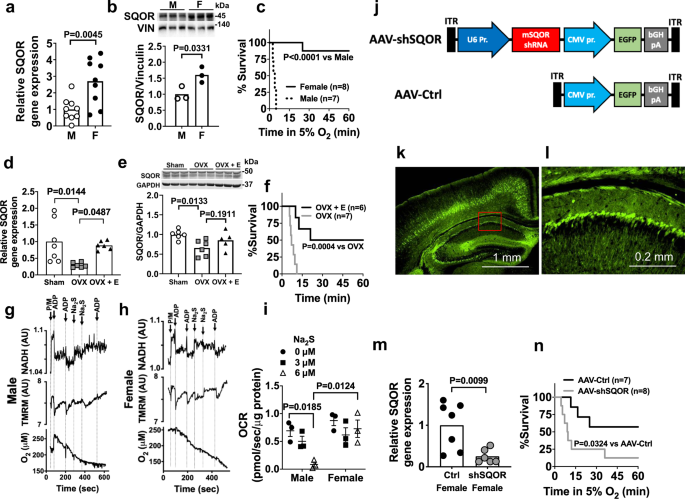
Compared
to the brains of 8-week old male CD-1 mice, the brains of age-matched
female CD-1 mice have ~3-fold and ~1.5 fold higher levels of SQOR mRNA
and protein, respectively (Fig. 2a, b).
To investigate the effects of SQOR levels on the ability of the murine
brain to tolerate acute oxygen shortage, we subjected CD-1 mice to
hypoxia using environmental chambers. The majority of female mice
tolerated breathing 5% O2 for at least 1 h, whereas all male mice died in less than 10 min (Fig. 2c).
Of note, higher SQOR levels in the brain of female mice appear to be
estrogen-dependent. Ovariectomy decreased mRNA and protein levels of
SQOR in the brain and abolished hypoxia tolerance in female mice,
whereas estrogen supplementation restored brain SQOR mRNA levels and
hypoxia tolerance and tended to restore SQOR protein levels in
ovariectomized female mice (Fig. 2d–f).
To
investigate the role of SQOR in sulfide catabolism and mitochondrial
respiration, we examined the impact of exogenous sulfide, which mimics
hypoxia, on mitochondrial respiration in suspensions of brain
mitochondria isolated from male and female mice. We used a custom-made
spectrophotometer38
to simultaneously measure the ADP-induced changes in NADH levels,
mitochondrial membrane potential (ΔΨm, measured with
tetramethylrhodamine, methylester, TMRM), and OCR before and after
addition of Na2S. In both male and female mice, in the
presence of pyruvate and malate (2.5 mM each), ADP (30 nmol) stimulated
oxidative phosphorylation, transiently decreased NADH and ΔΨm, and
increased OCR in brain mitochondria (Fig. 2g–i). The addition of Na2S
at 3 or 6 µM (final concentration) inhibited the ADP-induced
respiratory stimulation in brain mitochondria from male mice (Fig. 2g, after 3rd ADP addition), but not from female mice (Fig. 2h,
after 3rd ADP addition). These observations suggest that higher levels
of SQOR in female brain mitochondria confer resistance to
hypoxia-induced sulfide accumulation and inhibition of mitochondrial
respiration.
To further investigate whether increased brain SQOR
levels are responsible for the relative resistance of female mice to
hypoxia, we used shRNA targeting mouse Sqor (shSQOR) to knock down SQOR. Adeno-associated virus vectors AAV-shSQOR or control (AAV-Ctrl) were administered at 1010
viral particles per cerebral hemisphere to newborn female CD-1 mice on
postnatal day 0 (P0), via intracerebroventricular (ICV) injection, as
described previously (Fig. 2j)39. Eight weeks after administration of the AAV, we observed widespread GFP expression in neurons throughout the brain (Fig. 2k, l).
Levels of SQOR mRNA, but none of the other enzymes in the
transsulfuration and sulfide oxidation pathways, were significantly
decreased throughout the brain of mice infected with AAV-shSQOR (Fig. 2m and Supplementary Fig. 5).
Compared to 8-week-old control female mice infected with AAV-Ctrl,
age-matched female mice with SQOR knockdown were more sensitive to
hypoxia induced by exposure to 5% O2 (Fig. 2n).
These results support the hypothesis that the higher levels of SQOR in
the brains of female mice resulted in a greater capacity to oxidize
sulfide and contribute to a greater tolerance to hypoxia.
Mice that lack SQOR in mitochondria exhibit higher sensitivity to hypoxia
To
elucidate the role of SQOR in mitochondrial bioenergetics during oxygen
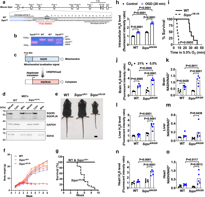
deprivation, we developed mice that lack SQOR in mitochondria. By using
CRISPR-Cas9 technology, we disrupted the translation start codon ATG of
the murine Sqor gene via a 14-bp deletion (Fig. 3a).
This change led to the initiation of translation starting at a
downstream ATG, which resulted in the production of SQOR protein that
lacked the N terminal mitochondrial localization sequence (SQOR∆N)
(Fig. 3c). We confirmed that SQOR∆N failed to localize to mitochondria in mouse embryonic fibroblasts obtained from Sqor∆N/∆N mice (Fig. 3d), although the lack of mitochondrial localization sequence did not affect protein stability of SQOR∆N (Supplementary Fig. 6a).
Sqor∆N/∆N mice were born normally according to the predicted Mendelian ratio. Although Sqor∆N/∆N
mice were indistinguishable from WT littermates before weaning, growth
of the homozygous mutant mice ceased around the weaning period (Fig. 3e). All Sqor∆N/∆N
mice gradually became emaciated, developed ataxia, and died within 10
weeks of age, whereas heterozygote mice were normal and similar to WT
mice (Fig. 3f, g).
To examine the role of SQOR in energy homeostasis and survival of neurons in hypoxia, we isolated primary cortical neurons from Sqor∆N/∆N and WT littermate embryos. Sqor∆N/∆N neurons had higher levels of intracellular sulfide compared to WT neurons (Fig. 3h),
suggesting that SQOR has a critical role in the catabolism of sulfide
in neurons even under normoxic conditions. In addition, the magnitude of
the oxygen and glucose deprivation (OGD)-induced increase in sulfide
levels was markedly greater in Sqor∆N/∆N compared to WT neurons. Reoxygenation after OGD decreased cell viability more markedly in Sqor∆N/∆N than in WT neurons (Supplementary Fig. 6b). Compared to 5-week-old WT mice, SqorΔN/ΔN littermates had a shorter survival when exposed to 5.5% O2 (Fig. 3i). When SqorΔN/ΔN and control mice breathed air, there were no differences in the levels of sulfide and persulfide or in the ratio of NADH/NAD+ in whole-brain tissue homogenates (Fig. 3j, k and Supplementary Fig. 6c). Although breathing 5.5% O2 for 3 min increased levels of sulfide and persulfide and the ratio of NADH/NAD+ in the brains of both genotypes, sulfide levels and the ratio of NADH/NAD+ were higher in the brains of Sqor∆N/∆N mice than in control mice. In contrast to the results observed in the brains of WT mice, breathing 5.5% O2 did not affect the levels of sulfide and persulfide or the ratio of NADH/NAD+ in the heart or liver of WT mice (Fig. 3l-o and Supplementary Fig. 6d). However, exposure to 5.5% O2 increased these metrics in the heart and liver of SqorΔN/ΔN
mice. These results indicate that mitochondrial SQOR prevents the
accumulation of sulfide and persulfide and decreased oxidative
phosphorylation induced by hypoxia not only in the brain but also in the
heart and liver.
Hypoxia-tolerant ground squirrels have a high capacity to catabolize sulfide in the brain
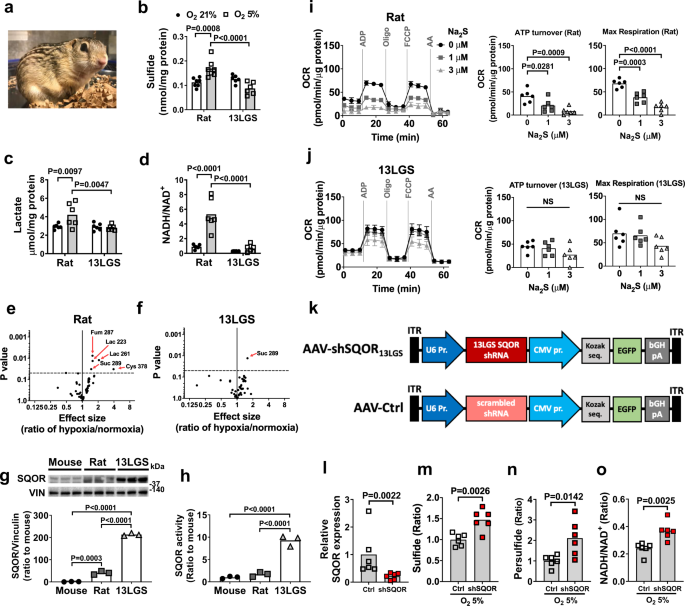
Several mammalian species including 13-lined ground (13LG) squirrels (Ictidomys tridecemlineatus) are highly resistant to oxygen deprivation (Fig. 4a)40,41,42.
To explore the relationship between sulfide metabolism and natural
hypoxia tolerance, we measured brain sulfide levels in hypoxia-resistant
13LG squirrels and hypoxia-sensitive Sprague-Dawley rats. Details of
the 13LG squirrel model are provided in the “Methods” section. Brain
sulfide levels were comparable between rats and 13LG squirrels breathing
room air (Fig. 4b). Breathing 5% O2 for 5 min decreased PaO2 to a similar extent in rats and squirrels (Supplementary Fig. 7a). Levels of sulfide and lactate and the ratio of NADH to NAD+ were increased in the brains of rats when breathing 5% O2 (Fig. 4b-d). Breathing 5% O2 also increased the levels of homocysteine, but not GSH, thiosulfate, and cysteine, in the brains of rats (Supplementary Fig. 7b). In contrast, exposure to 5% O2 did not affect levels of sulfide, lactate, GSH, homocysteine, thiosulfate, and cysteine and the ratio of NADH to NAD+ in the brains of 13LG squirrels (Fig. 4b-d and Supplementary Fig. 7b).
Metabolomic analysis using GC-MS revealed increases in the levels of
lactate, fumarate, succinate, and cysteine in the brains of rats
following acute hypoxia, while only succinate was increased in the
brains of hypoxic 13LG squirrels (Fig. 4e, f and Supplementary Tables 1 and 2).
These observations indicate that 13LG squirrel brains do not accumulate
sulfide under hypoxia and are resistant to hypoxia-induced impairment
of sulfide metabolism and oxidative phosphorylation.
To
investigate the mechanisms responsible for the tolerance of 13LG
squirrels to the effects of cerebral hypoxia, we measured SQOR levels in
the brains of mice, rats, and 13LG squirrels. SQOR protein levels in
the brains of 13LG squirrels were ~100-fold greater than that in the
brains of mice and rats (Fig. 4g). SQOR enzyme activity, measured as the capacity of brain tissue homogenates to oxidize sulfide using coenzyme Q1 as an electron acceptor43, was also markedly higher in 13LG squirrels than in mice and rats (Fig. 4h).
Protein levels of TST, a sulfide oxidation enzyme downstream of SQOR,
were also modestly higher in the brains of 13LG squirrels compared to
rats and mice (Supplementary Fig. 7c).
In contrast, the abundance and activities of transsulfuration pathway
enzymes were comparable between mice, rats, and 13LG squirrels
(Supplementary Fig. 7c, d).
To
examine the effect of higher SQOR levels and activity on brain
mitochondrial function, we measured OCR in isolated brain mitochondria
of rats and 13LG squirrels at baseline and in the presence of increasing
doses of sulfide (mimicking the effects of hypoxia). Sodium sulfide
inhibited oxygen consumption in mitochondria isolated from the brains of
rats in a dose-dependent manner. The ability of sodium sulfide to
decrease oxygen consumption was attenuated in mitochondria isolated from
the brains of 13LG squirrels, indicating a resistance of 13LG squirrel
brain mitochondria to the inhibitory effects of sulfide on oxidative
phosphorylation (Fig. 4i, j).
To
investigate the role of SQOR in the ability of 13LG squirrels to
tolerate brain hypoxia, we designed and produced an AAV containing an
shRNA targeting 13LG squirrel Sqor (AAV-shSQOR13LGS). This shRNA was the best of four candidate sequences at decreasing the level of Sqor in myoblasts isolated from 13LG squirrels (Supplementary Fig. 7e and Supplementary Table 3).
We packaged shRNA in the AAV9, which, compared to serotypes AAV2, 4,
and 8, exhibited the highest transfection efficiency in 13LG squirrel
brains (Supplementary Fig. 7f). We administered AAV-shSQOR13LGS or a control AAV with a scrambled sequence (AAV-Ctrl) into the cerebral ventricles of 13LG squirrels at 2.5 × 10¹¹ viral particles per hemisphere (Fig. 4k). Two weeks after AAV infection, brain SQOR mRNA was decreased in squirrels that were infected with AAV-shSQOR13LGS compared to those infected with AAV-Ctrl (Fig. 4l). Control and SQOR knockdown 13LG squirrels were anesthetized and ventilated with 5% O2
for 5 min. Compared to control animals, the brains of SQOR knockdown
squirrels had increased levels of sulfide and persulfide (Fig. 4m, n). After exposure to 5% O2, the ratio of NADH/NAD+ was higher in the brains of SQOR knockdown squirrels than in the brains of control squirrels (Fig. 4o).
The results show that silencing SQOR results in increased levels of
sulfide and persulfide and decreased oxidative phosphorylation in the
brain of squirrels in response to hypoxia. While there was some
variability in the efficiency of SQOR knockdown among animals, there was
a negative linear correlation between SQOR mRNA level and the ratio of
NADH/NAD+, whereas there was a positive linear correlation between brain sulfide levels and NADH/NAD+ ratio (Supplementary Fig. 7g).
These observations indicate that the higher level of SQOR in the brain
mitochondria of 13 LG squirrels contributes to the resistance of these
animals to acute hypoxia.
SQOR facilitates ATP production in neuronal cells
To
examine the effects of increased sulfide oxidation on mitochondrial ETC
function, SQOR was expressed in the human neuroblastoma cell line
SH-SY5Y. The cells expressed very low levels of SQOR when transfected
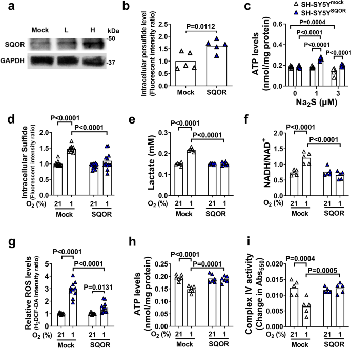
with a control plasmid (Fig. 5a). Expression of SQOR in SH-SY5Y cells incubated in 21% O2
resulted in increased intracellular concentrations of persulfide,
indicating that SQOR expression enhanced sulfide oxidation (Fig. 5b). Expression of SQOR did not affect ATP levels in isolated mitochondria incubated in 21% O2 (Fig. 5c). However, when incubated with Na2S (a sulfide donor) in 21% O2,
mitochondria isolated from cells expressing SQOR exhibited markedly
increased ATP levels compared to mitochondria isolated from control
cells. These observations suggest that sulfide oxidation by SQOR
contributes to cellular ATP production under normoxic conditions.
When SQOR-expressing and control SH-SY5Y cells were incubated in 1% O2,
SQOR prevented the hypoxia-induced increase in sulfide, lactate, and
ROS levels, and prevented the hypoxia-induced increase in the NADH/NAD+ ratio (Fig. 5d–g). In addition, expression of SQOR inhibited hypoxia-induced decreases in ATP production and complex IV activity (Fig. 5h, i).
These results indicate that enhanced sulfide oxidation by SQOR
expression may prevent sulfide accumulation and the impairment of
mitochondrial ATP production during oxygen shortage.
Neuron-specific SQOR expression improves survival in hypoxia
To determine whether enhanced sulfide oxidation confers resistance to oxygen deprivation in vivo, we used an AAV encoding mouse Sqor (AAV-SQOR) under the hSYN1 promoter to express SQOR specifically in neurons of CD-1 mice (Fig. 6a).
In the brains of CD-1 mice treated with AAV-SQOR, the mRNA and protein
levels of SQOR, but none of the other proteins that metabolize sulfide,
were increased (Fig. 6b, c and Supplementary Fig. 8a and b). Because male CD-1 mice are more sensitive to hypoxia than female mice (Fig. 3c),
male and female mice were treated with different concentrations of
oxygen. When an 8-week-old male or female mice breathed 5.5% or 4.5% O2,
respectively, mice that expressed SQOR in the brain exhibited better
survival rates compared to control mice infected with AAV-GFP (Fig. 6d, e).
Because
the effects of SQOR expression on indices of oxidative phosphorylation
were more robust in male than in female mice presumably due to the lower
(baseline) brain levels of SQOR in males (Fig. 3a, b),
only male mice were used in the following experiments. SQOR expression
prevented the hypoxia-induced increase in the levels of sulfide and
persulfide and the ratio of NADH/NAD+ in the brain of male mice (Fig. 6f–h).
Metabolomic analysis was used to investigate the effects of hypoxia on
metabolites in the brain. Neuron-specific SQOR expression mitigated the
hypoxia-induced increase in the ratios of lactate/pyruvate and
succinate/fumarate in the brain of male mice that breathed 5.5% O2 (Fig. 6I, j and Supplementary Fig. 8c, Supplementary Tables 4 and 5).
To
further define the effects of enhanced sulfide oxidation on oxygen
deprivation specifically in the brain, we examined the impact of SQOR
expression on brain injury induced by global cerebral ischemia and
reperfusion. Male mice were subjected to global cerebral ischemia
induced by 20 min of bilateral carotid artery occlusion (2VO) followed
by reperfusion. In control mice that were infected with AAV-GFP and
subjected to 2VO and reperfusion, a number of Fluoro-Jade B
(FJB)-positive dead neurons were observed in the hippocampal CA1 and CA3
regions (Fig. 6k–m) and cerebral cortex (Fig. 6n, o).
In mice expressing SQOR in neurons, only a few FJB-positive neurons
were found in the hippocampus and cortex after 2VO and reperfusion. The
number of viable neurons counted in H&E-stained brain sections
showed a reciprocal trend of the number of FJB-positive dead neurons in
these brain regions (Supplementary Fig. 9). After 2VO and reperfusion, mice expressing SQOR exhibited better neurological function (Fig. 6p) and survival rate compared to control mice (survival rate at day 3, 9/9 vs 6/12, respectively, P < 0.05).
Taken together, these results suggest that neuron-specific SQOR
expression prevents sulfide accumulation and impairment of mitochondrial
respiration in the brain, improves survival in severe hypoxia, and
prevents ischemic brain injury and death after global cerebral ischemia.
Sulfide scavenging prevents ischemic brain injury
The
mechanism by which enhanced sulfide oxidation by SQOR increases ATP
production is unknown. The predominant effect of sulfide oxidation may
be to directly provide electrons to the ETC. Alternatively, the major
effect of sulfide oxidation may be to prevent sulfide-induced inhibition
of mitochondrial complex IV (Fig. 5i).
Arguing against the first possibility, oxidation of sulfide as a direct
source of electrons is energetically unfavorable, especially during
oxygen shortage. Compared to oxidation of NADH or succinate (the
canonical electron donors), sulfide oxidation requires three times more
oxygen for the same electron transfer in the ETC30.
We, therefore, hypothesized that the beneficial effects of
SQOR-mediated sulfide catabolism during oxygen shortage are
predominantly mediated by the avoidance of sulfide accumulation, rather
than the provision of electrons to ETC from oxidizing sulfide. To test
this hypothesis, we examined the effects of pharmacological scavengers
of sulfides on SH-SY5Y cells incubated in 21% or 1% O2. Both
the highly specific sulfide fluoroprobe/scavenger HSip-1 and the
broad-spectrum scavenger hydroxocobalamin dose-dependently prevented the
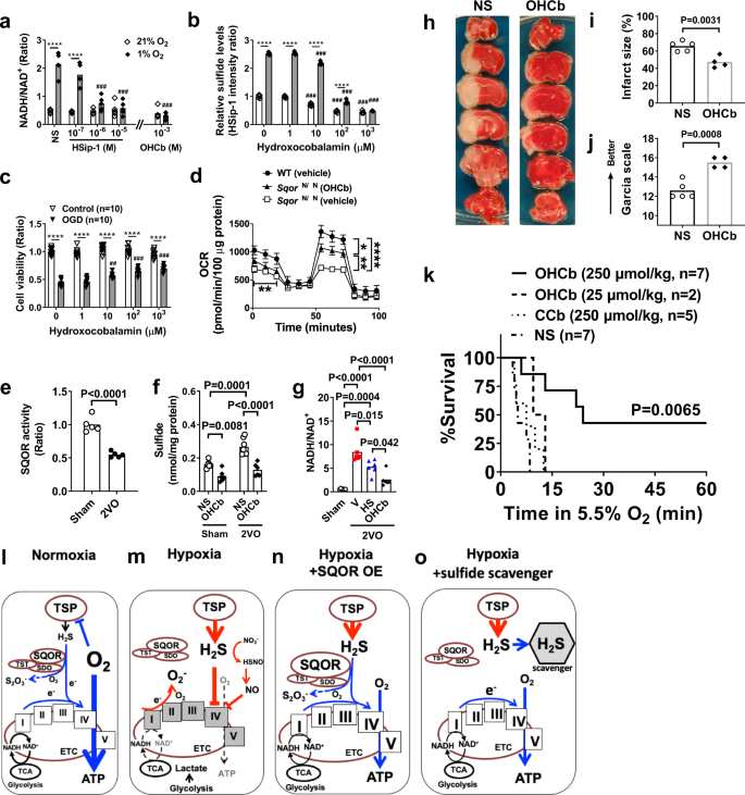
increase in the ratio of NADH/NAD+ induced by 1% O2 (Fig. 7a). Hydroxocobalamin attenuated the low oxygen-induced increase in SH-SY5Y intracellular sulfide levels (Fig. 7b), and improved survival of cells subjected to oxygen and glucose deprivation (Fig. 7c) in a dose-dependent manner. Although increased sulfide levels observed in primary cortical neurons of SqorΔN/ΔN mice (Fig. 3h) were associated with decreased oxygen consumption in isolated brain mitochondria obtained from SqorΔN/ΔN mice, scavenging sulfide by hydroxocobalamin partially restored OCR (Fig. 7d).
These results suggest that scavenging sulfide can restore mitochondrial
energy homeostasis even when sulfide oxidation is impaired.
In
mice, global cerebral ischemia induced by 2VO decreased SQOR activity
in the brain, leading to increased sulfide levels and the NADH/NAD+ ratio (Fig. 7e–g). Administration of sulfide scavengers prevented sulfide accumulation and the increase in the NADH/NAD+
ratio in the brain during global cerebral ischemia. Furthermore,
treatment with hydroxocobalamin at 10 min after the onset of focal brain
ischemia markedly attenuated ischemic brain injury and neurological
dysfunction after permanent middle cerebral artery occlusion (MCAO)
without reperfusion (Fig. 7h–j) and dose-dependently improved survival when mice were exposed to 5.5% O2 (Fig. 7k).
In contrast, cyanocobalamin, a synthetic vitamin B12 that scavenges
ROS, but not sulfide, did not improve the survival of hypoxic mice44 (Fig. 7k).
To further examine the ability of sulfide scavengers to protect against
the effects of cerebral ischemia, mice were subjected to a more
clinically relevant model of transient (60 min) MCAO and reperfusion and
treated with the highly specific sulfide scavenger SS-2045
45 min after the onset of ischemia. Treatment with SS-20 during
ischemia markedly decreased the infarct size and improved neurological
function at 48 h after reperfusion without changing CBF (Supplementary
Fig. 10).
These results suggest that the prevention of sulfide accumulation is
sufficient to maintain energy homeostasis and neuronal survival during
acute oxygen deprivation in the brain.