Changing stroke rehab and research worldwide now.Time is Brain! trillions and trillions of neurons that DIE each day because there are NO effective hyperacute therapies besides tPA(only 12% effective). I have 523 posts on hyperacute therapy, enough for researchers to spend decades proving them out. These are my personal ideas and blog on stroke rehabilitation and stroke research. Do not attempt any of these without checking with your medical provider. Unless you join me in agitating, when you need these therapies they won't be there.

What this blog is for:

My blog is not to help survivors recover, it is to have the 10 million yearly stroke survivors light fires underneath their doctors, stroke hospitals and stroke researchers to get stroke solved. 100% recovery. The stroke medical world is completely failing at that goal, they don't even have it as a goal. Shortly after getting out of the hospital and getting NO information on the process or protocols of stroke rehabilitation and recovery I started searching on the internet and found that no other survivor received useful information. This is an attempt to cover all stroke rehabilitation information that should be readily available to survivors so they can talk with informed knowledge to their medical staff. It lays out what needs to be done to get stroke survivors closer to 100% recovery. It's quite disgusting that this information is not available from every stroke association and doctors group.

Showing posts with label brain repair. Show all posts

Monday, August 10, 2026

Regenerative Astrocytes Repair Brain Damage

 A competent? doctor and hospital WOULD INSURE HUMAN TESTING OCCURS! Do you have competence among in your stroke medical 'professionals'?

Do you prefer your doctor, hospital and board of director's incompetence NOT KNOWING? OR NOT DOING? Your choice; let them be incompetent or demand action!

Regenerative Astrocytes Repair Brain Damage

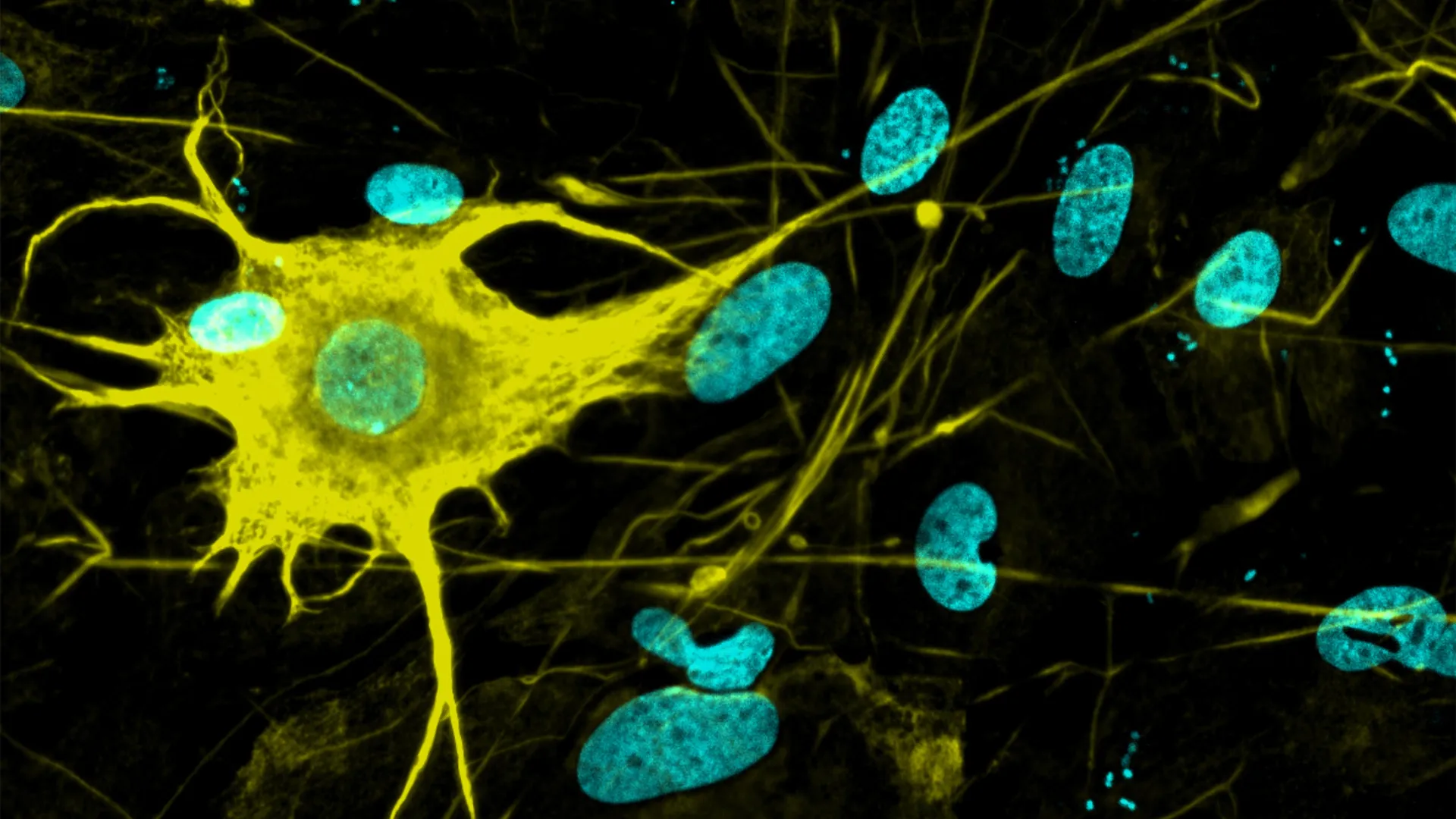
Summary: Researchers discovered a previously unknown mechanism through which the adult mammalian brain repairs itself following focal injuries or autoimmune damage. Using two-photon microscopy and longitudinal gene mapping in living mouse models, researchers identified a specialized population of “regenerative” astrocytes capable of repopulating damaged brain regions.

Rather than relying solely on classical cell body division at the site of injury, these specialized astrocytes situated along the lesion perimeter send newly formed daughter cell nuclei gliding long distances through their star-shaped cellular extensions. These migrating cell nuclei repopulate the depleted lesion zone, re-establishing functional astrocyte networks.

This discovery overturns long-held assumptions regarding the limited regenerative capacity of adult glial networks, revealing molecular signaling pathways that could serve as therapeutic targets for traumatic brain injury and autoimmune conditions such as neuromyelitis optica spectrum disorder (NMOSD).

Key Facts

  • Overturning Dogma on Glial Regeneration: Demonstrates that the adult central nervous system possesses a previously unrecognized capability to replace lost astrocytes and restore damaged tissue architecture.
  • Mechanism of Long-Distance Nuclear Migration: Specialized regenerative astrocytes remain at the lesion boundary and send newly generated cell nuclei gliding across long astrocytic extensions into the depleted injury core.
  • Rebuilding Functional Glial Networks: Astrocytes perform vital homeostatic functions, including nutrient supply to neurons, blood flow regulation via end-feet, and extracellular ion balance, making their network reconstruction essential for neuronal survival.
  • Targeted Clinical Applications: Holds therapeutic relevance for neurotraumatic brain injuries and rare autoimmune conditions like neuromyelitis optica spectrum disorder (NMOSD), where autoantibodies selectively destroy astrocytes.
  • Molecular Targets for Therapeutics: The team identified specific genes and signaling pathways temporarily activated during nuclear migration, providing potential targets for pharmacological interventions to accelerate brain repair.

Source: University of Zurich

The brain evidently can regenerate itself better than previously assumed after injuries or certain autoimmune diseases. Using a mouse model, researchers at the University of Zurich have demonstrated that special supporting and nourishing cells repopulate damaged areas of the brain by initially sending only newly formed cell nuclei there.

Glial cells are supporting and nourishing cells in the brain. Star-shaped glial cells called astrocytes are vital to the functioning of neurons. They supply the nerve cells with nutrients, help to regulate blood flow and keep brain tissue healthy.

It had long been assumed that when astrocytes are lost – as happens, for instance, in brain injuries or autoimmune diseases such as rare neuromyelitis optica spectrum disorder, in which the body’s own antibodies destroy these cells – the adult brain cannot fully replace them.

Regenerative astrocytes repair damaged tissue

A new study by co-lead authors Marina Herwerth and Matthias Wyss from the Institute of Pharmacology and Toxicology at the University of Zurich (UZH) has now overturned that assumption: their research team headed by Bruno Weber discovered a specialized group of “regenerative” astrocytes in the brains of living mice that step in on the perimeter of the damaged area of the brain to rebuild the cells.

“The findings of our study reveal a previously unknown ability of the adult brain to repair itself. They point toward new ways of supporting recovery from ailments involving the loss of astrocytes,” Weber says.

Only cell nuclei migrate

The researchers used two-photon microscopy to observe the brains of living mice in real time over a period of several weeks and mapped which genes switch on in which areas of the brain. This way they were able to identify the special astrocytes that take care of rebuilding injured tissue. But those cells don’t just divide, they also perform a remarkable feat: “they send the newly formed nuclei of their daughter cells gliding across long distances to repopulate the damaged area of the brain and knit the astrocyte network back together,” Weber explains.

Starting points for targeted regeneration

The discovery of how adult brain cell nuclei migrate through the long star-shaped extensions of astrocytes to injured tissue expands comprehension of how the brain organizes and regenerates itself after certain injuries. If those mechanisms can be selectively activated, that could help to more effectively repair damaged brain tissue, restore astrocyte networks and thus improve recovery after certain brain disorders.

“We were able to identify numerous genes and signaling pathways that are temporarily activated during repair. They could serve as starting points in the future for influencing post-disease and -injury regeneration processes,” Weber stresses.

Key Questions Answered:

Q: How do “regenerative” astrocytes differ from standard cell division during tissue repair?

A: Instead of whole cells migrating or simply dividing locally, these specialized astrocytes remain at the perimeter of the damaged area. They divide and send the newly formed nuclei of their daughter cells gliding long distances through their extended cellular processes directly into the injured zone to rebuild the network.

Q: What conditions cause the loss of astrocytes in the adult brain?

A: Astrocytes are lost during traumatic brain injuries, strokes, and specific neuroinflammatory or autoimmune conditions, most notably Neuromyelitis Optica Spectrum Disorder (NMOSD), where the body’s immune system produces autoantibodies that target and destroy astrocytes.

Q: How was this nuclear migration observed in real time?

A: Researchers at the University of Zurich used in vivo two-photon microscopy in living mouse models over several weeks. This allowed them to track living cells, observe nuclear movement through astrocytic branches, and map corresponding gene expression changes as the tissue repaired itself.

Editorial Notes:

  • This article was edited by a Neuroscience News editor.
  • Journal paper reviewed in full.
  • Additional context added by our staff.

About this neuroscience research news

Author: Kurt Bodenmueller
Source: University of Zurich
Contact: Kurt Bodenmueller – University of Zurich
Image: The image is credited to Institute of Pharmacology and Toxicology, University of Zurich

Original Research: Open access.
Focal astrocyte loss reveals nuclear translocation during lesion repopulation” by Marina Herwerth, Matthias T. Wyss, Nicola B. Schmid, Anna Lasne, Jacqueline Condrau, Luca Ravotto, José María Mateos Melero, Andres Kaech, Gustav Bredell, Carolina Thomas, Rachel Kim, Petra Kukanja, Vladyslav L. Korobeynyk, Christine Stadelmann, Thomas Misgeld, Jeffrey L. Bennett, Sebastian Jessberger, Aiman S. Saab, Shane A. Liddelow & Bruno Weber. Nature Neuroscience
DOI:10.1038/s41593-026-02354-5

Thursday, June 18, 2026

Scientists find way to continue natural method to repair brains


How long will it take for your incompetent? doctor to drive the research that solves this for humans and creates protocols that deliver this solution? Just why can't your doctor accomplish that?

Laziness? Incompetence? Or just don't care? NO leadership? NO strategy? Not my job? Not my Problem!

Scientists find way to continue natural method to repair brains

A team of Japanese and German scientists said its recent discoveries could lead to medicine that promotes and prolongs reparations to brain functions among victims of strokes and other diseases.

The researchers uncovered a mechanism that prompts recovery of brain functions in the early stages after the onset of the disease. They also identified a protein that causes the reparability to wane and eventually halt.

And they developed a drug that suppresses the workings of that protein in mice.

The group, including researchers from the Institute of Science Tokyo and the Tokyo Metropolitan Institute of Medical Science, plans to apply the findings to humans to ease the aftereffects of brain diseases.

Their research results were published online May 13 in Nature, a British science journal.

Infarction and other diseases of the brain cause neurological symptoms, such as loss of limb mobility and speech impairment.

Dead brain cells cannot be restored.

However, part of the lost brain functions can be recovered through rehabilitation, although the reparability is lost after a while. The science world does not understand exactly why this occurs.

The researchers on the team set their sights on microglia, a class of immune cells that work in the brain.

In mouse experiments, they found a mechanism whereby a brain injury prompts the microglia to begin generating nourishing substances that encourage brain tissue repair.

The microglia continued making the nourishing substances for about a month in mice that suffered a brain stroke. But production of the substances eventually stopped, and the reparative functions were lost in two months.

The researchers found that a protein called ZFP384 works to prevent the microglia from generating those nourishing substances.

In mice disabled from making ZFP384, the nourishing substances continued to be produced one month following the stroke, and the neurological symptoms improved, according to the team.

The researchers developed a drug that inhibits the workings of ZFP384 and administered it to mice one week and one month following the onset of a stroke.

They found the neurological symptoms improved and the brain functions continued to recover.

The scientists said the microglia-created nourishing substances encouraged the recovery of synaptic connections and other features of the nervous system.

SAME MECHANISM IN HUMANS?

Among humans with brain infarction, the scholars found abundant microglia of the type that generates the nourishing substances one week following the disease.

Similar microglia, however, decreased with time in terms of abundance, leading to a loss of reparative functions. The researchers also found that more ZFP384 was being generated.

The scientists said this likely represents the same sort of change that took place in mice, and they are hoping to develop therapeutic drugs that work in humans as well.

“We have presented this novel therapeutic concept of relying on the natural reparability that is inherent in the brain and making it last longer,” said Jun Tsuyama, a Science Tokyo junior associate professor of neuroscience, one of the leading members of the research team.

“We hope to help realize medical treatment where you don’t have to give up hopes for post-stroke functional recovery with no lingering aftereffects,” he said.

The research article can be viewed at (https://www.nature.com/articles/s41586-026-10480-0). 

Friday, April 3, 2026

USC study identifies brain rewiring mechanism that may aid stroke recovery

 But you didn't create a useable protocol that repeatedly delivers recovery! This did nothing that will help survivors! You're fired!

USC study identifies brain rewiring mechanism that may aid stroke recovery

Findings from an international ENIGMA collaboration reveal that stroke survivors with severe motor impairment show signs of brain “youthfulness” in undamaged regions, suggesting compensatory neuroplasticity.

Peer-Reviewed Publication

Keck School of Medicine of USC

In a new study published in The Lancet Digital Health, scientists at the USC Mark and Mary Stevens Neuroimaging and Informatics Institute (Stevens INI) have discovered that the brains of people who experience severe physical impairment after a stroke may reorganize themselves in unexpected ways, showing signs of “younger” brain structure in undamaged regions as they adapt to injury.

The international research effort is part of the Enhancing NeuroImaging Genetics through Meta-Analysis (ENIGMA) Stroke Recovery Working Group, which analyzed brain scans from more than 500 stroke survivors across 34 research sites in eight countries. Using deep learning models trained on tens of thousands of MRI scans, the researchers estimated the “brain age” of different regions in each hemisphere to see how stroke damage affects brain structure and recovery.

“We found that larger strokes accelerate aging in the damaged hemisphere but paradoxically make the opposite side of the brain appear younger,” said Hosung Kim, PhD, associate professor of research neurology at the Keck School of Medicine of USC and co-senior author of the study. “This pattern suggests the brain may be reorganizing itself, essentially rejuvenating undamaged networks to compensate for lost function.”

The research team used an advanced form of artificial intelligence known as a graph convolutional network to predict the biological age of 18 brain regions from MRI data. The difference between a person’s predicted brain age and their actual chronological age, known as the brain-predicted age difference (brain-PAD), served as a sensitive marker of neural health.

When the team associated these measurements with motor performance scores, they found a striking pattern: stroke survivors with severe movement deficits, even after more than 6 months of rehabilitation, showed younger-than-expected brain age in regions opposite the lesion, particularly within the frontoparietal network, a key system involved in motor planning, attention, and coordination.

“These findings suggest that when stroke damage leads to greater movement loss, undamaged regions on the opposite side of the brain may adapt to help compensate,” Kim explained. “We saw this in the contralesional frontoparietal network, which showed a more ‘youthful’ pattern and is known to support motor planning, attention, and coordination. Rather than indicating full recovery of movement, this pattern may reflect the brain’s attempt to adjust when the damaged motor system can no longer function normally. This gives us a new way to see neuroplasticity that traditional imaging could not capture.”

The study was conducted through ENIGMA, a global alliance that unites data from more than 50 countries to better understand the brain across diseases. Researchers harmonized MRI data and clinical measures across dozens of cohorts to build the world’s largest stroke neuroimaging dataset of its kind.

“By pooling data from hundreds of stroke survivors worldwide and applying cutting-edge AI, we can detect subtle patterns of brain reorganization that would be invisible in smaller studies. These findings of regionally differential brain aging in chronic stroke could eventually guide personalized rehabilitation strategies,” said Arthur W. Toga, PhD, director of the Stevens INI and Provost Professor at USC.

The team plans to expand their work to include longitudinal studies tracking patients from the acute to chronic stages of stroke recovery. By observing how patterns of brain aging and reorganization develop over time, clinicians might be able to customize interventions based on each patient’s unique neural adaptation process, ultimately improving recovery outcomes and quality of life in the near future.

Learn more about associations between contralesional neuroplasticity and motor impairment by viewing this video made by the Stevens INI.

About the study

The study, “Deep learning prediction of MRI-based regional brain age reveals contralesional neuroplasticity associated with severe motor impairment in chronic stroke: A worldwide ENIGMA study,” was funded by the National Institutes of Health (NIH) grant R01 NS115845 and supported by international collaborators from institutions including the University of British Columbia, Monash University, Emory University, and the University of Oslo.

For more information on the ENIGMA Stroke Recovery Working Group, visit https://enigma.ini.usc.edu.

Disclaimer: AAAS and EurekAlert! are not responsible for the accuracy of news releases posted to EurekAlert! by contributing institutions or for the use of any information through the EurekAlert system.

Wednesday, November 26, 2025

Your Brain Quietly Rewires Itself at 9, 32, 66 and 83

 Ask your competent? doctor if this rewiring will get you recovered from your stroke. I passed 66 3 years ago and I don't think anything occurred, my spasticity didn't get better at all. Cure my spasticity and I'll easily recover my lost motor functions. 

As compared to this idiotic idea of do nothingism of Dr. William M. Landau on spasticity? I completely disagree with that bogus idea! Comeuppance is going to be a bitch for him. 

His statement from here:

Spasticity After Stroke: Why Bother? Aug. 2004 

The latest here: 

Your Brain Quietly Rewires Itself at 9, 32, 66 and 83

Summary: Researchers identified five major phases of human brain wiring that unfold from birth to old age, marked by four major turning points at ages 9, 32, 66, and 83. Childhood and adolescence are periods of rapid reorganization, while adulthood brings a long plateau of structural stability.

Beginning in the mid-60s, gradual declines in connectivity begin to reshape brain networks, accelerating into highly localized wiring in late aging. These findings reveal that the brain doesn’t change steadily over time but instead shifts through distinct eras that may influence learning, vulnerability, and resilience.

Key Facts

  • Lifespan Rewiring: MRI data revealed five structural brain epochs defined by shifts in neural connectivity at ages 9, 32, 66, and 83.
  • Peak Efficiency: Neural efficiency rises throughout adolescence and peaks around age 32, marking the strongest rewiring shift of the lifespan.
  • Aging Transition: After midlife, brain networks gradually weaken their global connectivity, increasing vulnerability to cognitive decline.

Source: University of Cambridge

Neuroscientists at the University of Cambridge have identified five “major epochs” of brain structure over the course of a human life, as our brains rewire to support different ways of thinking while we grow, mature, and ultimately decline.  

A study led by Cambridge’s MRC Cognition and Brain Sciences Unit compared the brains of 3,802 people between zero and ninety years old using datasets of MRI diffusion scans, which map neural connections by tracking how water molecules move through brain tissue.

In a study published in Nature Communications, scientists say they detected five broad phases of brain structure in the average human life, split up by four pivotal “turning points” between birth and death when our brains reconfigure.

Childhood brain “topology” runs from birth until a turning point at the age of nine, when it transitions to the adolescent phase – an era that lasts right up to the age of 32, on average.

Our early thirties see the brain’s neural wiring shift into adult mode. This is the longest era, which lasts over three decades. A third turning point around age 66 marks the start of an “early aging” phase of brain architecture. Finally, the “late aging” brain takes shape at around 83 years old.

“We know the brain’s wiring is crucial to our development, but we lack a big picture of how it changes across our lives and why,” said Dr Alexa Mousley, a Gates Cambridge Scholar who led the research. “This study is the first to identify major phases of brain wiring across a human lifespan.”

“These eras provide important context for what our brains might be best at, or more vulnerable to, at different stages of our lives. It could help us understand why some brains develop differently at key points in life, whether it be learning difficulties in childhood, or dementia in our later years.”

From infancy through childhood, our brains are defined by “network consolidation”, as the wealth of synapses – the connectors between neurons – overproduced in a baby’s brain are whittled down, with the more active ones surviving.

Across the whole brain, connections rewire in the same pattern from birth until about nine years old.

Meanwhile, grey and white matter grow rapidly in volume, so that cortical thickness – the distance between outer grey matter and inner white matter – reaches a peak, and cortical folding, the characteristic ridges on the outer brain, stabilises.

By the first turning point at nine years old, the brain is experiencing a step-change in cognitive capacity, as well as an increased risk of mental health disorders.

The second “epoch” of the brain, the adolescence era, sees white matter continue to grow in volume, so organisation of the brain’s communications networks is increasingly refined, as measured by the diffusion of water in the scans.

This era is defined by the efficiency of connections both within specific regions as well as rapid communication right across the whole brain, which is related to enhanced cognitive performance.

“Neural efficiency is as you might imagine, well connected by short paths, and the adolescent era is the only one in which this efficiency is increasing,” said Mousley.

These developments peak in the early thirties, on average, which is the “strongest topological turning point” of the entire lifespan say researchers.

“Around the age of 32, we see the most directional changes in wiring and largest overall shift in trajectory, compared to all the other turning points,” said Mousley.

“While puberty offers a clear start, the end of adolescence is much harder to pin down scientifically. Based purely on neural architecture, we found that adolescent-like changes in brain structure end around the early thirties.”

At age 32, the longest era, that of adulthood, begins. Brain architecture stabilises compared to previous phases – with no major turning points for thirty years. This corresponds with a “plateau in intelligence and personality” based on other studies, say researchers.

They also found “segregation” is more noticeable during this epoch, as regions slowly start to become more compartmentalised.

The turning point at age 66 is far milder, and not defined by any major structural shifts, although researchers still found meaningful changes to the pattern of brain networks on average at around this age.

“The data suggest that a gradual reorganisation of brain networks culminates in the mid-sixties,” said Mousley. “This is probably related to aging, with further reduced connectivity as white matter starts to degenerate.

“This is an age when people face increased risk for a variety of health conditions that can affect the brain, such as hypertension.”  

The last turning point comes around age 83, and the final brain structure epoch is entered. While data is limited for this era, the defining feature is a shift from global to local, as whole brain connectivity declines even further, with increased reliance on certain regions.      

“Looking back, many of us feel our lives have been characterised by different phases. It turns out that brains also go through these eras,” added senior author Prof Duncan Astle, Professor of Neuroinformatics at Cambridge.

“Many neurodevelopmental, mental health and neurological conditions are linked to the way the brain is wired. Indeed, differences in brain wiring predict difficulties with attention, language, memory, and a whole host of different behaviours”

“Understanding that the brain’s structural journey is not a question of steady progression, but rather one of a few major turning points, will help us identify when and how its wiring is vulnerable to disruption.”

Funding: The research was supported by the Medical Research Council, Gates Foundation and Templeton World Charitable Foundation.

Key Questions Answered:

Q: What did researchers discover about brain wiring across the lifespan?

A: They identified five major epochs of brain structure, each defined by distinct patterns of neural wiring and four major turning points.

Q: When do these turning points occur?

A: Ages 9, 32, 66, and 83, marking transitions between childhood, adolescence, adulthood, early aging, and late aging.

Q: Why are these findings important?

A: They reveal when the brain is most adaptable — or vulnerable — and could help explain learning differences, mental health risk, and neurodegeneration.

Editorial Notes:

  • This article was edited by a Neuroscience News editor.
  • Journal paper reviewed in full.
  • Additional context added by our staff.

About this neurodevelopment and brain aging research news

Author: Fred Lewsey
Source: University of Cambridge
Contact: Fred Lewsey – University of Cambridge
Image: The image is credited to Neuroscience News

Sunday, July 13, 2025

Researchers grow 400+ brain cell types—a leap for Alzheimer’s and Parkinson’s research

 You lost hundreds of millions of neurons, think your competent? doctor can ensure research produces the quantity needed? And the types you need? AND get them connected?

Researchers grow 400+ brain cell types—a leap for Alzheimer’s and Parkinson’s research

Date:
July 12, 2025
Source:
ETH Zurich
Summary:
Scientists at ETH Zurich have broken new ground by generating over 400 types of nerve cells from stem cells in the lab, far surpassing previous efforts that produced only a few dozen. By systematically experimenting with combinations of morphogens and gene regulators, the researchers replicated the vast diversity of neurons found in the human brain. This breakthrough holds major promise for studying neurological diseases like Alzheimer’s and Parkinson’s, creating more accurate models for drug testing, and eventually even enabling neuron replacement therapies.
Share:
FULL STORY

Nerve cells are not just nerve cells. Depending on how finely we distinguish, there are several hundred to several thousand different types of nerve cell in the human brain according to the latest calculations. These cell types vary in their function, in the number and length of their cellular appendages, and in their interconnections. They emit different neurotransmitters into our synapses and, depending on the region of the brain - for example, the cerebral cortex or the midbrain - different cell types are active.

When scientists produced nerve cells from stem cells in Petri dishes for their experiments in the past, it was not possible to take their vast diversity into account. Until now, researchers had only developed procedures for growing a few dozen different types of nerve cell in vitro. They achieved this using genetic engineering or by adding signalling molecules to activate particular cellular signalling pathways. However, they never got close to achieving the diversity of hundreds or thousands of different nerve cell types that actually exists.

"Neurons derived from stem cells are frequently used to study diseases. But up to now, researchers have often ignored which precise types of neuron they are working with," says Barbara Treutlein, Professor at the Department of Biosystems Science and Engineering at ETH Zurich in Basel. However, this is not the best approach to such work. "If we want to develop cell culture models for diseases and disorders such as Alzheimer's, Parkinson's and depression, we need to take the specific type of nerve cell involved into consideration."

Systematic screening was the key to success

Treutlein and her team have now successfully produced over 400 different types of nerve cell. In doing so, the scientists have paved the way for more precise basic neurological research with cell culture experiments.

The ETH researchers achieved this by working with a culture of human induced pluripotent stem cells that had been generated from blood cells. In these cells, they used genetic engineering to activate certain neuronal regulator genes and treated the cells with various morphogens, a special class of signalling molecules. Treutlein and her team took a systematic approach, using seven morphogens in different combinations and concentrations in their screening experiments. This resulted in almost 200 different sets of experimental conditions.

Morphogens

Morphogens are messengers that are known from research into embryonic development. They are not distributed uniformly within an embryo but occur in a variety of concentrations forming spatial patterns. In this way, they define the position of cells within the embryo, for example whether a cell is near the body axis or in the back, abdomen, head or torso. Accordingly, morphogens help to determine what grows where in the embryo.

The researchers used various analyses to prove that they had produced over 400 different types of nerve cell in their experiment. They examined the RNA (and therefore genetic activity) at the level of individual cells, as well as the external appearance of cells and their function: for example, which type of cell appendage they had in which quantities and which electric nerve impulses they emitted.

The researchers then compared their data with information from databases of neurons from the human brain. By doing this, they were able to identify the types of nerve cell that had been created, such as those found in the peripheral nervous system or brain cells and the part of the brain they come from, whether they perceive pain, cold or movement, and so on.

In-vitro neurons for active ingredient research

Treutlein clarifies that they are still a long way off producing all types of nerve cell that exist in vitro. Nonetheless, the researchers now have access to a much larger number of different cell types than they had before.

They would like to use in-vitro nerve cells to develop cell culture models for studying serious neurological conditions, including schizophrenia, Alzheimer's, Parkinson's, epilepsy, sleep disorders and multiple sclerosis. Cell culture models of this kind are also of great interest in pharmaceutical research for testing the effects of new active compounds in cell cultures without animal testing, with the ultimate aim of one day being able to cure these conditions.

In the future, the cells could also be used for cell replacement therapy, which involves replacing sick or dead nerve cells in the brain with new human cells.

But there is a challenge to overcome before this can happen: the researchers often produced a mixture of multiple different types of nerve cell in their experiments. They are now working to optimise their method so that each experimental condition only produces one specific cell type. They already have some initial ideas as to how this might be achieved.

Story Source:

Materials provided by ETH Zurich. Note: Content may be edited for style and length.


Journal Reference:

  1. Hsiu-Chuan Lin, Jasper Janssens, Benedikt Eisinger, Philipp Hornauer, Ann-Sophie Kroell, Malgorzata Santel, Maria Pascual-Garcia, Ryoko Okamoto, Kyriaki Karava, Zhisong He, Marthe Priouret, Manuel Schröter, J. Gray Camp, Barbara Treutlein. Human neuron subtype programming via single-cell transcriptome-coupled patterning screens. Science, 2025; 389 (6756) DOI: 10.1126/science.adn6121

Cite This Page:

ETH Zurich. "Researchers grow 400+ brain cell types—a leap for Alzheimer’s and Parkinson’s research." ScienceDaily. ScienceDaily, 12 July 2025. <www.sciencedaily.com/releases/2025/07/250711224316.htm>.

Explore More

from ScienceDaily

RELATED STORIES


Monday, July 7, 2025

CRISPR Delivers RNA to Repair Neurons Right Where It’s Needed

 This should trigger massive rejoicing in the stroke medical world. We can have our researchers directly repair our damaged neurons. But nothing will occur! THERE IS NO LEADERSHIP IN STROKE! You're screwed, so don't have a stroke is the only valid response. 

CRISPR Delivers RNA to Repair Neurons Right Where It’s Needed

Summary: Researchers have developed a new CRISPR-based technology that transports RNA to exact locations within neurons, where it can trigger repair and regrowth, offering hope for treating neurological diseases and injuries. Unlike traditional CRISPR tools that edit DNA, this system repurposes CRISPR-Cas13 to act like a “mailman,” carrying RNA to damaged sites using built-in molecular zip codes.

In lab tests, the technique, called CRISPR-TO, boosted neurite growth by up to 50% in just 24 hours, marking a major step forward in spatial RNA medicine. This breakthrough may enable safer, more effective RNA-based treatments for conditions like ALS, spinal cord injuries, and neurodegenerative disorders.

Key Facts:

  • CRISPR-TO System: Cas13 delivers RNA to precise neural sites using molecular zip codes.
  • Enhanced Regrowth: Promoted 50% greater neurite growth in injured neurons.
  • New Class of Medicine: Introduces “spatial RNA medicine” for targeted cellular repair.

Source: Stanford

When a neuron in our body gets damaged, segments of RNA produce proteins that can help repair the injury. But in neurological disorders such as ALS and spinal muscular atrophy, or following spinal cord injuries, the mechanisms for moving life-essential RNA to injured sites within the cell fail. As a result, RNA molecules can’t get to where they are needed and damage becomes permanent.

Researchers at Stanford have developed a technology for transporting RNA to specific locations within a neuron, where it can repair and even regrow parts of the cell.

This shows a neuron.
Typically, CRISPR is used to slice and edit genetic code, but in this case the researchers didn’t want to make any changes. Credit: Neuroscience News

Their work, supported by the National Institutes of Health, forms the foundation for a new class of therapeutics that the researchers are calling “spatial RNA medicine,” which they hope will lead to treatments for neurological diseases as well as traumatic injuries.

“For the first time, we’ve harnessed the power of CRISPR technology to create a precise spatial ‘zip code’ that delivers RNA molecules exactly where they’re needed within cells,” said Stanley Qi, an associate professor of bioengineering and senior author on the paper published May 21 in Nature.

“Imagine being able to specifically target damaged sites within a neuron, repairing them, and promoting their regrowth – this is what our technology achieves.”

A CRISPR-based mailman

In recent years, researchers have realized that the distribution of RNA within a cell – where specific molecules are located – may be just as important as what they are capable of doing.

An individual neuron can be over a meter long, and aging, injury, and mutations can all disrupt its ability to transport the tiny RNA over such a distance.

“Therapeutic RNA can’t help if it doesn’t get to where it’s needed,” Qi said. “We wanted to create a technology that could reliably move RNA to where it needs to function.”

Qi and his colleagues used a version of the gene-editing tool CRISPR, called CRISPR-Cas13, to target individual pieces of RNA (unlike the more widely known CRISPR-Cas9, which targets DNA).

Typically, CRISPR is used to slice and edit genetic code, but in this case the researchers didn’t want to make any changes. They simply wanted to move the existing RNA to a new place within the cell.

“Cas13 naturally acts like a pair of scissors, but we engineered it to act like a mailman instead,” Qi said.

“Then we can tell it to carry the RNA from one precise location to another.”

The researchers paired Cas13 with specific localization signals that act as addresses, instructing the Cas13 where to deliver the RNA. Each location within the cell has its own address molecule, so the researchers can direct the RNA to various locations by adding different molecules to the cell.

Qi and his colleagues used their technology, which they are calling CRISPR-TO, to screen dozens of pieces of RNA and see if any of them would help neurons to grow.

They added CRISPR-TO to mouse brain neurons in a petri dish, where it carried the RNA molecules to the tips of neurites – fingerlike protrusions that form synapses and connect to other neurons.

They found several promising candidates, including one RNA molecule that increased neurite growth by as much as 50% over a 24-hour period.

“We are discovering more RNA targets that could promote neurite outgrowth and regeneration,” said Mengting Han, a postdoctoral scholar in Qi’s lab and lead author on the paper.

“We’ve added a new tool to the CRISPR toolbox, using it to control RNA localization inside the cell. This has never been achieved before and, importantly, it opens new therapeutic directions for treating neurodegenerative diseases.”

Safer, more effective RNA medicine

The researchers are using CRISPR-TO to screen additional RNA molecules to determine which ones will be most effective at repairing injured neurons in the brains of mice, as well as in human neurons.

“We are at the beginning of understanding how spatial organization of RNA benefits brain repair,” Qi said.

“We hope our technology will help people figure out which RNAs will be the biggest players for better therapeutics.”

Currently, the researchers are using CRISPR-TO to move endogenous RNAs – RNA molecules that are naturally produced within the cell. But it could also be used to provide precise control over RNA-based medicines, making them both safer and more efficient, Qi said.

“This potential excites us tremendously,” Qi said.

“It’s not enough for a molecule to just be in the cell. We need it to be in the right location at the right time. With our precise, programmable technology, you can target any RNA in any type of cell and bring it to the site of need in the body.”

Funding: This work was funded by the National Science Foundation, the National Institutes of Health, the National Center for Research Resources, the Stanford School of Medicine Dean’s Postdoctoral Fellowship, and the American Heart Association Postdoctoral Fellowship.

About this CRISPR and neuroscience research news

Author: Chloe Dionisio
Source: Stanford
Contact: Chloe Dionisio – Stanford
Image: The image is credited to Neuroscience News

Original Research: Open access.
Clonal tracing with somatic epimutations reveals dynamics of blood ageing” by Stanley Qi et al. Nature

Wednesday, April 26, 2023

Progress in application of adult endogenous neurogenesis in brain injury repair

Which way is your doctor incompetent about this? NOT KNOWING or NOT DOING?

Wednesday, April 14, 2021

Synthetic hydrogel to repair circuitry in severe TBI

Since we have NO LEADERSHIP IN STROKE, there is no one who will look at this and say. 'Yes, this is in rats and for TBI but we should do the research and test this in humans for stroke. Nothing will happen.

Synthetic hydrogel to repair circuitry in severe TBI


At a cost of $38 billion a year, an estimated 5.3 million people are living with a permanent disability related to traumatic brain injury in the United States today, according to the Centers for Disease Control and Prevention. The physical, mental and financial toll of a TBI can be enormous, but new research provides promise.

In a new study, researchers have demonstrated the long-term benefits of a hydrogel, which they call "brain glue," for the treatment of traumatic brain injury. The new study provides evidence that not only does the gel protect against loss of brain tissue after a severe injury, but it also might aid in functional neural repair.

Brain damage following significant TBI commonly results in extensive tissue loss and long-term disability. There currently are no clinical treatments to prevent the resulting cognitive impairments or tissue loss.

Reported in Sciences Advances, the new finding is the first to provide visual and functional evidence of the repair of brain neural circuits involved in reach-to-grasp movement in brain glue-implanted animals following severe TBI.

"Our work provides a holistic view of what's going on in the recovery of the damaged region while the animal is accomplishing a specific reach-and-grasp task," said the lead author.

Created in 2017, brain glue was designed to mimic the structure and function of the meshwork of sugars that support brain cells. The gel contains key structures that bind to basic fibroblast growth factor and brain-derived neurotrophic factor, two protective protein factors that can enhance the survival and regrowth of brain cells after severe TBI.

In a prior short-term study, the team showed that brain glue significantly protected brain tissue from severe TBI damage. In this new research, to harness the neuroprotective capacity of the original, they further engineered the delivery surface of protective factors to help accelerate the regeneration and functional activity of brain cells. After 10 weeks, the results were apparent.

"Animal subjects that were implanted with the brain glue actually showed repair of severely damaged tissue of the brain," said the author. "The animals also elicited a quicker recovery time compared to subjects without these materials."

To measure the glue's effectiveness, the team used a tissue-clearing method to make brain tissue optically transparent, which allowed them to visually capture the immediate response of cells in the reach-to-grasp circuit using a 3D imaging technique.

"Because of the tissue-clearing method, we were able to obtain a deeper view of the complex circuitry and recovery supported by brain glue," said the author. "Using these methods along with conventional electrophysiological recordings, we were able to validate that brain glue supported the regeneration of functional neurons in the lesion cavity."

The author pointed out that the RTG circuit is evolutionarily similar in rats and humans. "The modulation of this circuit in the rat could help speed up clinical translation of brain glue for humans," the author said.

https://news.uga.edu/brain-glue-helps-repair-circuitry-in-severe-tbi/


https://advances.sciencemag.org/content/7/10/eabe0207

http://sciencemission.com/site/index.php?page=news&type=view&id=publications%2Fengineered-glycomaterial&filter=22