Changing stroke rehab and research worldwide now.Time is Brain! trillions and trillions of neurons that DIE each day because there are NO effective hyperacute therapies besides tPA(only 12% effective). I have 523 posts on hyperacute therapy, enough for researchers to spend decades proving them out. These are my personal ideas and blog on stroke rehabilitation and stroke research. Do not attempt any of these without checking with your medical provider. Unless you join me in agitating, when you need these therapies they won't be there.

What this blog is for:

My blog is not to help survivors recover, it is to have the 10 million yearly stroke survivors light fires underneath their doctors, stroke hospitals and stroke researchers to get stroke solved. 100% recovery. The stroke medical world is completely failing at that goal, they don't even have it as a goal. Shortly after getting out of the hospital and getting NO information on the process or protocols of stroke rehabilitation and recovery I started searching on the internet and found that no other survivor received useful information. This is an attempt to cover all stroke rehabilitation information that should be readily available to survivors so they can talk with informed knowledge to their medical staff. It lays out what needs to be done to get stroke survivors closer to 100% recovery. It's quite disgusting that this information is not available from every stroke association and doctors group.

Showing posts with label DTI. Show all posts

Saturday, July 17, 2021

Predicting functional motor potential in chronic stroke patients using diffusion tensor imaging

Not even close to being useful. Recovery predictions right now are just predictions of 100% recovery failure and this is even worse. Predicting 'potential' motor something or other rather than actual motor recovery. 

And yet the mentors and senior researchers were OK with this crapola.

Predicting functional motor potential in chronic stroke patients using diffusion tensor imaging

 Robert Lindenberg, 1 *
 Lin L. Zhu, 1
Theodor Ru¨ber,1,2
and Gottfried Schlaug1 *
1 Department of Neurology, Neuroimaging and Stroke Recovery Laboratories, Beth Israel Deaconess Medical Center/Harvard Medical School, Boston, Massachusetts
2 Department of Epileptology, Bonn University Hospital, Bonn, Germany

Abstract:

 Electrophysiological and neuroimaging studies suggest that the integrity of ipsilesional and inter-hemispheric motor circuits is important for motor recovery after stroke. However, the extent to which each of these tracts contributes to the variance in outcome remains unclear. We examined whether diffusion tensor imaging (DTI)-derived measures of corticospinal and transcallosal tracts predict motor improvement in an experimental neurorehabilitation trial. 15 chronic stroke patientsreceived bihemispheric transcranial direct current stimulation and simultaneous physical/occupational therapy for five consecutive days. Motor impairment was assessed prior to and after the intervention.At baseline, the patients underwent DTI; probabilistic fiber tracking was used to reconstruct the pyramidal tract (PT), alternate descending motor fibers (aMF), and transcallosal fibers connecting primary motor cortices (M1-M1). Ipsilesional corticospinal tracts (PT, aMF) and M1-M1 showed significantly decreased fractional anisotropy (FA) and increased directional diffusivities when compared to age-matched healthy controls. Partial correlations revealed that greater gains in motor function were related to higher FA values and lower directional diffusivities of transcallosal and ipsilesional corticospinal tracts. M1-M1 diffusivity had the greatest predictive value. An additional slice-by-slice analysisof FA values along the corticospinal tracts demonstrated that the more the ipsilesional FA profiles of patients resembled those of healthy controls, the greater their functional improvement. In conclusion,our study shows that DTI-derived measures can be used to predict functional potential for subsequent motor recovery in chronic stroke patients. Diffusivity parameters of individual tracts and tract combinations may help in assessing a patient’s individual recovery potential and in determining optimal neurorehabilitative interventions.
 Hum Brain Mapp 00:000–000, 2011

Keywords:
 alternate motor fibers; corticospinal tract; directional diffusivity; fractional anisotropy;inter-hemispheric interaction; motor impairment; pyramidal tract; transcallosal fibers

Thursday, June 3, 2021

Relationship Between Motor Function, DTI, and Neurophysiological Parameters in Patients with Stroke in the Recovery Rehabilitation unit

So what are you doing to change these failure to recover predictions? What protocols will change this to recovery?

Relationship Between Motor Function, DTI, and Neurophysiological Parameters in Patients with Stroke in the Recovery Rehabilitation unit 

Abstract

Objectives

We investigated the relationship between pyramidal tract evaluation indexes (i.e., diffusion tensor imaging, transcranial magnetic stimulation (TMS)-induced motor-evoked potential (MEP), and central motor conduction time (CMCT) on admission to the recovery rehabilitation unit) and motor functions at discharge in patients with ischemic or hemorrhagic stroke.

Materials and Methods

Seventeen patients were recruited (12 men; 57.9 ± 10.3 years). The mean fractional anisotropy (FA) values of the right and left posterior limbs of the internal capsule were estimated using a computer-automated method. We determined the ratios of FA values in the affected and unaffected hemispheres (rFA), TMS-induced MEP, and the ratios of CMCT in the affected and unaffected hemispheres (rCMCT) and examined their association with motor functions (Fugl–Meyer Assessment (FMA) and Action Research Arm Test (ARAT)) at discharge.

Results

Higher rFA values of the posterior limb of the internal capsule on admission to the recovery rehabilitation unit led to a better recovery of upper limb function (FMA: r = 0.78, p < 0.001; ARAT: r = 0.74, p = 0.001). Patients without MEP had poorer recovery of upper limb function than those with MEP (FMA: p < 0.001; ARAT: p = 0.001). The higher the rCMCT, the poorer the recovery of upper limb function (ARAT: r = −0.93, p < 0.001). However, no association was observed between the pyramidal tract evaluation indexes and recovery of lower limb motor function.

Conclusions

Evaluating the pyramidal tract is useful for predicting upper limb function prognosis, but not for lower limb function prognosis.


Sunday, May 30, 2021

Predicting functional motor potential in chronic stroke patients using diffusion tensor imaging

Providing no protocols that will recover this predicted motor potential, THIS IS ABSOLUTELY FUCKING USELESS. 

Predicting functional motor potential in chronic stroke patients using diffusion tensor imaging

 2012, Human Brain Mapping
 Robert Lindenberg,1*
 Lin L. Zhu,1
Theodor Ru¨ber,1,2
and Gottfried Schlaug 1*
1 Department of Neurology, Neuroimaging and Stroke Recovery Laboratories, Beth Israel Deaconess Medical Center/Harvard Medical School, Boston, Massachusetts
2 Department of Epileptology, Bonn University Hospital, Bonn, Germany
r r

Abstract:

 Electrophysiological and neuroimaging studies suggest that the integrity of ipsilesional andinter-hemispheric motor circuits is important for motor recovery after stroke. However, the extent towhich each of these tracts contributes to the variance in outcome remains unclear. We examined whether diffusion tensor imaging (DTI)-derived measures of corticospinal and transcallosal tracts predict motor improvement in an experimental neurorehabilitation trial. 15 chronic stroke patients received bihemispheric transcranial direct current stimulation and simultaneous physical/occupational therapy for five consecutive days. Motor impairment was assessed prior to and after the intervention.At baseline, the patients underwent DTI; probabilistic fiber tracking was used to reconstruct the pyramidal tract (PT), alternate descending motor fibers (aMF), and transcallosal fibers connecting primary motor cortices (M1-M1). Ipsilesional corticospinal tracts (PT, aMF) and M1-M1 showed significantly decreased fractional anisotropy (FA) and increased directional diffusivities when compared to age-matched healthy controls. Partial correlations revealed that greater gains in motor function were related to higher FA values and lower directional diffusivities of transcallosal and ipsilesional corticospinal tracts. M1-M1 diffusivity had the greatest predictive value. An additional slice-by-slice analysis of FA values along the corticospinal tracts demonstrated that the more the ipsilesional FA profiles of patients resembled those of healthy controls, the greater their functional improvement. In conclusion,our study shows that DTI derived measures can be used to predict functional potential for subsequent motor recovery in chronic stroke patients. Diffusivity parameters of individual tracts and tract combinations may help in assessing a patient’s individual recovery potential and in determining optimal neurorehabilitative interventions.
 Hum Brain Mapp 00:000–000, 2011
.
 V
C
 2011 Wiley-Liss, Inc.

Wednesday, December 16, 2020

White Matter Integrity Predicts Electrical Stimulation (tDCS) and Language Therapy Effects in Primary Progressive Aphasia

 You'll have to ask your doctor if they have both the TBS and DTI machines. YOUR RESPONSIBILTY to get those machines in if you need them. Your hospital will do nothing.

White Matter Integrity Predicts Electrical Stimulation (tDCS) and Language Therapy Effects in Primary Progressive Aphasia

First Published December 14, 2020 Research Article 

Transcranial direct current stimulation (tDCS), in conjunction with language therapy, improves language therapy outcomes in primary progressive aphasia (PPA). However, no studies show whether white matter integrity predicts language therapy or tDCS effects in PPA.

We aimed to determine whether white matter integrity, measured by diffusion tensor imaging (DTI), predicts written naming/spelling language therapy effects (letter accuracy on trained and untrained words) with and without tDCS over the left inferior frontal gyrus (IFG) in PPA.

Thirty-nine participants with PPA were randomly assigned to tDCS or sham condition, coupled with language therapy for 15 daily sessions. White matter integrity was measured by mean diffusivity (MD) and fractional anisotropy (FA) in DTI scans before therapy. Written naming outcomes were evaluated before, immediately after, 2 weeks, and 2 months posttherapy. To assess tDCS treatment effect, we used a mixed-effects model with treatment evaluation and time interaction. We considered a forward model selection approach to identify brain regions/fasciculi of which white matter integrity can predict improvement in performance of word naming.

Both sham and tDCS groups significantly improved in trained items immediately after and at 2 months posttherapy. Improvement in the tDCS group was greater and generalized to untrained words. White matter integrity of ventral language pathways predicted tDCS effects in trained items whereas white matter integrity of dorsal language pathways predicted tDCS effects in untrained items.

White matter integrity influences both language therapy and tDCS effects. Thus, it holds promise as a biomarker for deciding which patients will benefit from language therapy and tDCS.

Access Options
 

Tuesday, July 25, 2017

Early Prediction of Delayed Ischemia and Functional Outcome in Acute Subarachnoid Hemorrhage - Role of Diffusion Tensor Imaging

Who the fuck cares about predicting recovery? The non-negotiable goal is 100% recovery for all stroke survivors. Research dollars could be much better spent getting patients recovered.  
http://stroke.ahajournals.org/content/48/8/2091?etoc=
Isabel Fragata, Marta Alves, Ana Luísa Papoila, Ana Paiva Nunes, Patrícia Ferreira, Nuno Canto-Moreira, Patrícia Canhão
This article requires a subscription to view the full text. If you have a subscription you may use the login form below to view the article. Access to this article can also be purchased.


Abstract

Background and Purpose—Diffusion tensor imaging (DTI) parameters are markers of cerebral lesion in some diseases. In patients with acute subarachnoid hemorrhage (SAH), we investigated whether DTI parameters measured at <72 hours might be associated with delayed cerebral ischemia (DCI) and with poor functional outcome at 3 months (modified Rankin Scale score ≥3).
Methods—DTI was performed in a prospective cohort of 60 patients with nontraumatic SAH at <72 hours. Association of fractional anisotropy and apparent diffusion coefficient values at <72 hours with the occurrence of DCI and outcome at 3 months was evaluated with logistic regression models, adjusting for known predictors of prognosis.
Results—At <72 hours after SAH, fractional anisotropy values at the cerebellum were associated with DCI occurrence (78% less odds of DCI for each 0.1 increase in fractional anisotropy; P=0.019). Early apparent diffusion coefficient values were not associated with DCI. After adjusting for confounding variables, an increase of 10 U in apparent diffusion coefficient at the frontal centrum semiovale corresponded to 15% increased odds of poor outcome (P=0.061).
Conclusions—DTI parameters at <72 hours post-SAH are independently associated with the occurrence of DCI and functional outcome. These preliminary results suggest the role of DTI parameters as surrogate markers of prognosis in nontraumatic SAH.

Motor Recovery Prediction With Clinical Assessment and Local Diffusion Homogeneity After Acute Subcortical Infarction

Who the fuck cares about predicting recovery? The non-negotiable goal is 100% recovery for all stroke survivors. Research dollars could be much better spent getting patients recovered. 
http://stroke.ahajournals.org/content/48/8/2121?etoc=
Gang Liu, Shuangquan Tan, Chao Dang, Kangqiang Peng, Chuanmiao Xie, Shihui Xing, Jinsheng Zeng
This article requires a subscription to view the full text. If you have a subscription you may use the login form below to view the article. Access to this article can also be purchased.


Abstract

Background and Purpose—Initial clinical assessment or conventional diffusion tensor imaging parameters alone do not reliably predict poststroke recovery of motor function. Recently, local diffusion homogeneity (LDH) has been proposed to represent the local coherence of water molecule diffusion and can serve as a complementary marker for investigating white matter alterations of the brain. We aimed to determine whether a combination of initial clinical assessment and LDH could predict motor recovery after acute subcortical infarction.
Methods—Standard upper extremity Fugl-Meyer assessment and diffusion tensor imaging were performed 1, 4, and 12 weeks after onset in 50 patients with subcortical infarction. Proportional recovery model residuals were used to assign patients to proportional recovery and poor recovery groups. Tract-based spatial statistics analysis was used to compare diffusion differences between proportional and poor recovery outcomes. Multivariate logistic regression model was used to identify the predictors of motor improvement within 12 weeks after stroke.
Results—The poor recovery group had lower LDH than the proportional recovery group, mainly in the ipsilesional corticospinal tract in the superior corona radiate and posterior limb of internal capsule 1 week after stroke (P<0.005; family-wise error corrected). Multivariate logistic regression analysis indicated that both initial Fugl-Meyer assessment and LDH in the ipsilesional corticospinal tract in the superior corona radiate and posterior limb of internal capsule were predictors of motor improvement within 12 weeks after stroke (G=47.22; P<0.001). Leave-one-out cross-validation confirmed a positive predictive value of 0.818, a negative predictive value of 0.833, and an accuracy of 0.824 (P<0.00 001; permutation test).
Conclusions—These results suggest that a combination of clinical assessment and LDH in the ipsilesional corticospinal tract in the acute phase can accurately predict resolution of upper limb impairment within 12 weeks after subcortical infarction.

Thursday, December 15, 2016

Researchers track severely brain injured woman to reveal biological process of recovery

The bolded line is why you shouldn't trust any diagnosis after a stroke from your doctor.
http://www.news-medical.net/news/20161209/Researchers-track-severely-brain-injuredc2a0woman-to-reveal-biological-process-of-recovery.aspx
A severely brain injured woman, who recovered the ability to communicate using her left eye, restored connections and function of the areas of her brain responsible for producing expressive language and responding to human speech, according to new research from Weill Cornell Medicine scientists.
The study, published Dec. 7 in Science Translational Medicine, began 21 months after Margaret Worthen suffered massive strokes, and her continuing recovery was tracked for nearly three years. The research signifies the first time that scientists have captured the restoration of communication of a minimally conscious patient by measuring aspects of brain structure and function before and after communication resumed. It also raises the question of whether other patients in chronic care facilities who appear to be minimally responsive or unresponsive may harbor organized, higher-level brain function.
"From the beginning of Margaret's attempt to communicate, through the course of our study, we were able to show reorganization of the areas of her brain responsible for expressive language, as well as an exceptionally large change in the correlation across the brain areas in response to human speech," said study lead author Daniel Thengone, the Fred Plum Fellow in Systems Neurology and Neuroscience in the Feil Family Brain and Mind Research Institute at Weill Cornell Medicine. Adds senior study author Dr. Nicholas D. Schiff, the Jerold B. Katz Professor of Neurology and Neuroscience in the Feil Family Brain and Mind Research Institute: "This is a unique demonstration of plastic change in the brain of an adult starting years after a severe brain injury. We showed a convergence of measurements over years and at multiple time points, revealing an evolving biological process of recovery."
For the first 21 months after Ms. Worthen's strokes, most doctors diagnosed her as being in a vegetative state, unable to speak and unaware of herself and her environment. Then, during her first visit with Dr. Schiff's team, doctors detected an ability to respond to their command to look down with her left eye. Her ability was initially intermittent, but over the course of a year she developed a one-way communication system. She was able to respond to yes and no questions, such as "Is your name Margaret?" or "Is your father's name Michael?" by moving her left eye down or up, but lacked a method to ask questions or use a brain-computer interface.
To measure the changes in Ms. Worthen's brain as she improved this form of communication, the researchers used a number of imaging tools. The main technique, called diffusion tensor imaging, uses structural information about the brain obtained by magnetic resonance imaging (MRI) to make measurements that enable scientists to infer connection between specific areas.
Dr. Schiff and his colleagues found evidence for two critical processes - most prominently, reconnection of Broca's area, the region of the brain located in the frontal lobe of the dominant hemisphere that is associated with language and speech. In addition, the two hemispheres of the patient's brain had increased their connectivity. The findings suggest that reconnection began within the left hemisphere and extended to the right, corresponding to the restoration of Ms. Worthen's ability to communicate.
The researchers also used functional MRI to measure blood flow changes in response to brain activity while Ms. Worthen listened to a human voice speaking. They found that the functional recovery of Broca's area over time corresponded with the change in structural connectivity. In addition, language-responsive brain regions showed increasing correlation across both hemispheres over time.
The restoration of Ms. Worthen's brain's expressive language system followed her caregivers' attempts to establish communication over the nearly three years of the study. Her recovered expressive language networks may also have triggered a restoration of Ms. Worthen's inner speech or inner dialogue, which could have reinforced those same networks, according to the study authors, which include Dr. Henning Voss, associate professor of physics in radiology at Weill Cornell Medicine, and Dr. Esteban Fridman, assistant professor of neuroscience at the Feil Family Brain and Mind Research Institute at Weill Cornell Medicine.
The findings also raise important questions about how clinicians care for patients who are diagnosed as in vegetative or minimally conscious state, Dr. Schiff said. "We looked carefully at Margaret, made an observation that there was a way to try to connect with her, and the world changed for her," he said. "Her life changed because now she wasn't being treated as if she may or may not be conscious. She was being treated as if she was there, and she was present."
Dr. Schiff's discovery that Ms. Worthen was able to communicate with her left eye "changed everything," according to her mother, Nancy Smith Worthen. For example, it opened the door to speech therapy, which enabled caregivers to assess whether or not she was experiencing pain, and for Mrs. Worthen to determine her daughter's wants or needs. Ms. Worthen was able to produce paintings with her mother's hand guiding her, which Mrs. Worthen credits with further stimulating her brain.
Ms. Worthen, who died of complications from pneumonia last year, went on to develop enough facility with a computer interface that she was able to attend her five-year college reunion and hold conversations with classmates, assisted by a speech therapist. Her story is among those included in "Rights Come to Mind: Brain Injury, Ethics, and the Struggle for Consciousness," by Dr. Joseph Fins, the E. William Davis Jr., MD, Professor of Medical Ethics and chief of the Division of Medical Ethics at Weill Cornell Medicine.
"When I would ask Margaret if she wanted to do this work with Dr. Schiff, she would always say yes," Mrs. Worthen said. "To have her brain be studied was the way she could do something in the world with her disability. And now, even after she's gone, she's still contributing. I'm just so proud of her."
Although the findings are from a single-subject study, scientists say they could apply to other patients based on the proposed mechanism and previous research findings. Ten years ago, team members reported similar changes of increased connections between the two cerebral hemispheres in Terry Wallis, a minimally conscious patient who spontaneously recovered speech 19 years after a traumatic brain injury. In addition, other studies have implicated injuries to the central thalamus such as those Ms. Worthen experienced in a general mechanism underlying impaired function in patients with disorders of consciousness.
Source:
Weill Cornell Medicine

Monday, September 12, 2016

New imaging technique will help understand muscle contracture after a stroke

How long before this is rolled out to your stroke hospital? Probably won't do one damn bit of good right now since there is no cure for spasticity. And a phone number for your doctor to call.
https://blog.neura.edu.au/2016/09/muscle-contracture-after-stroke/#sthash.G6k2GnGc.dpbs


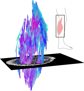
Cutting-edge imaging technologies are being used to understand what causes muscle contracture after a stroke and how we can improve treatments.
dti_gastrocnemius
Example of a 3D reconstruction of the muscle fibre structure of a human calf muscle (medial gastrocnemius) using diffusion tensor imaging.
We believe a lot of fundamental questions about muscles have yet to be answered, but that may be about to change with the use of diffusion tensor imaging (DTI).
DTI is an MRI-based imaging technique typically used to measure white matter in the brain. Recent advancements in imaging technology have allowed researchers, like ourselves, to more accurately measure muscle fibres as well.
A recent study we conducted revealed that DTI produces more reliable images and minimises the effect of errors typically found in ultrasound images. This is because muscles have complex 3D shapes, so 3D methods such as DTI have many advantages over the more conventional 2D techniques like ultrasound imaging.
We’ll put this information to good use in a new clinical trial aimed at understanding the cause of muscle contracture (stiffening of muscles) in stroke. Contracture is a common cause of disability in people who have had a stroke and, at present, the mechanisms that cause contracture are poorly understood. Additionally, current intervention strategies to treat contracture have been shown to be ineffective. Clearly, there is a need to improve our current understanding of contracture so that it can be treated effectively.
We are seeking volunteers to participate in the study. Participants could be people of any age who have had a stroke and have some physical disability, or healthy people aged over 60. Participation in the study involves coming to NeuRA and having an MRI scan. The visit, including the scan, takes around two hours.
If you are interested in finding out more about the study, please call NeuRA on 02 9399 1832 and ask to speak with Arkiev D’Souza, or send an email to a.dsouza@neura.edu.au with your enquiry.
- See more at: https://blog.neura.edu.au/2016/09/muscle-contracture-after-stroke/#sthash.G6k2GnGc.dpuf

Thursday, August 11, 2016

Can Neurological Biomarkers of Brain Impairment Be Used to Predict Poststroke Motor Recovery? A Systematic Review

Does no one else think this is fucking stupid to try to predict recovery without even acknowledging that even knowing this doesn't do survivors one damn bit of good because no protocols exist to map recovery to damaged areas? Everyone in stroke is completely flying blind.
http://nnr.sagepub.com/content/early/2016/08/05/1545968316662708.abstract

  1. Bokkyu Kim, MS1
  2. Carolee Winstein, PhD1⇑
  1. 1University of Southern California, Los Angeles, CA, USA
  1. Carolee Winstein, Motor Behavior and Neurorehabilitation Laboratory, Division of Biokinesiology and Physical Therapy, University of Southern California, 1540 Alcazar Street, CHP 155, Los Angeles, CA 90089, USA. Email: winstein@usc.edu

Abstract

Background. There is growing interest to establish recovery biomarkers, especially neurological biomarkers, in order to develop new therapies and prediction models for the promotion of stroke rehabilitation and recovery. However, there is no consensus among the neurorehabilitation community about which biomarker(s) have the highest predictive value for motor recovery. Objective. To review the evidence and determine which neurological biomarker(s) meet the high evidence quality criteria for use in predicting motor recovery. Methods. We searched databases for prognostic neuroimaging/neurophysiological studies. Methodological quality of each study was assessed using a previously employed comprehensive 15-item rating system. Furthermore, we used the GRADE approach and ranked the overall evidence quality for each category of neurologic biomarker. Results. Seventy-one articles met our inclusion criteria; 5 categories of neurologic biomarkers were identified: diffusion tensor imaging (DTI), transcranial magnetic stimulation (TMS), functional magnetic resonance imaging (fMRI), conventional structural MRI (sMRI), and a combination of these biomarkers. Most studies were conducted with individuals after ischemic stroke in the acute and/or subacute stage (~70%). Less than one-third of the studies (21/71) were assessed with satisfactory methodological quality (80% or more of total quality score). Conventional structural MRI and the combination biomarker categories ranked “high” in overall evidence quality. Conclusions. There were 3 prevalent methodological limitations: (a) lack of cross-validation, (b) lack of minimal clinically important difference (MCID) for motor outcomes, and (c) small sample size. More high-quality studies are needed to establish which neurological biomarkers are the best predictors of motor recovery after stroke. Finally, the quarter-century old methodological quality tool used here should be updated by inclusion of more contemporary methods and statistical approaches.

Monday, July 11, 2016

White Matter Changes Persist After Concussion

Followup research needed to see if this would be similar for stroke and what can be done to correct it. But this won't occur since we have NO stroke leadership to go to or a stroke strategy to update. Your children and grandchildren will continue to be screwed. 

White Matter Changes Persist After Concussion


Even though they no longer had symptoms, young athletes who had a concussion still had changes in the white matter of their brains half a year after their injury, researchers found.
In a single-center case-control study, high school and college athletes with a concussion showed decreased mean diffusivity -- a marker of white matter changes -- on diffusion tensor imaging at 6 months compared with healthy matched controls, according to Melissa Lancaster, PhD, of the Medical College of Wisconsin in Milwaukee, and colleagues.
That could have implications for managing concussions and determining recovery in athletes who have experienced a sports-related concussion, they reported at the American Academy of Neurology's Sports Concussion Conference in Chicago.
"Athletes may still experience long-term brain changes even after they feel they have recovered from the injury," Lancaster said in a statement. "Additional research is needed to determine how these changes relate to long-term outcomes."
Recent neuroimaging studies have suggested that sports-related concussion is associated with acute neuroanatomical and neurophysiological changes, and that white matter tracts are especially vulnerable to concussion. Yet the duration of those effects is not clear.
Lancaster and colleagues therefore conducted diffusion tensor imaging (DTI) and diffusion kurtosis tensor imaging (DKTI) on 17 high school and college football players, mean age 17.4 years, who had a concussion, and compared the scans with those from 18 matched athlete controls, mean age of 17.7, who did not have a concussion.
The researchers also assessed concussion symptoms, balance, and cognition at three time points: 24 hours after concussion, 8 days later, and 6 months later.
Overall, they saw no differences between the two groups in terms of self-reported concussion symptoms, cognition, or balance 6 months after the injury.
But there was widespread decreased mean diffusivity in those with a concussion compared with controls at that time point -- which was similar to the acute findings at 24 hours and 8 days, the researchers said.
Also, those with more severe symptoms at the time of concussion were more likely to have alterations in white matter 6 months later.
"Despite normalization of clinical symptoms, concussed athletes showed significant white matter alterations 6 months post-injury that were related to initial symptom severity ratings," Lancaster and colleagues concluded. "These findings have implications for determination of recovery following sports-related concussion and concussion management."
There were no differences, however, on DKTI, which is an emerging technique for evaluating the microstructural environment of the brain. The reasons for that are not clear, the researchers said, and further study is needed.
Additional study is also needed as to what, exactly, the changes seen on DTI actually mean, and whether or not they have implications for long-term recovery from concussion, as well as its long-term impact.
The hunt is still on for biomarkers that could help determine recovery, as well as for biomarkers that can help better diagnose concussion.
The study was supported by the National Center for Advancing Translational Sciences at the NIH, the U.S. Army Medical Research and Materiel Command, and the NFL-GE Head Health Challenge.
The authors disclosed having no financial relationships with industry.

Tuesday, October 27, 2015

Changes in structural integrity are correlated with motor and functional recovery after post-stroke rehabilitation.

No clue what this means so go ask your doctor or stroke association to clarify how this will help your recovery. Or is this once again describing something but not putting out a stroke protocol to actually help stroke survivors?
http://europepmc.org/abstract/med/26484696

School of Occupational Therapy, College of Medicine, National Taiwan University, Taipei, Taiwan.
Highlight Terms
Diffusion tensor imaging (DTI) studies indicate the structural integrity of the ipsilesional corticospinal tract (CST) and the transcallosal motor tract, which are closely linked to stroke recovery. However, the individual contribution of these 2 fibers on different levels of outcomes remains unclear. Here, we used DTI tractography to investigate whether structural changes of the ipsilesional CST and the transcallosal motor tracts associate with motor and functional recovery after stroke rehabilitation.Ten participants with post-acute stroke underwent the Fugl-Meyer Assessment (FMA), the Wolf Motor Function Test (WMFT), the Functional Independence Measure (FIM), and DTI before and after bilateral robotic training.All participants had marked improvements in motor performance, functional use of the affected arm, and independence in daily activities. Increased fractional anisotropy (FA) in the ipsilesional CST and the transcallosal motor tracts was noted from pre-treatment to the end of treatment. Participants with higher pre-to-post differences in FA values of the transcallosal motor tracts had greater gains in the WMFT and the FIM scores. A greater improvement on the FMA was coupled with increased FA changes along the ipsilesional CST.These findings suggest 2 different structural indicators for post-stroke recovery separately at the impairment-based and function-based levels.