Changing stroke rehab and research worldwide now.Time is Brain! trillions and trillions of neurons that DIE each day because there are NO effective hyperacute therapies besides tPA(only 12% effective). I have 523 posts on hyperacute therapy, enough for researchers to spend decades proving them out. These are my personal ideas and blog on stroke rehabilitation and stroke research. Do not attempt any of these without checking with your medical provider. Unless you join me in agitating, when you need these therapies they won't be there.

What this blog is for:

My blog is not to help survivors recover, it is to have the 10 million yearly stroke survivors light fires underneath their doctors, stroke hospitals and stroke researchers to get stroke solved. 100% recovery. The stroke medical world is completely failing at that goal, they don't even have it as a goal. Shortly after getting out of the hospital and getting NO information on the process or protocols of stroke rehabilitation and recovery I started searching on the internet and found that no other survivor received useful information. This is an attempt to cover all stroke rehabilitation information that should be readily available to survivors so they can talk with informed knowledge to their medical staff. It lays out what needs to be done to get stroke survivors closer to 100% recovery. It's quite disgusting that this information is not available from every stroke association and doctors group.

Showing posts with label researcher question. Show all posts

Monday, June 9, 2025

The Essential Fat Most Diets Are Missing (And Why It Matters for Aging Well)

 Now if researchers would tell us the EXACT amount of dairy fat to consume.

The Essential Fat Most Diets Are Missing (And Why It Matters for Aging Well)

A newly discovered essential fatty acid, C15:0, may help your cells resist aging,

[in-fluh-mey-shuhn] noun

Your body’s response to an illness, injury or something that doesn’t belong in your body (like germs or toxic chemicals).

Learn More
inflammation, and
[burn-out] noun

Physical or emotional exhaustion from chronic stress.

Learn More
burnout
.

Your ability to age with energy, clarity, and

[ri-zil-yuhns] noun

The ability to recover quickly from stress or setbacks.

Learn More
resilience relies on your daily habits and also your cellular resilience. And a little-known fatty acid, C15:0, is emerging as one of the most promising tools for supporting long-term cellular health.

Once overlooked, C15:0, also known as pentadecanoic acid, has now been recognized by scientists as the first essential fatty acid discovered in nearly a century. Recent research shows it may play a critical role in helping cells stay strong, reduce inflammation, and resist age-related decline.

In other words: C15:0 isn’t just another wellness trend. It could be the nutritional missing link in your

[lon-jev-i-tee] noun

Living a long life; influenced by genetics, environment, and lifestyle.

Learn More
longevity toolkit.

Why Cellular Resilience Matters More Than Ever

Every function in your body, from sleep and metabolism to memory and immunity, starts with how well your cells perform. But as we age, our cells become more susceptible to disruption. They lose structural integrity.

[mahy-tuh-kon-dree-uh] noun

Organelles in cells responsible for producing energy (ATP), often called the powerhouse of the cell.

Learn More
Mitochondria (your cellular energy engines) sputter. Inflammatory signals ramp up. Repair processes slow down.

This is the biological backdrop of aging, and where C15:0 comes in.

In peer-reviewed research published in Nutrients and Metabolites, scientists found that C15:0 supports:

  • Strenghtened cell membranes, reducing susceptibility to oxidative stress
  • Improved mitochondrial efficiency, which may boost daily energy
  • Decreased inflammatory cytokines like IL-6 and TNF-α
  • Activated longevity-related pathways, including AMPK and PPAR signaling

These mechanisms aren’t just theoretical. They translate into real-world benefits, like better sleep, improved metabolic health, and more vitality over time. Like other nutrients, your body’s C15:0 levels decrease with time. And now, researchers have proposed a new term to describe the signs and symptoms associated with C15:0 deficiency, Cellular Fragility Syndrome (CFS). 

“When our cells don’t have enough C15:0, they become fragile and age faster,” says Dr. Stephanie Venn-Watson, the veterinarian-turned-researcher who first uncovered the fatty acid’s power while studying aging dolphins for the U.S. Navy and reporting her findings in the journal Nature’s Scientific Reports.

The Discovery Story: Dolphins, Data, and a Surprising Breakthrough

C15:0 might not be mainstream yet, but its path to scientific recognition is anything but ordinary.

While working with the U.S. Navy to monitor dolphin health, Dr. Venn-Watson noticed something surprising: dolphins with higher levels of C15:0 aged better. Their cells stayed stronger. Their energy levels held up. That sparked more research and eventually a bold new hypothesis: that declining C15:0 levels may be linked to Cellular Fragility Syndrome (CFS).

Although not terminology formally recognized by conventional Western medicine, CFS symptoms mirror many of the frustrations adults begin to feel in midlife and beyond, including fatigue, brain fog, poor sleep, and slower recovery, but stem from one root issue: weakened cells.

Why Modern Diets May Be Starving Your Cells

Historically, humans consumed trace amounts of C15:0 through full-fat dairy and certain fish. But in recent decades, dietary shifts have caused those sources to drop off. Full-fat dairy became demonized. Plant-based and ultra-processed diets took center stage. And just like that, a key nutrient quietly slipped away. (Well, your competent? doctor had the dietician put dairy fat in the diet protocol, right?  NO? So, you DON'T have a functioning stroke doctor, do you?

dairy fat (31 posts to April 2016)

Dairy fat from milk, butter, and cheese could actually PREVENT a heart attack September 2021)

This isn’t the first time this has happened. Omega-3s followed a similar arc in the early 2000s, with deficiency linked to mood, memory, and cardiovascular issues before they were embraced as a staple of healthy aging. Now, C15:0 is having its moment.

What C15:0 Can Do for Longevity

In side-by-side comparisons with known longevity compounds like metformin and rapamycin, C15:0 demonstrated similar protective activity in human cell models, particularly around inflammation and cellular repair.

Supplementation studies, report:

  • Improved cholesterol and liver enzyme markers
  • Improved gut
    [mahy-kroh-bahy-ohm] noun

    The community of microorganisms (bacteria, viruses, fungi) living in a particular environment, especially the gut.

    Learn More
    microbiome
  • Healthier red blood cell profiles

Importantly, this isn’t about “

[bahy-oh-hak-ing] noun

Optimizing biology using science, technology, and lifestyle changes.

Learn More
biohacking” or quick fixes. C15:0 supports your body’s natural ability to heal, stabilize, and age well at the level of the cell.

A Fat That Speaks to Your Nervous System

What makes C15:0 truly unique isn’t just what it does for your cells, it’s how it communicates across your entire body.

Scientists recently discovered that one of its natural metabolites, pentadecanoylcarnitine, acts as a full endocannabinoid, meaning it can bind to and activate the body’s cannabinoid receptors (sometimes called your body’s “natural bliss” network) much like compounds produced by your own brain.

In a 2022 Scientific Reports study, researchers found that this C15:0-derived molecule:

  • Activated CB1 and CB2 cannabinoid receptors (the same receptors involved in regulating pain, mood, inflammation, and sleep)
  • Showed
    [an-tee-in-flam-uh-tawr-ee] adjective

    Reducing inflammation, which contributes to better overall health.

    Learn More
    anti-inflammatory
    effects across multiple disease-relevant cell systems
  • Influenced serotonin and histamine pathways tied to mental health and immune function

These effects suggest that C15:0 may help support not just physical vitality but emotional resilience as well.

For adults navigating hormonal shifts, chronic stress, or sleep challenges, this could be a game-changer. The ability to nourish your endocannabinoid system, which plays a key role in homeostasis, through diet or supplementation represents a new frontier in longevity and well-being.

How to Know If You’re Deficient in C15:0

C15:0 levels naturally decline with age, and today’s low-fat and dairy-free diets aren’t helping. Since this essential fatty acid is only found in trace amounts in certain animal-based foods, many people—especially those following plant-based or low-fat protocols—may unknowingly fall short.

Here’s how to spot a possible deficiency:

  • Blood levels below 0.2% of total circulating fatty acids may indicate low C15:0
  • High red cell distribution width (RDW), a common blood test marker, has been associated with C15:0 deficiency and increased oxidative stress
  • Non-specific symptoms may include low energy, disrupted sleep, chronic inflammation, slower recovery, or brain fog, signs that your cellular health may be struggling

You can ask your doctor for a fatty acid profile comprehensive test to see if your levels are low. There are some at-home testing options, as well. 

What to Look for in a C15:0 Supplement

Unlike food-based sources, where dosage and absorption vary, supplements offer a precise, stable, and consistent delivery of the fatty acid. If you’re considering a C15:0 supplement, quality and bioavailability matter. When selecting a supplement:

  • Look for third-party testing to ensure purity, potency, and absence of contaminants
  • Seek traceability and ingredient transparency
  • Check for published studies supporting its safety and efficacy
  • C15:0 in its purest supplemental form is FA15, a patented, vegan-friendly ingredient found in Fatty15.

And of course, if you’re on medications or managing chronic conditions, speak with your healthcare provider before adding anything new.

Thursday, September 19, 2024

Coffee could be more than a morning pick-me-up, according to new research

 Don't these researchers follow research at all? 

Not mentioning either Parkinsons or dementia prevention and is it really the caffeine?

Coffee May Lower Your Risk of Dementia Feb. 2013 

And this: Coffee's Phenylindanes Fight Alzheimer's Plaque December 2018

How coffee protects against Parkinson’s Aug. 2014 

Benefits Aren't Just From Caffeine December 2018

The latest here:

Coffee could be more than a morning pick-me-up, according to new research

A morning cup of coffee may do more than just perk you up, according to new research.

Moderate amounts of caffeine intake — defined as about three cups of coffee or tea a day — were associated with a lower risk of developing cardiometabolic multimorbidity, said the study’s lead author, Dr. Chaofu Ke, associate professor of epidemiology and biostatistics at Soochow University in Suzhou, China.

Cardiometabolic multimorbidity, or CM, is the coexistence of at least two cardiometabolic diseases such as coronary heart disease, stroke and type 2 diabetes.

“Coffee and caffeine consumption may play an important protective role in almost all phases of CM development,” Ke said.

Researchers analyzed data from about 180,000 people in the UK Biobank, a large biomedical database and research resource that follows people long-term. Those involved did not have cardiometabolic diseases at the outset.

The information included the participants’ self-reported caffeine consumption through coffee or black or greentea and the cardiometabolic diseases they developed through their primary care data, hospital records and death certificates, according to the study published Tuesday in the Endocrine Society’s Journal of Clinical Endocrinology & Metabolism.

Moderate caffeine consumers had a reduced risk of new onset cardiometabolic multimorbidity. The risk was reduced by 48.1% if they had three cups a day, or 40.7% if they had 200 to 300 milligrams of caffeine daily, compared with people who didn’t drink or drank less than one cup, Ke said.

What do you think? Join 114 others in the comments

The study had a large sample size and used multiple biomarkers to support the findings, making it a strong look at how caffeine affects heart health, said Dr. Gregory Marcus, associate chief of cardiology for research and professor of medicine at the University of California, San Francisco. He was not involved in the research.

“These observations add to the growing body of evidence that caffeine, and commonly consumed natural substances that contain caffeine such as tea and coffee, may enhance cardiovascular health,” Marcus said in an email.

What researchers don’t know

The methodology is strong and the results line up with existing data about caffeine and heart health, but there are still questions about the extent of the connection between caffeine and heart health, Marcus said.

“It is important to emphasize that, while these data suggest a relationship between caffeine, tea, and coffee and a reduced risk of a combination of cardiovascular diseases, we need to be careful before we infer true causal effects,” Marcus said.

Because the study is observational, it can only show a connection between caffeine and heart health, he said. Other factors may actually be the cause of the improved heart health, he added.

“It remains possible that the apparent protective effects do not truly exist at all and that the positive associations are all explained by some as yet unknown or unmeasured true determining factor,” Marcus added.

“For example, perhaps those more likely to consume these substances also tend to have a healthier diet or to be more physically active.”

The study also didn’t take into consideration the impact of caffeine from carbonated beverages or energy drinks, meaning that researchers can’t say whether those substances would also have a positive effect, Ke said.

Should you start drinking coffee?

Plenty of literature shows a benefit from caffeine consumption.

Several studies have suggested a lower risk of diabetes, Marcus said. And contrary to popular wisdom, drinking caffeine in coffee is associated with experiencing a lower risk of abnormal heart rhythms, he added, pointing to his and others’ research.

But much of that research is observational, and one study showed a mixed result, with more caffeine linked to additional daily step counts but less sleep, Marcus said.

Although the new study should provide comfort to those who already have a coffee or tea habit, it isn’t necessarily a sign to start a regular caffeine routine, Marcus said.

“It is also important to mention that more is not necessarily better,” he said.

“Even if caffeine, coffee, and tea in the amounts described in this study … are indeed healthy, there is also strong evidence that high-dose caffeine, particularly when included in artificial concoctions like energy drinks, may actually cause harmful and even dangerous heart rhythm problems.”

Clarification: A previous version of this story listed high blood pressure as a cardiometabolic disease. It has been removed from the list of such diseases to reflect the scope of the study.

Correction: A previous version of this story misstated the sample size of data collected from the UK Biobank and how many cups of coffee reduced risk.

Sunday, February 4, 2024

Spider venom molecule meets benchmarks to treat heart attack and stroke

 There is a venom library. What the fuck are our stroke researchers doing with this to help with stroke recovery? There is already this out there:

Snake Venom Helps Hydrogels Stop the Bleeding

Intravenous Ancrod for Treatment of Acute Ischemic Stroke

Biting back - snake venom contains toxic clotting factors


Snake Venom Could Hold Key To Alzheimer’s Breakthrough


Metrion taps Venomtech’s venom library for ion channel modulator discovery

The latest here:

Spider venom molecule meets benchmarks to treat heart attack and stroke

A spider venom molecule being investigated by a University of Queensland team has met critical benchmarks towards becoming a treatment for heart attack and stroke.

Associate Professor Nathan Palpant and Professor Glenn King from UQ's Institute for Molecular Bioscience have previously shown that the drug candidate Hi1a protects cells from the damage caused by heart attack and stroke.

Dr Palpant said a subsequent study has put the drug through a series of preclinical tests designed to mimic real-life treatment scenarios.

These tests are a major step towards helping us understand how Hi1a would work as a therapeutic – at what stage of a heart attack it could be used and what the doses should be.

We established that Hi1a is as effective at protecting the heart as the only cardioprotective drug to reach Phase 3 clinical trials, a drug that was ultimately shelved due to side effects.

Importantly, we found that Hi1a only interacts with cells in the injured zone of the heart during an attack and doesn't bind to healthy regions of the heart – reducing the chance of side effects."

Dr. Nathan Palpant, Associate Professor, UQ's Institute for Molecular Bioscience

Professor King, who recently won the Prime Minister's Prize for Innovation for developing the world's first insecticides from spider venom, discovered Hi1a in the venom of the K'gari funnel web spider.

"Hi1a could reduce damage to the heart and brain during heart attacks and strokes by preventing cell death caused by lack of oxygen," Professor King said.

"Our testing and safety studies from independent contract research organisations has provided evidence that Hi1a could be an effective and safe therapeutic."

Infensa Bioscience, a company co-founded by the researchers, raised $23 million in 2022 to develop Hi1a for commercial purposes.

Infensa CEO and UQ researcher, Associate Professor Mark Smythe, said cardiovascular disease is the leading cause of death globally.

"Most deaths from cardiovascular disease are caused by heart attacks and strokes, yet there are no drugs on the market that prevent the damage they cause," Dr Smythe said.

"An effective drug to treat heart attacks would have worldwide impact, providing a breakthrough to improve the lives of millions of individuals living with heart disease."

The research team included Dr Meredith Redd from IMB as well as Dr Melissa Reichelt and Dr Yusuke Yoshikawa from UQ's School of Biomedical Sciences.

The study was published in the world's leading cardiac journal The European Heart Journal.

Source:
Journal reference:

Redd, M. A., et al. (2023). Acid-sensing ion channel 1a blockade reduces myocardial injury in rodent models of myocardial infarction. European Heart Journal. doi.org/10.1093/eurheartj/ehad793.

Revolutionary 3D-Printed Brain Tissue Mimics Human Function

Which built brain should our researchers be using?. I expect our researchers to be using the best one.

Nearly complete human brain grown in US lab: scientist August 2015

Multiregional brain on a chip  Jan 2017
Draper Laboratory developing “Brain-on-a-Chip”  October 2012

"Alzheimer's-in-a-Dish" Docs Win Top Smithsonian Ingenuity Award Nov. 2015 

A patient’s budding cortex — in a dish?  June 2015 


,Cell cultures in petri dishes open new doors to brain research  April 2017

Scientists create 3D-printed brain-like tissue from stem cells July 2017

3D Mini-Brains Accelerate Research for Repairing Brain Function December 2017

 The latest here:

Revolutionary 3D-Printed Brain Tissue Mimics Human Function

Summary: Researchers developed the world’s first 3D-printed brain tissue that grows and behaves similarly to natural brain tissue, marking a significant leap forward for neurological and neurodevelopmental disorder research.

This novel 3D-printing technique uses a horizontal layering approach and a softer bio-ink, allowing neurons to interconnect and form networks akin to human brain structures.

The ability to precisely control cell types and arrangements provides unparalleled opportunities to study brain functions and disorders in a controlled environment, offering new avenues for drug testing and understanding brain development and diseases like Alzheimer’s and Parkinson’s.

Key Facts:

  1. The 3D-printed brain tissue can form networks and communicate through neurotransmitters, similar to human brain interactions.
  2. This new printing method allows for precise control over cell types and arrangements, surpassing the capabilities of traditional brain organoids.
  3. The technique is accessible to many labs, not requiring special equipment or culture methods, and can significantly impact the study of various neurological conditions and treatments.

Source: University of Wisconsin

A team of University of Wisconsin–Madison scientists has developed the first 3D-printed brain tissue that can grow and function like typical brain tissue.

It’s an achievement with important implications for scientists studying the brain and working on treatments for a broad range of neurological and neurodevelopmental disorders, such as Alzheimer’s and Parkinson’s disease.

“This could be a hugely powerful model to help us understand how brain cells and parts of the brain communicate in humans,” says Su-Chun Zhang, professor of neuroscience and neurology at UW–Madison’s Waisman Center.

This shows a brain.
“Our tissue stays relatively thin and this makes it easy for the neurons to get enough oxygen and enough nutrients from the growth media,” Yan says. Credit: Neuroscience News

“It could change the way we look at stem cell biology, neuroscience, and the pathogenesis of many neurological and psychiatric disorders.”

Printing methods have limited the success of previous attempts to print brain tissue, according to Zhang and Yuanwei Yan, a scientist in Zhang’s lab. The group behind the new 3D-printing process described their method today in the journal Cell Stem Cell.

Instead of using the traditional 3D-printing approach, stacking layers vertically, the researchers went horizontally. They situated brain cells, neurons grown from induced pluripotent stem cells, in a softer “bio-ink” gel than previous attempts had employed.

“The tissue still has enough structure to hold together but it is soft enough to allow the neurons to grow into each other and start talking to each other,” Zhang says.

The cells are laid next to each other like pencils laid next to each other on a tabletop.

“Our tissue stays relatively thin and this makes it easy for the neurons to get enough oxygen and enough nutrients from the growth media,” Yan says.

The results speak for themselves — which is to say, the cells can speak to each other. The printed cells reach through the medium to form connections inside each printed layer as well as across layers, forming networks comparable to human brains.

The neurons communicate, send signals, interact with each other through neurotransmitters, and even form proper networks with support cells that were added to the printed tissue.

“We printed the cerebral cortex and the striatum and what we found was quite striking,” Zhang says. “Even when we printed different cells belonging to different parts of the brain, they were still able to talk to each other in a very special and specific way.”

The printing technique offers precision — control over the types and arrangement of cells — not found in brain organoids, miniature organs used to study brains. The organoids grow with less organization and control.

“Our lab is very special in that we are able to produce pretty much any type of neurons at any time. Then we can piece them together at almost any time and in whatever way we like,” Zhang says.

“Because we can print the tissue by design, we can have a defined system to look at how our human brain network operates. We can look very specifically at how the nerve cells talk to each other under certain conditions because we can print exactly what we want.”

That specificity provides flexibility. The printed brain tissue could be used to study signaling between cells in Down syndrome, interactions between healthy tissue and neighboring tissue affected by Alzheimer’s, testing new drug candidates, or even watching the brain grow.

“In the past, we have often looked at one thing at a time, which means we often miss some critical components. Our brain operates in networks. We want to print brain tissue this way because cells do not operate by themselves. They talk to each other. This is how our brain works and it has to be studied all together like this to truly understand it,” Zhang says.

“Our brain tissue could be used to study almost every major aspect of what many people at the Waisman Center are working on. It can be used to look at the molecular mechanisms underlying brain development, human development, developmental disabilities, neurodegenerative disorders, and more.”

The new printing technique should also be accessible to many labs. It does not require special bio-printing equipment or culturing methods to keep the tissue healthy, and can be studied in depth with microscopes, standard imaging techniques and electrodes already common in the field.

The researchers would like to explore the potential of specialization, though, further improving their bio-ink and refining their equipment to allow for specific orientations of cells within their printed tissue..

“Right now, our printer is a benchtop commercialized one,” Yan says. “We can make some specialized improvements to help us print specific types of brain tissue on-demand.”

Funding: This study was supported in part by NIH-NINDS (NS096282, NS076352, NS086604), NICHD (HD106197, HD090256), the National Medical Research Council of Singapore (MOH-000212, MOH-000207), Ministry of Education of Singapore (MOE2018-T2-2-103), Aligning Science Across Parkinson’s (ASAP-000301), the Bleser Family Foundation, and the Busta Foundation.

About this neurotech research news

Author: Emily Leclerc
Source: University of Wisconsin
Contact: Emily Leclerc – University of Wisconsin
Image: The image is credited to Neuroscience News

Original Research: Open access.
“3D bioprinting of human neural tissues with functional connectivity” by Su-Chun Zhang et al. Cell Stem Cell


Abstract

3D bioprinting of human neural tissues with functional connectivity

Highlights

  • Functional human neural tissues assembled by 3D bioprinting
  • Neural circuits formed between defined neural subtypes
  • Functional connections established between cortical-striatal tissues
  • Printed tissues for modeling neural network impairment

Summary

Probing how human neural networks operate is hindered by the lack of reliable human neural tissues amenable to the dynamic functional assessment of neural circuits. We developed a 3D bioprinting platform to assemble tissues with defined human neural cell types in a desired dimension using a commercial bioprinter.

The printed neuronal progenitors differentiate into neurons and form functional neural circuits within and between tissue layers with specificity within weeks, evidenced by the cortical-to-striatal projection, spontaneous synaptic currents, and synaptic response to neuronal excitation.

Printed astrocyte progenitors develop into mature astrocytes with elaborated processes and form functional neuron-astrocyte networks, indicated by calcium flux and glutamate uptake in response to neuronal excitation under physiological and pathological conditions.

These designed human neural tissues will likely be useful for understanding the wiring of human neural networks, modeling pathological processes, and serving as platforms for drug testing.

Saturday, May 13, 2023

Accessing the brain with soft deployable electrocorticography arrays

When are our researchers going to find and interpret the signals between neurons that cause neuroplasticity to happen?  Or are they already using these and we don't know about it yet?


Accessing the brain with soft deployable electrocorticography arrays

Abstract

Soft robotics facilitates the deployment of large radial electrode arrays on the brain cortex through small craniotomies.

Get full access to this article

View all available purchase options and get full access to this article.


Monday, April 24, 2023

Alzheimer's Proteins Reduced by Sleep Drug

 This really tells us nothing until further research is completed and we find out if it reduces the Alzheimer's risk or clears up Alzheimer's symptoms. And impaired persons need to be in the research.

 Your doctor - IF COMPETENT - needs to ensure further research is completed. 

Your risk of dementia, has your doctor told you of this?

1. A documented 33% dementia chance post-stroke from an Australian study?   May 2012.

2. Then this study came out and seems to have a range from 17-66%. December 2013.`    

3. A 20% chance in this research.   July 2013.

4. Dementia Risk Doubled in Patients Following Stroke September 2018 

The latest here:

Alzheimer's Proteins Reduced by Sleep Drug

Both amyloid and tau levels fell in early trial

A photo of a mature woman sitting on the edge of her bed in a dark room.

Suvorexant (Belsomra), a dual orexin receptor antagonist approved for insomnia, reduced levels of tau phosphorylation and amyloid beta, a small clinical trialopens in a new tab or window showed.

The ratio of phosphorylated tau-threonine-181 (p-tau-181) to unphosphorylated tau-threonine-181 decreased 10% to 15% in cognitively normal adults treated with suvorexant 20 mg compared with placebo, reported Brendan Lucey, MD, MSCI, of Washington University School of Medicine in St. Louis, and co-authors.

Amyloid-beta levels fell 10% to 20% compared with placebo starting 5 hours after suvorexant administration, the researchers wrote in Annals of Neurologyopens in a new tab or window.

"This is a small, proof-of-concept study," Lucey said in a statement. "We don't yet know whether long-term use is effective in staving off cognitive decline, and if it is, at what dose and for whom."

"Still, these results are very encouraging," he added. "This drug is already available and proven safe, and now we have evidence that it affects the levels of proteins that are critical for driving Alzheimer's disease."

In recent years, researchers have moved closer to understanding the complex relationshipopens in a new tab or window between sleep and Alzheimer's disease. In earlier work, Lucey and colleagues reported that older adults who had less slow-wave sleep had higher levels of brain tauopens in a new tab or window. Other studies have shown that sleep apnea was tied to higher tau burdenopens in a new tab or window.

But what's been called the chicken-and-egg questionopens in a new tab or window by Alzheimer's researcher Ron Petersen, MD, PhD, of the Mayo Clinic in Rochester, Minnesota, still hasn't been answered: "Is it that your sleep is disrupted and the Alzheimer's proteins build up -- or are the Alzheimer's proteins being deposited in the brain, disrupting sleep, and that's where the cycle gets initiated?"

Orexin is a wake-promoting neuropeptide. Evidence supports a role for the orexin system in the development of Alzheimer's pathology, Lucey and co-authors noted. In mouse models, dual orexin receptor antagonists have been shown to decrease soluble amyloid-beta levelsopens in a new tab or window and amyloid plaques.

Lucey and colleagues recruited 38 people ages 45 to 65 with no cognitive impairment to undergo a 2-night sleep study, randomizing them to suvorexant 10 mg (13 people), suvorexant 20 mg (12 people), or placebo (13 people). They assessed cerebrospinal fluid (CSF) via intrathecal lumbar catheter every 2 hours for 36 hours, starting 1 hour before suvorexant or placebo was given.

Participants mostly were women (68.4%) and white (78.9%). All were in good general health and had no clinical sleep or neurologic disease.

Suvorexant 10 mg did not show a statistically significant effect on p-tau-181 or amyloid compared with placebo. Neither dose of suvorexant significantly increased total sleep time, sleep efficiency, time in non-rapid eye movement (REM) sleep, or time in REM sleep over placebo. Suvorexant did not decrease phosphorylation at tau-serine-202 or tau-threonine-217.

At 24 hours after the first dose, p-tau-181 increased but amyloid levels remained low in the 20-mg group. Levels of both proteins fell again after suvorexant 20 mg was administered on the second night.

Suvorexant's action may extend beyond sleep induction at night, Lucey and colleagues observed. The response of CSF tau and amyloid to suvorexant without a significant change in sleep suggests that different mechanistic pathways may be involved, they noted.

"If we can lower amyloid every day, we think the accumulation of amyloid plaques in the brain will decrease over time," Lucey said. "If you can reduce tau phosphorylation, potentially there would be less tangle formation and less neuronal death."

The researchers have funding for additional trials to answer some outstanding questions, Lucey stated at a press conference. "Do we see similar changes in these biomarkers or these proteins when these drugs are given for months? That's one question that we have," he said.(My question is: 'Does it prevent Alzheimers? That seems important to answer.)

The group also will study cognitively unimpaired people with biomarker evidence of amyloid pathology and "see if we see similar changes, which would suggest potential larger studies could then be done as secondary prevention for Alzheimer's disease," he added.

The findings were limited by the study's small sample size. In addition, two other dual orexin receptor antagonists -- lemborexant (Dayvigo) and daridorexant (Quviviq) -- recently received FDA approval to treat insomnia. Future studies should test whether these drugs show the same effects on amyloid and tau, Lucey noted.

  • Judy George covers neurology and neuroscience news for MedPage Today, writing about brain aging, Alzheimer’s, dementia, MS, rare diseases, epilepsy, autism, headache, stroke, Parkinson’s, ALS, concussion, CTE, sleep, pain, and more. Follow

Disclosures

This study was funded by the National Institutes of Health and the BrightFocus Foundation.

Lucey has consulted for Merck, which makes suvorexant. Co-authors disclosed relationships with C2N Diagnostics.

Primary Source

Annals of Neurology

Source Reference: opens in a new tab or windowLucey BP, et al "Suvorexant acutely decreases tau phosphorylation and Aβ in the human CNS" Ann Neurol 2023; DOI: 10.1002/ana.26641.

Monday, February 6, 2023

Blobs of human brain planted in rats offer new treatment hope

Which built brain should our researchers be using? I expect our researchers to be using the best one. Shades of Christine O’Donnell famously insisting that scientists were putting human brains into mice.

Nearly complete human brain grown in US lab: scientist.

Multiregional brain on a chip  Jan 2017


Draper Laboratory developing “Brain-on-a-Chip”  October 2012


"Alzheimer's-in-a-Dish" Docs Win Top Smithsonian Ingenuity Award Nov. 2015 

A patient’s budding cortex — in a dish?  June 2015 


Cell cultures in petri dishes open new doors to brain research  April 2017


Blobs of human brain planted in rats offer new treatment hope

Scientists suggest patient’s own cells could be grown in the lab and used to repair stroke or trauma injuries

A white rat
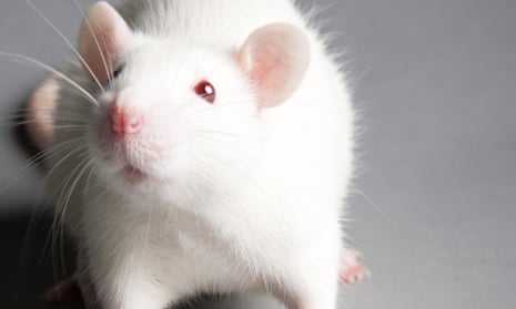
In the study, human brain tissue was transplanted into the brains of adult rats. Photograph: Zoonar GmbH/Alamy
Science correspondent
Fri 3 Feb 2023 04.57 ESTLast modified on Fri 3 Feb 2023 05.14 EST


  • Blobs of human brain tissue have been transplanted into the brains of rats in work that could pave the way for new treatments for devastating brain injuries.

    The groundbreaking study showed that the “human brain organoids” – sesame seed-sized balls of neurons – were able to integrate into the rat brain, linking up with their blood supplies and communicating with the rat neurons.

    The team behind the work suggest that eventually doctors might be able to grow blobs of brain tissue from a patient’s own cells in the lab and use them to repair brain injuries caused by stroke or trauma.

    “This is incredibly exciting to me as a physician,” said Isaac Chen, a physician and assistant professor of neurosurgery at the University of Pennsylvania.

    The study is the latest in the rapidly growing and ethically complex field of brain organoids. Scientists have shown that when cultivated in the right conditions, neurons begin to form tiny brain-like structures, allowing scientists to investigate developmental conditions such as autism and a wide range of basic neuroscience questions.

    The new work is the first demonstration that the lab-grown brain tissue can be successfully implanted into an injury site to repair an adult brain, suggesting there could be future clinical applications.

    Chen and colleagues grew human brain organoids in a dish until they were about 1.5mm in diameter. The balls of tissue were then transplanted into the brains of adult rats that had sustained injuries to their visual cortex. Within three months, the grafted organoids had integrated with their host’s brain, hooking up with the blood supply, expanding to several times the initial volume and sending out projections that linked up with the rat’s neurons, according to the study published in Cell Stem Cell.

    “We were not expecting to see this degree of functional integration so early,” says Chen. “[This] suggests that neural tissue transplantation in the adult mammalian brain, especially one that has been disrupted with some sort of injury, really is a viable path forward for neural repair.”

    The scientists did not assess whether the implants improved how well the rats were able to function, but tests showed that the human neurons fired off electrical signals when the rats were exposed to flashing lights. Chen said that this supported the idea that organoids could act as “blank processing units” that the brain could absorb and use to rebuild itself after injury.

    “By rationally introducing these engineered processing units to specific areas of the injured brain, we think that the increased computational capacity of those areas would result in sufficient restoration of brain networks to restore neurological function,” said Chen.

    In theory, personalised brain organoids could be created in the lab from a patient’s own cells, although Chen predicted that clinical applications would be at least five to 10 years away. “We are at the very beginning of this journey,” he said.

    skip past newsletter promotion

    Dr Serena Barral, a lecturer in developmental neuroscience at UCL, who was not involved in the work, described it as an “incredible” demonstration of the sheer adaptability of neurons. “There’s a lot of information that’s in the DNA itself and that allows the neurons to work wherever they are – whether they’re in a plastic box in the lab or in the brain,” she said.

    She added that in future clinical applications, the degree to which the brain could be repaired was likely to depend on which functions had been lost.

    “The visual cortex is more simple, but if you think about replacing the areas that are important for speech, mathematical calculations, thinking – that could be a little bit more tricky because there are a lot of abilities of the brain that are grown with experience,” she said.

    Monday, March 21, 2022

    Effects of Involuntary and Voluntary Exercise in Combination with Acousto-Optic Stimulation on Adult Neurogenesis in an Alzheimer's Mouse Model

    I want to know how in hell you would ever translate involuntary treadmill running in mice to a human model. With no answer this research is useless.

    Effects of Involuntary and Voluntary Exercise in Combination with Acousto-Optic Stimulation on Adult Neurogenesis in an Alzheimer's Mouse Model

    Abstract

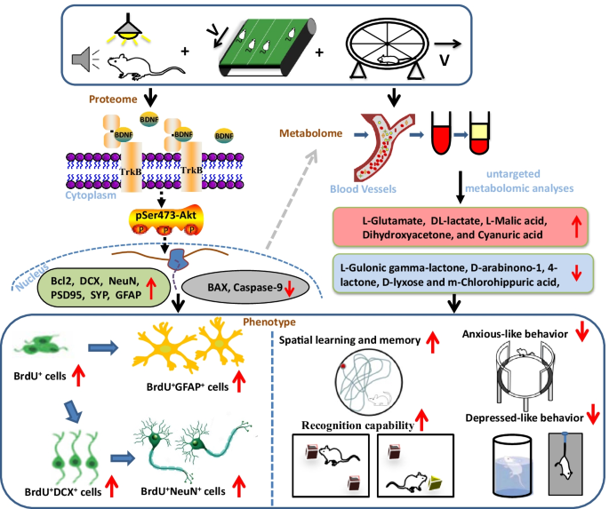
    Single-factor intervention, such as physical exercise and auditory and visual stimulation, plays a positive role on the prevention and treatment of Alzheimer’s disease (AD); however, the therapeutic effects of single-factor intervention are limited. The beneficial effects of these multifactor combinations on AD and its molecular mechanism have yet to be elucidated. Here, we investigated the effect of multifactor intervention, voluntary wheel exercise, and involuntary treadmill running in combination with acousto-optic stimulation, on adult neurogenesis and behavioral phenotypes in a mouse model of AD. We found that 4 weeks of multifactor intervention can significantly increase the production of newborn cells (BrdU+ cells) and immature neurons (DCX+ cells) in the hippocampus and lateral ventricle of Aβ oligomer-induced mice. Importantly, the multifactor intervention could promote BrdU+ cells to differentiate into neurons (BrdU+ DCX+ cells or BrdU+ NeuN+ cells) and astrocytes (BrdU+GFAP+ cells) in the hippocampus and ameliorate Aβ oligomer-induced cognitive impairment and anxiety- and depression-like behaviors in mice evaluated by novel object recognition, Morris water maze tests, elevated zero maze, forced swimming test, and tail suspension test, respectively. Moreover, multifactor intervention could lead to an increase in the protein levels of PSD-95, SYP, DCX, NeuN, GFAP, Bcl-2, BDNF, TrkB, and pSer473-Akt and a decrease in the protein levels of BAX and caspase-9 in the hippocampal lysates of Aβ oligomer-induced mice. Furthermore, sequencing analysis of serum metabolites revealed that aberrantly expressed metabolites modulated by multifactor intervention were highly enriched in the biological process associated with keeping neurons functioning and neurobehavioral function. Additionally, the intervention-mediated serum metabolites mainly participated in glutamate metabolism, glucose metabolism, and the tricarboxylic acid cycle in mice. Our findings suggest the potential of multifactor intervention as a non-invasive therapeutic strategy for AD to anti-Aβ oligomer neurotoxicity.

    Graphical abstract

    This is a preview of subscription content, access via your institution.

    Data Availability

    The data generated during the current study are available from the corresponding author on reasonable request.