Changing stroke rehab and research worldwide now.Time is Brain! trillions and trillions of neurons that DIE each day because there are NO effective hyperacute therapies besides tPA(only 12% effective). I have 523 posts on hyperacute therapy, enough for researchers to spend decades proving them out. These are my personal ideas and blog on stroke rehabilitation and stroke research. Do not attempt any of these without checking with your medical provider. Unless you join me in agitating, when you need these therapies they won't be there.

What this blog is for:

My blog is not to help survivors recover, it is to have the 10 million yearly stroke survivors light fires underneath their doctors, stroke hospitals and stroke researchers to get stroke solved. 100% recovery. The stroke medical world is completely failing at that goal, they don't even have it as a goal. Shortly after getting out of the hospital and getting NO information on the process or protocols of stroke rehabilitation and recovery I started searching on the internet and found that no other survivor received useful information. This is an attempt to cover all stroke rehabilitation information that should be readily available to survivors so they can talk with informed knowledge to their medical staff. It lays out what needs to be done to get stroke survivors closer to 100% recovery. It's quite disgusting that this information is not available from every stroke association and doctors group.

Showing posts with label not useful. Show all posts

Friday, January 24, 2020

Gait impairment in neurological disorders: a new technological approach

Notice that 'improve' and not yet sufficient are what you get from gait rehab. So you are a one person guinea pig in an unregistered clinical trial. This times 10 million yearly stroke survivors.   And our fucking failures of stroke associations, along with your stroke doctors and stroke hospital are OK with doing nothing to solve that.  That is the tyranny of low expectations you have to fight against. Screaming may be required.

Gait impairment in neurological disorders: a new technological approach

 Roberta Semprini,MD
a
Patrizio Sale, MD
a1
Calogero Foti,MD
b
Massimo Fini,MD
a
Marco Franceschini MD
a
a
IRCCS San Raffaele Pisana, Rome, Italy
b
Physical and Rehabilitation Medicine, Public HealthDepartment, Tor Vergata University, Rome, ItalyCorresponding author:Marco FranceschiniIRCCS San Raffaele Pisana Via della Pisana, 235Rome, ItalyE-mail: marco.franceschini@sanraffaele.it

Summary

Gait recovery is considered one of the main objectives of rehabilitation interventions in neurological disabilities, as restricted movement can significantly reduce an individualʼs ability to take part in normal activities of daily living. Locomotor training has been shown to improve gait rehabilitation. Studies have recently been published on the use of robots and other devices in patients with gait disabilities, particularly in the rehabilitation of the lower limbs. However, analysis of the recent literature reveals a relative paucity of strong methodological studies. The evidence that is available, while strong, is not yet sufficient to allow definite conclusions to be drawn regarding the efficacy of these devices. From these considerations, it is clear that validated and standardized methods need to be adopted for each of the different systems available. This would help to clarify the indications for and correct use of robotic devices in the different neurological disorders.

Monday, July 8, 2019

Baptist Health Paducah stroke program receives national quality award

Big fucking whoopee.

  'Care' NOT RESULTS. 

I hate when they do this chest thumping thing for nothing proven useful for survivors.


Baptist Health Paducah stroke program receives national quality award

PADUCAH —  The Baptist Health Paducah stroke program joined an elite group by being honored with The Get With the Guidelines® Silver Plus Quality Achievement Award for meeting national guidelines for stroke care, as outlined by the American Heart Association/American Stroke Association.
 In addition, the hospital received the associations’ Target: Stroke Honor Roll Elite for meeting stroke quality measures that reduce the time between hospital arrival and treatment with the clot-busting drug known as tPA. If given intravenously in the first three hours after the start of stroke symptoms, tPA has been shown to significantly reduce the effects of stroke and lessen the chance of permanent disability.
 “This award says we strive for the best possible care for our patients,” said neurologist Joseph Ashburn, MD, stroke services director. “While meeting the minimum requirements for a stroke center is considered acceptable, we at Baptist Health believe the people of our region deserve nothing less than the very best medicine has to offer. We are always moving forward to take it to the next level.”
 Baptist Health Paducah became the region’s first certified primary stroke center in 2009. Since then, the administration of tPA has increased from 9 percent of total stroke patients to 75 percent for eligible patients.
 Neuroscience coordinator Chapman Offutt, RN, said the hospital is 100 percent compliant with a door to tPA administration time of less than 60 minutes, and met the goal of 50 percent or more of tPA patients receiving the drug less than 45 minutes after arrival.  
“This is a huge accomplishment, given the steps that must be completed prior to administering the medication,” Offutt said. “We have developed a very rapid and efficient process to accomplish these goals.”

Tuesday, May 28, 2019

Putting the “Sensory” Into Sensorimotor Control: The Role of Sensorimotor Integration in Goal-Directed Hand Movements After Stroke

But is this better than the Margaret Yekutiel  book about this from 2001, 'Sensory Re-Education of the Hand After Stroke'? The objective should have been to update the sersorimotor recovery protocol with the newest research results.  Not whatever the fuck this review was for.  Lots of big words in here but I see nothing that even remotely looks like useful rehab interventions. And the conclusion is more research needed.


Putting the “Sensory” Into Sensorimotor Control: The Role of Sensorimotor Integration in Goal-Directed Hand Movements After Stroke

  • 1Neuroscience Graduate Program, Graduate Division of Biological and Biomedical Sciences, Emory University, Atlanta, GA, United States
  • 2Department of Rehabilitation Medicine, Laney Graduate School, Emory University, Atlanta, GA, United States
  • 3Department of Neurology, Emory University, Atlanta, GA, United States
  • 4Department of Radiology and Imaging Sciences, School of Medicine, Emory University, Atlanta, GA, United States
Integration of sensory and motor information is one-step, among others, that underlies the successful production of goal-directed hand movements necessary for interacting with our environment. Disruption of sensorimotor integration is prevalent in many neurologic disorders, including stroke. In most stroke survivors, persistent paresis of the hand reduces function and overall quality of life. Current rehabilitative methods are based on neuroplastic principles to promote motor learning that focuses on regaining motor function lost due to paresis, but the sensory contributions to motor control and learning are often overlooked and currently understudied. There is a need to evaluate and understand the contribution of both sensory and motor function in the rehabilitation of skilled hand movements after stroke. Here, we will highlight the importance of integration of sensory and motor information to produce skilled hand movements in healthy individuals and individuals after stroke. We will then discuss how compromised sensorimotor integration influences relearning of skilled hand movements after stroke. Finally, we will propose an approach to target sensorimotor integration through manipulation of sensory input and motor output that may have therapeutic implications.

Introduction

Goal-directed movements of the hand are required to perform most tasks of daily living, such as tying a shoe, buttoning a shirt, and typing, among others. These highly coordinated voluntary movements involve interacting with and manipulating objects in the environment and rely on sensorimotor integration. Sensorimotor integration is the ability to incorporate sensory inputs that provide information about one’s body and the external environment to inform and shape motor output (Wolpert et al., 1998). More specifically, sensory inputs for goal-directed hand movements provide information in an egocentric reference frame detailing location, size, weight, and shape of an object. In addition, kinematic information about the hand and upper extremity, including the trajectory needed to interact with the object, is provided. Successful integration of information contributes to generating the most efficient motor plan to execute a given task. Additionally, ongoing sensory feedback during motor performance refines the motor plan to optimize current and future performance. This process of sensorimotor integration is often disrupted in neurological disorders, such as stroke.
Stroke is defined as infarction of central nervous system tissue attributable to ischemia, based on neuropathological, neuroimaging, and/or clinical evidence of permanent injury (Sacco et al., 2013). Stroke is the fourth leading cause of death and remains the number one leading cause of long-term adult disability (Benjamin et al., 2017). Furthermore, the loss of productivity after stroke currently costs the United States an average of $33.9 billion per year and is expected to reach $56 billion by 2030 (Ovbiagele et al., 2013), making stroke a public health crisis. A primary contributor to persistent disability after stroke is incomplete motor recovery (Lai et al., 2002). Spontaneous biological recovery of motor function occurs during the first months after stroke (Cramer, 2008), underlying a current emphasis on intensive early intervention, although results are often mixed and complex (Bernhardt et al., 2017a). Despite intensive therapy, upper extremity impairment resolves up to 70% of baseline function for a given patient with some patients showing even less recovery than predicted (Winters et al., 2015). Most stroke survivors are left with a limited ability to perform skilled hand movements necessary for daily functioning (Lang et al., 2013). To reduce disability after stroke, there is a need to improve our understanding of the neuronal network physiology necessary to regain skilled functional hand use.
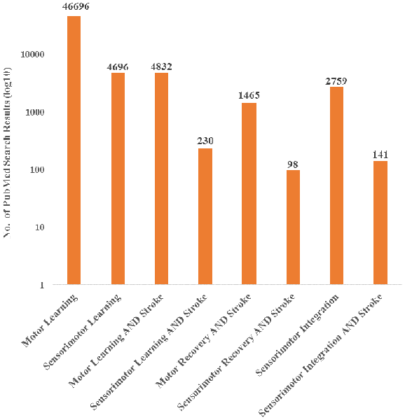
Currently, the field has primarily investigated motor deficits and motor learning with limited consideration of the role of sensory information, even though it is recognized that integration of sensory information is a critical component of motor control (Borich et al., 2015; Bolognini et al., 2016). Furthermore, evidence has shown that sensory input is important for recovery after stroke. In a systematic review, Meyer et al. found that across six studies, the extent of deficits in proprioception and light touch of the arm and hand were significantly related to recovery after stroke (Meyer et al., 2014). Despite evidence that sensory input is a critical component to motor execution, research nomenclature has been primarily focused on motor characteristics post-stroke and has therefore not capitalized fully on the information a sensorimotor perspective could provide. This observation is supported by a literature search showing an emphasis towards motor recovery and learning after stroke, over sensorimotor recovery and learning, with limited focus on sensorimotor integration (Figure 1). While it is possible that authors may use these terms interchangeably, the literature search terminology suggests that there is potential bias towards motor contributions. Therefore, there is an important gap in our understanding of the contributions of sensorimotor integration to recovery.
FIGURE 1
www.frontiersin.org Figure 1. PubMed search results for both motor and sensorimotor aspects of learning and stroke recovery. More publications focused on motor learning and recovery than on both motor and sensory components of learning and recovery. Furthermore, there were a relatively small number of publications involving sensorimotor integration and stroke compared to sensorimotor integration overall. “Sensorimotor,” “Sensori-motor,” and “Sensory motor” were all used to ensure differences in terminology did not affect the search results. Additionally, “Sensory motor” and “Sensory-motor” produced the same search results.
In the following brief review, we will highlight the importance of processing and integrating sensory and motor information that underlies skill performance and learning with an emphasis on skilled hand movements in stroke. We will focus primarily on three cortical regions: primary motor cortex (M1), posterior parietal cortex (PPC) and primary somatosensory cortex (S1) while briefly mentioning other cortical and subcortical brain areas also involved in sensorimotor integration. These brain regions are highlighted due to our focus on the integration of sensory and motor information at the level of the cortex, but also because these cortical areas receive blood supply from the middle cerebral artery (MCA), which is the most common type of stroke (Walcott et al., 2014). Furthermore, all three brain regions contribute to the corticospinal tract (CST) that provide necessary contributions to executing and controlling skilled hand movements routinely used in daily life. It should be noted that strokes occur in other brain regions but usually have less of an impact on sensorimotor integration underlying goal-directed, skilled hand movements and are outside the primary scope of this review article.
In the first section of this review article, we will discuss the role of sensorimotor integration via M1, PPC, and S1 in normal, skilled hand movements. We will then discuss how sensorimotor integration is affected by stroke and how impaired sensorimotor integration can impact relearning of skilled hand movements. Last, we propose an approach to target sensorimotor integration by manipulating sensory input and restricting motor output that may have therapeutic implications for stroke recovery.

More at link. 

Redundancy Among Parameters Describing the Input-Output Relation of Motor Evoked Potentials in Healthy Subjects and Stroke Patients

I got absolutely nothing out of this that would help survivors recover. 

Redundancy Among Parameters Describing the Input-Output Relation of Motor Evoked Potentials in Healthy Subjects and Stroke Patients

Claire Kemlin1, Eric Moulton1, Sara Leder2, Marion Houot3, Sabine Meunier1, Charlotte Rosso1,2† and Jean-Charles Lamy1*†
  • 1Institut du Cerveau et de la Moelle épinière, ICM, Inserm U 1127, CNRS UMR 7225, Sorbonne Université, Paris, France
  • 2APHP, Urgences Cérébro-Vasculaires, Hôpital de la Pitié Salpêtrière, Paris, France
  • 3AP-HP, Department of Neurology, Hôpital de la Pitié-Salpêtrière, Centre of excellence of neurodegenerative disease (CoEN), Institute of Memory and Alzheimer's Disease (IM2A), ICM, CIC Neurosciences, Paris, France
Background: Transcranial magnetic stimulation (TMS) is widely used to probe corticospinal excitability through Motor Evoked Potential (MEP) amplitude measurements. The input-output (I/O) curve is a sigmoid-shaped relation between the MEP amplitude at incremented TMS intensities. The aim of this study was to examine the relationships between seven parameters derived from the sigmoid function.
Methods: Principal Component Analysis and Spearman's rank correlation matrices were used to determine if the seven I/O curve parameters capture similar or, conversely, different aspects of the corticospinal excitability in 24 healthy subjects and 40 stroke survivors with a hand motor impairment.
Results: Maximum amplitude (MEPmax), peak slope, area under the I/O curve (AUC), and MEP amplitude recorded at 140% of the resting motor threshold showed strong linear relationships with each other (ρ > 0.72, p < 0.001). Results were found to be similar in healthy subjects and in both hemispheres of stroke patients. Our results did not support an added benefit of sampling entire I/O curves in both healthy subjects and stroke patients, with the exception of S50, the stimulus intensity needed to obtain half of MEPmax amplitude.
Conclusions: This demonstrates that MEP elicited at a single stimulus intensity allows to capture the same characteristics of the corticospinal excitability as measured by the AUC, MEPmax and the peak slope, which may be of interest in both clinical and research settings. However, it is still necessary to plot I/O curves if an effect or a difference is expected at S50.

Introduction

Transcranial magnetic stimulation (TMS) is widely used to probe corticospinal excitability in both healthy subjects and in a broad range of neuropsychiatric conditions. A common approach from basic research to pivotal clinical trials is to compare recruitment curves of TMS-induced motor evoked potentials (MEPs) between groups of subjects or before and after different types of interventions aimed at promoting brain plasticity (i.e., pharmacotherapy or non-invasive brain stimulation).
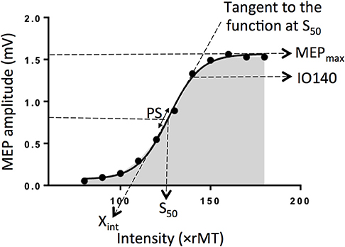
The input-output (I/O) relation in the corticospinal pathway is assessed by plotting MEP amplitude vs. stimulus intensity and fitting the data with the following sigmoid function equation (1–4):
, where MEP(s) is the MEP amplitude at the stimulation intensity s, MEPmax is the maximum MEP amplitude, S50 is the stimulus intensity needed to obtain 50% of MEPmax amplitude, and m is the slope parameter of the sigmoid function, i.e., the global slope of the function (Figure 1). Three additional parameters can be derived from the I/O curve: (1) the peak slope (PS), i.e., the instantaneous slope of the ascending limb of the curve at S50, which reflects the recruitment gain of motoneurons and is given by the formula: PS = m x MEPmax/4, (2) the x-intercept (Xint) of the tangent at S50, and (3) the area under the I/O curve (AUC) usually calculated using the trapezoidal area method (5).
FIGURE 1
www.frontiersin.orgFigure 1. Electrophysiological parameters extracted from an example of an input output curve (I/O curve) fitted by a sigmoid function. Are shown the following variables: Xint: X intercept, IO140: Motor Evoked Potential amplitude recorded at 140% rMT, PS, peak slope; AUC, area under the I/O curve in gray; S50, stimulus intensity needed to obtain 50% of the maximum response; MEPmax, maximum value of the sigmoid function.
To date, the inter-dependency between all these parameters are not fully understood. Indeed, although the PS depends on both m parameter and MEPmax, it does not mean these three parameters are correlated together. Same for Xint, which depends on m parameter and S50. The question arises whether these variables capture similar or, conversely, different aspects of the corticospinal excitability and if so, how each of them relates to one other. To clarify the interdependency between these parameters, we estimated I/O curves from the dominant hemisphere of healthy volunteers and performed Principal Component Analyses (PCA) in addition to correlation matrices to summarize the most important linear relationships between variables. PCA is a tool capable of summarizing the most important linear relationships between variables and computing synthetic variables from the original variables named principal components (PCs). PCA provides a visual and geometric representation of the correlation matrix (6, 7). In a second step, to test whether our results could be extrapolated to patients suffering from neurological conditions, we performed the same analyses on data collected in both the affected and unaffected hemispheres of stroke patients given that this population represents the most frequent brain damaged disease worldwide. Indeed, the sigmoid function has been previously shown to be a reliable method to plot IO curve in stroke patients (4).

More at link. 

Tuesday, January 29, 2019

Not all saturated fats are equal when it comes to heart health

More reasons for YOUR DOCTOR to come up with diet protocols.

You need to know EXACT amounts per body weight and sex. No guessing allowed.

You may have to call the stroke hospital president and DEMAND accountability from the doctors and therapists.

THIS IS YOUR DOCTORS' RESPONSIBILITY!

Unless you think you can decipher this on your own.  You need all these diet protocols. Good luck.  

For stroke prevention; for dementia prevention; for cognitive improvement; for cholesterol reduction; for plaque removal; for Parkinsons prevention; for inflammation reduction; for blood pressure reduction.


Not all saturated fats are equal when it comes to heart health


Newswise | January 28, 2019
The type of saturated fats we eat can affect our risk of a heart attack, according to a study published in the International Journal of Cardiology. People whose diets contain relatively little palmitic and stearic acid—saturated fats composed of 16 or more carbon atoms (longer-chain saturated fats) that are typically found in meats—and eat plant-based proteins instead have decreased chances of myocardial infarction. Moreover, individuals who eat more saturated fats with 14 or fewer carbon atoms (shorter-chain saturated fats) that are typically found in dairy products have lower risk of myocardial infarction.

"Our analysis of the diets of large groups of individuals in two countries over time shows that the type of saturated fats we consume could affect our cardiovascular heath," explained lead investigator Ivonne Sluijs, PhD, Julius Center for Health Sciences and Primary Care, University Medical Center Utrecht, Utrecht University, Utrecht, the Netherlands.
The study investigated whether saturated fats with chains varying in length from 4 to 18 carbon atoms are associated with the risk of developing a myocardial infarction. Data from approximately 75,000 people in the UK and Denmark were analyzed. Of these two groups, nearly 3,500 people experienced myocardial infarction in the period between the study's initial outreach and follow-up 13 years later (in Denmark) and 18 years later (in the UK).
"We found that eating relatively little of the longer chained saturated fatty acids and consuming plant-based proteins instead was associated with a lowered risk. Substitution of those saturated fats with other energy sources such as carbohydrates did not affect the risk to develop myocardial infarction," said Dr. Sluijs. Although diets vary by nationality and other factors, the most frequently consumed saturated fat is palmitic acid, with 16 carbon atoms, followed by stearic acid, with 18 carbon atoms, both of which are found in meat products. Consumption of saturated fats that have shorter carbon atom chains and are present in dairy products is less prevalent.
Since the 1960s, when diets high in saturated fat were linked to elevated "bad" low-density lipoprotein (LDL) cholesterol and coronary heart disease, dietary guidelines recommended restricting saturated fatty acids across the board. In recent years, research studies have raised some questions about what was considered established evidence. Inconsistent findings have pointed to the possibility that different types of saturated fats have different effects on cholesterol levels and the development of coronary heart disease. Despite the fact that their study's findings support this hypothesis, Dr. Sluijs and her fellow investigators recommend proceeding with caution before changing dietary guidelines:(Look at that laziness; guidelines NOT protocols.)
"Our study only allowed us to draw conclusions on the level of associations between saturated fatty acids and the development of myocardial infarction. We do not know whether those fatty acids are actually the cause of differences between the occurrences of myocardial infarction we observed. To further explore this, we need experiments in which the consumption of saturated fatty acids is more controlled and, for instance, compared with consumption of unsaturated fatty acids," she noted.
"The study is applaudable for its large size, prospective cohort study design, and detailed assessment of diet and lifestyle factors. In addition, it is among the few studies that specifically examined individual saturated fatty acids in relation to coronary heart disease risk and compared with different macronutrients," commented Jun Li, MD, PhD, and Qi Sun, MD, ScD, both at the Harvard T.H. Chan School of Public Health, Boston, MA, USA, in an accompanying editorial. They also noted a few limitations of the study and thus called for cautious interpretation of the overall null results for the primary saturated fatty acids.
Dr. Li and Dr. Sun advise that shifts in fat intake should align with the recommended healthy dietary patterns, which emphasize limited intakes of red and processed meat and added sugars, lower salt intake, replacement of refined grains with whole grains, and higher consumption of fruits and vegetable (So you want us to completely guess what to do? Good to know these MDS and PhDs are so not helpful.)