Changing stroke rehab and research worldwide now.Time is Brain! trillions and trillions of neurons that DIE each day because there are NO effective hyperacute therapies besides tPA(only 12% effective). I have 523 posts on hyperacute therapy, enough for researchers to spend decades proving them out. These are my personal ideas and blog on stroke rehabilitation and stroke research. Do not attempt any of these without checking with your medical provider. Unless you join me in agitating, when you need these therapies they won't be there.

What this blog is for:

My blog is not to help survivors recover, it is to have the 10 million yearly stroke survivors light fires underneath their doctors, stroke hospitals and stroke researchers to get stroke solved. 100% recovery. The stroke medical world is completely failing at that goal, they don't even have it as a goal. Shortly after getting out of the hospital and getting NO information on the process or protocols of stroke rehabilitation and recovery I started searching on the internet and found that no other survivor received useful information. This is an attempt to cover all stroke rehabilitation information that should be readily available to survivors so they can talk with informed knowledge to their medical staff. It lays out what needs to be done to get stroke survivors closer to 100% recovery. It's quite disgusting that this information is not available from every stroke association and doctors group.

Showing posts with label metabolic changes. Show all posts

Friday, August 2, 2024

Perturbation-based estimation of within-stride cycle metabolic cost

 If your doctor and therapists can't use this to identify and correct your walking imperfections, then you need to find competent ones.

Perturbation-based estimation of within-stride cycle metabolic cost

Abstract

Metabolic cost greatly impacts trade-offs within a variety of human movements. Standard respiratory measurements only obtain the mean cost of a movement cycle, preventing understanding of the contributions of different phases in, for example, walking. We present a method that estimates the within-stride cost of walking by leveraging measurements under different force perturbations. The method reproduces time series with greater consistency (r = 0.55 and 0.80 in two datasets) than previous model-based estimations (r = 0.29). This perturbation-based method reveals how the cost of push-off (10%) is much smaller than would be expected from positive mechanical work (~ 70%). This work elucidates the costliest phases during walking, offering new targets for assistive devices and rehabilitation strategies.

Introduction

Metabolic cost is a critical measure used to characterize movement behavior [1,2,3]. Healthy walkers naturally adopt an energetically optimal stride cycle, for example, by walking with a step length [4] and knee flexion angle [5] that minimizes metabolic cost. Pathologies like stroke and cerebral palsy alter patients’ walking stride resulting in increases to metabolic cost by 60 to 300% [6, 7]. Such increases in metabolic cost correlate to drastic reductions in people’s mobility and overall quality of life [8, 9]. If we understand how stride cycle phases contribute to metabolic cost, therapies and devices may be better optimized to improve mobility (Fig. 1A).
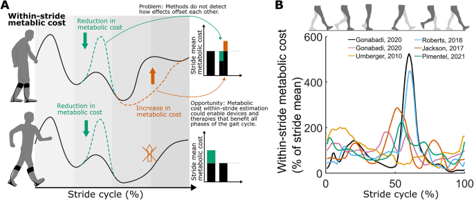
Fig. 1
figure 1

Motivation. (A). Limitation of assessing stride-mean metabolic cost using breath-by-breath measurements. The upper figure illustrates an intervention resulting in a cost reduction (depicted in green) during push-off and a cost increase (depicted in brown) during swing. The stride-mean metabolic cost (displayed in bars) does not enable differentiation of these effects. The lower section of the figure illustrates how comprehending the costs associated with various phases could facilitate the enhancement of interventions. (B). Limited consistency between estimations of within-stride metabolic cost using model-based methods. The mean correlation between estimations is 0.29 (95% confidence interval (CI) = 0.03–0.43) [16, 19, 38, 41, 42]

Measurements of metabolic cost are too slow to detect the contributions of different stride phases. Current methods to calculate energy from oxidative reactions include measuring respiratory CO2 production by ingesting water with a radioisotope (‘doubly labelled water method’), measuring oxidative heat production using a chamber (‘direct calorimetry’), and measuring O2 consumption from respiration (‘indirect calorimetry’) [10]. Indirect calorimetry is the fastest and most commonly used method for measuring metabolic cost during locomotion; however, it still requires averaging several minutes of breaths to be reliable [11,12,13]. A typical walking stride lasts about one second meaning current methods can only measure the mean metabolic cost following a bout of steady-state walking. Experiments that approximated the cost of the swing phase by recording cyclical leg swinging [14] and by measuring blood flow from injected microspheres in animals that are then sacrificed [15] suggest that the stride-mean metabolic cost does not necessarily represent the contributions of individual phases (‘within-stride metabolic cost’).

Several model-based methods of estimating within-stride metabolic cost have been proposed but remain inconclusive. Umberger developed a set of equations to estimate metabolic cost from muscle parameters and used this to produce the first estimation of within-stride metabolic cost from a forward simulation of walking [16]. Other groups used EMG-driven simulations [17] or equations based on joint kinetics instead of muscle parameters [18]. However, when comparing those methods to each other, their estimations of within-stride metabolic cost are relatively inconsistent (Pearson correlation: r = 0.29, n = 6 estimations, Fig. 1B) [19]. Currently, there is no way to validate these model-based estimations for within-stride metabolic cost since measurements from indirect calorimetry only obtain a stride mean. This motivates the development of an alternative method to estimate within-stride metabolic cost that is supported by indirect validation approaches.

We hypothesized that applying a set of perturbations creates a set of instances of the behavior where the differences in the time series between each perturbed instance can be attributed to the different magnitudes and timings of the applied perturbation. By applying perturbations repeatedly to a specific part of the gait cycle for several minutes, we can induce changes in the stride-mean metabolic cost as well as in the biomechanical time series (e.g., kinematics, kinetics, and muscle activations) [19,20,21]. We postulated the variation across the set of perturbed walking strides would be representative of the fluctuations in metabolic cost within the stride cycle so long as the set contained a large number of different perturbations. If true, this would enable a method to extract key features of within-stride metabolic cost. Our approach is inspired by prior studies that utilized ankle perturbations to assess time series of joint impedance during the stance phase [22, 23] as well as studies that used elastic bands and added mass to estimate the cost of stance and swing phases [24, 25]. To the best of our knowledge, using of a perturbation-based approach for estimating within-stride metabolic cost time series is novel.

Using this concept, extraction of within-stride behaviour from a collection of perturbed instances, we developed an alternative method to estimate within-stride metabolic cost that we refer to as our ‘perturbation-based method’. Our method estimates within-stride metabolic cost using measurements from a set of perturbed walking strides. We then evaluated our method’s ability to consistently reproduce model-based estimates of within-stride metabolic cost.

More at link.

Tuesday, July 10, 2018

Using Imaging Techniques to Assess the Effects of a Stroke

Wrong, wrong, wrong. You should be using imaging to diagnose all the damage(dead and dying neurons), then map protocols to recover from such damage. This is so fucking easy to understand, why isn't it being done? No protocols is not a valid answer. Lots of words here but nothing useful for recovery.
https://www.news-medical.net/whitepaper/20180626/Using-Imaging-Techniques-to-Assess-the-Effects-of-a-Stroke.aspx

Cerebral ischaemia

Cerebral ischaemia is a type of stroke causing high rates of disability and mortality1. It occurs when blood vessels serving the brain become blocked or burst. This prevents the affected area of the brain from receiving sufficient oxygen and nutrients to meet its metabolic demands.
© sfam_photo/Shutterstock.com
Consequently, cells in the affected area cannot work properly and the bodily functions for which they are responsible are impaired. Cerebral ischaemia can affect a specific localised area of the brain or be more widespread.
Symptoms of cerebral ischaemia may be fleeting if the interruption of blood supply to the brain is restored quickly, but if oxygen deprivation is prolonged the affected brain tissue will die causing irreversible brain damage. Cerebral ischaemia symptoms include sight disturbances, dizziness, loss of co-ordination, muscle weakness or paralysis, and difficulty speaking.

Changes in the brain after cerebral ischaemia

Cerebral ischaemia results in changes in energy usage, disruption to the metabolism of neurotransmitters and lipids and alterations to protein synthesis. In addition, intracellular cytosolic calcium concentration increases, as does the level of damaging free radicals. It is these metabolic, biochemical and ionic perturbations that ultimately cause neuronal death. Understanding the precise nature of these changes may therefore help in treatments to minimise the debilitating  consequences of a stroke. However, since such a wide range of metabolites is affected, leading to disruption of several complex pathways, unravelling the effects of cerebral ischaemia is not straightforward.
Furthermore, it appears that the pathological changes of stroke may be even more far-reaching than initially thought. Research indicates that metabolic changes occur not only in the area of the brain that has become ischaemic but may also induce secondary effects in other areas of the brain. Such functional impairment of the brain at a location removed from the injury site is known as diaschisis, which appears to be a form of shock response.
Defining and understanding the metabolic disturbances caused by oxygen and nutrient deprivation have this been identified as key to the development of treatments to minimise morbidity in patients suffering cerebral ischaemia2. With the possibility of diaschisis, the physiological effects of cerebral ischaemia must be evaluated across the brain and not just in the ischaemic area.
Magnetic resonance imaging and positron emission tomography have successfully measured remote metabolic changes after cerebral ischaemia. Such techniques have provided insight into the more far-reaching effects of stroke in areas of the brain far-removed from the ischaemic area. However, although cerebellar diaschisis has been well studied3, there are few data relating to the less common interhemispheric diaschisis. It remains unclear whether ischaemic damage in one hemisphere can lead to diaschisis in another hemisphere.

Measuring metabolic changes

Metabolomics is the study of biochemical pathways through the measurement of the metabolites. By determining the relative changes in concentration of substrate and product metabolites, it can be inferred whether specific pathways have been up or down-regulated. This methodology has been widely used across a range of disciplines to investigate the effects of drugs or contaminants.
Nuclear magnetic resonance spectroscopy detects molecules based on their chemical shift on application of a magnetic field. Since different types of molecules give different spectral peaks, it can measure the many different components in a complex mixture. It has been widely used to investigate the pathological mechanisms of cerebral ischaemia, but has not to study the effects on biochemical processes within the brain.

Investigating inter-hemisphere effects of cerebral ischemia

A recent study investigated metabolite changes after cerebral artery occlusion in both the left and right cerebral hemispheres of rats4. In order to simultaneously measure the quantities of numerous metabolites, 1H nuclear magnetic resonance (1H NMR) spectroscopy was employed for the analysis using a Bruker AVANCE III 600 MHz NMR spectrometer. Characteristic changes in metabolites indicated that biochemical changes had occurred in both the ischaemic and contralateral cerebral hemispheres.
In the ischaemic cerebral hemisphere, the changes in metabolites after cerebral ischaemia indicated an increase in anaerobic glycolysis, a perturbation of choline metabolism, neuronal cell damage and neurotransmitter imbalance. In the contralateral hemisphere, an increase in anaerobic glycolysis was also observed, along with changes in energy metabolism and alteration in the balance of neurotransmitters. Since the two cerebral hemispheres are connected by a large mass of neural fibres called the corpus callosum, it is likely that this is the means by which ischaemic damage in one hemisphere can initiate metabolic changes in the other hemisphere.
These findings indicate that interhemispheric diaschisis can indeed occur after cerebral ischaemia. Furthermore, they support cerebral metabolic analysis as valuable tool for understanding the biochemical mechanisms of cerebral ischaemia and its effects on distant areas of the brain.

References

  1. Anuncibay-Soto B, et al. Neuroprotection by salubrinal treatment in global cerebral ischemia. Neural Regen Res 2016;11:1744‑1745.
  2. Yang M, et al. NMR analysis of the rat neurochemical changes induced by middle cerebral artery occlusion. Talanta 2012;88:136‑144.
  3. Madai VI, et al. Crossed cerebellar diaschisis after stroke: can perfusion-weighted MRI show functional inactivation? J Cereb Blood Flow Metab 2011;31:1493‑1500.
  4. Ruan L, et al. Metabolite changes in the ipsilateral and contralateral cerebral hemispheres in rats with middle cerebral artery occlusion. Neural Regen Res 2017;12(6):931-937.

Monday, August 24, 2015

Effects of handrail hold and light touch on energetics, step parameters, and neuromuscular activity during walking after stroke

A couple of important points from this, your doctor should  be able to determine which 60% successfully walk again in a community setting. 

Effects of handrail hold and light touch on energetics, step parameters, and neuromuscular activity during walking after stroke


T. IJmker12*, C. J. Lamoth3, H. Houdijk12, M. Tolsma2, L. H. V. van der Woude3, A. Daffertshofer1 and P. J. Beek1
1 MOVE Research Institute Amsterdam, Department of Human Movement Sciences, Faculty of Behavioural and Movement Sciences, VU University Amsterdam, van der Boechorststraat 9, Amsterdam, 1081 BT, The Netherlands
2 Heliomare Rehabilitation, Research and Development, Relweg 51, Wijk aan Zee, 1949 EC, The Netherlands
3 University of Groningen, University Medical Center Groningen, Center for Human Movement Sciences, Center for Rehabilitation, Antonius Deusinglaan 1, Groningen, 9713AV, The Netherlands
For all author emails, please log on.
Journal of NeuroEngineering and Rehabilitation 2015, 12:70  doi:10.1186/s12984-015-0051-3
The electronic version of this article is the complete one and can be found online at: http://www.jneuroengrehab.com/content/12/1/70

Received:5 February 2015
Accepted:26 June 2015
Published:23 August 2015
© 2015 IJmker et al.

Open Access This article is distributed under the terms of the Creative Commons Attribution 4.0 International License (http://creativecommons.org/licenses/by/4.0/), which permits unrestricted use, distribution, and reproduction in any medium, provided you give appropriate credit to the original author(s) and the source, provide a link to the Creative Commons license, and indicate if changes were made. The Creative Commons Public Domain Dedication waiver (http://creativecommons.org/publicdomain/zero/1.0/) applies to the data made available in this article, unless otherwise stated.

Abstract

Background

Holding a handrail or using a cane may decrease the energy cost of walking in stroke survivors. However, the factors underlying this decrease have not yet been previously identified. The purpose of the current study was to fill this void by investigating the effect of physical support (through handrail hold) and/or somatosensory input (through light touch contact with a handrail) on energy cost and accompanying changes in both step parameters and neuromuscular activity. Elucidating these aspects may provide useful insights into gait recovery post stroke.

Methods

Fifteen stroke survivors participated in this study. Participants walked on a treadmill under three conditions: no handrail contact, light touch of the handrail, and firm handrail hold. During the trials we recorded oxygen consumption, center of pressure profiles, and bilateral activation of eight lower limb muscles. Effects of the three conditions on energy cost, step parameters and neuromuscular activation were compared statistically using conventional ANOVAs with repeated measures. In order to examine to which extent energy cost and step parameters/muscle activity are associated, we further employed a partial least squares regression analysis.

Results

Handrail hold resulted in a significant reduction in energy cost, whereas light touch contact did not. With handrail hold subjects took longer steps with smaller step width and improved step length symmetry, whereas light touch contact only resulted in a small but significant decrease in step width. The EMG analysis indicated a global drop in muscle activity, accompanied by an increased constancy in the timing of this activity, and a decreased co-activation with handrail hold, but not with light touch. The regression analysis revealed that increased stride time and length, improved step length symmetry, and decreased muscle activity were closely associated with the decreased energy cost during handrail hold.

Conclusion

Handrail hold, but not light touch, altered step parameters and was accompanied by a global reduction in muscle activity, with improved timing constancy. This suggests that the use of a handrail allows for a more economic step pattern that requires less muscular activation without resulting in substantial neuromuscular re-organization. Handrail use may thus have beneficial effects on gait economy after stroke, which cannot be accomplished through enhanced somatosensory input alone.
Keywords:
Energy cost; Stroke; Neuromuscular function; Gait; Balance support

Background

Regaining the ability to walk independently is an important goal in the rehabilitation of stroke survivors. Only 60 % of all stroke survivors eventually attain this goal to the level of community walking [1]. An important limiting factor in this regard is the substantial metabolic cost of hemiparetic gait, which can be more than two times larger than in healthy subjects [2]–[4], and which is predictive of community ambulation [5]. We have previously shown that an increased (metabolic) effort to control balance contributes to this decreased gait economy [6], and that this cost can be reduced considerably by providing balance support in the form of a handrail or cane [7].
Using a handrail or cane may have biomechanical and/or somatosensory advantages that could facilitate balance control. Biomechanically, the use of a handrail or cane increases the base of support, resulting in greater margins of stability, and enables one to generate corrective forces via the hands to compensate for perturbations [8]. Apart from this biomechanical advantage, the use of a handrail or cane may provide additional somatosensory (tactile and proprioceptive) information about body orientation and movement relative to the point of contact [8], [9]. This may reduce sensory noise/uncertainty and might therefore lead to better balance control [9], [10]. There is experimental support that, even in the absence of additional biomechanical support, the mere contact of fingertips or hand with a stable support surface can decrease the excursion of the center of mass during standing and walking [9]–[13]. This decrease matched that observed with firm handrail hold in healthy participants and stroke survivors. This suggests that enhanced somatosensory information may add to the mechanical stabilization through holding a handrail, which in turn may result in a decreased energy cost of walking after stroke.
To unravel the factors underlying the differential effects of handrail hold and light touch on the energy cost of walking, it is imperative to investigate which gait parameters alter in line with metabolic changes and which neuromuscular modifications might engender these effects. In stroke survivors, handrail or cane use yields increased stride length and time as well as decreased cadence and step width and variability [14], [15]. These changes may be linked to an improved gait efficiency through a more optimal step length/frequency combination [16], [17], and lower step-to-step transition costs with a smaller step width [18]. Using a handrail or cane may also improve gait symmetry [19], [15], which may also contribute to enhanced gait economy [20].
Effects of holding a handrail or cane have also been examined in terms of changes in neuromuscular control as reflected in altered amplitude and timing of muscle activation. Some studies reported decreases in EMG burst duration and a decrease in amplitude of several lower limb muscles during cane use [21], [22]. Furthermore, a decrease in the variability of EMG profiles of the lower leg muscles has been found as a result of handrail support, which indicates a more consistent timing of muscle activity possibly relating to increased (lateral) gait stability [23], [24]. Reduced EMG amplitude and more accurate timing of muscle activity may reflect improved economy [25]. In contrast, other studies reported no effect of firm handrail hold or light touch contact with a cane on muscle activity [26], [27], while light touch contact has even been shown to result in higher activation amplitudes than force contact [12], [26].
As of yet, it is unclear whether somatosensory and/or biomechanical aspects of handrail or cane use affect the energy cost of walking after stroke, nor whether altered step parameters and/or altered neuromuscular control are responsible for this effect. Our research aims were therefore twofold: 1) to compare the effects of light touch contact with a handrail and firm handrail hold on the energy cost of walking, step parameters, and muscle activity (in terms of amplitude and timing) in stroke survivors, and 2) to examine which changes in step parameters and muscle activity are associated with the observed changes in energy cost. To evaluate changes in muscle activation amplitude and timing we used a principal component analysis (PCA), since this method allows for studying patterns of multivariate muscle activation instead of looking at isolated muscle activities alone. 

More at link.