Changing stroke rehab and research worldwide now.Time is Brain! trillions and trillions of neurons that DIE each day because there are NO effective hyperacute therapies besides tPA(only 12% effective). I have 523 posts on hyperacute therapy, enough for researchers to spend decades proving them out. These are my personal ideas and blog on stroke rehabilitation and stroke research. Do not attempt any of these without checking with your medical provider. Unless you join me in agitating, when you need these therapies they won't be there.

What this blog is for:

My blog is not to help survivors recover, it is to have the 10 million yearly stroke survivors light fires underneath their doctors, stroke hospitals and stroke researchers to get stroke solved. 100% recovery. The stroke medical world is completely failing at that goal, they don't even have it as a goal. Shortly after getting out of the hospital and getting NO information on the process or protocols of stroke rehabilitation and recovery I started searching on the internet and found that no other survivor received useful information. This is an attempt to cover all stroke rehabilitation information that should be readily available to survivors so they can talk with informed knowledge to their medical staff. It lays out what needs to be done to get stroke survivors closer to 100% recovery. It's quite disgusting that this information is not available from every stroke association and doctors group.

Showing posts with label does nothing. Show all posts

Tuesday, July 14, 2026

CT vs MRI: Assessing Stroke and Dementia Risk in White Matter Disease


My incompetent? doctor told me I had a bunch of white matter hyperintensities but never showed me them on any scan, so I don't know the size, location or any intervention needed, because my doctor knew nothing and did nothing. I have zero cognitive impairment and I'm 20 years out.

CT vs MRI: Assessing Stroke and Dementia Risk in White Matter Disease

 Patients with incidentally discovered white matter disease on computed tomography have a higher risk for future stroke or dementia than those with disease detected only on magnetic resonance imaging. White matter disease (WMD) detected on computed tomography (CT) identified patients at greater risk for future stroke or dementia than WMD detected only on magnetic resonance imaging (MRI), according to study results published in Neurology. Covert cerebrovascular disease (CCD), which includes covert brain infarction (CBI) and WMD, is frequently identified incidentally during neuroimaging in patients without a history of stroke or dementia. Although MRI is generally considered more sensitive for detecting these abnormalities, CT remains the most commonly used neuroimaging modality in routine clinical practice. Researchers therefore compared incidentally discovered CCD detected on CT and MRI to determine whether findings from each modality differed in their association with subsequent neurologic outcomes. Researchers conducted a retrospective cohort study of adults aged 50 years and older who underwent both head CT and brain MRI within a 30-day period between 2009 and 2022. They excluded patients with a prior history of stroke or dementia, as well as those with major neurologic symptoms suggestive of acute stroke. They used natural language processing to identify CBI and WMD from radiology reports and classify WMD severity. These findings highlight the importance of modality-specific interpretation of CCD in clinical practice and research. The analysis included 18,628 participants with a mean age of 64.9 years; 59.1% were women. The cohort was racially and ethnically diverse, with 41.4% identifying as non-Hispanic White, 32.7% as Hispanic, 12.0% as Asian or Pacific Islander, and 11.3% as African American. Cardiovascular risk factors were common, including hypertension (63.3%), hypercholesterolemia (71.3%), diabetes (30.3%), and a history of tobacco use (44.9%). The prevalence of CBI was similar across imaging modalities, occurring in 6.3% of CT scans and 6.1% of MRI scans. Overall agreement for CBI presence or absence was 91.6%. Among patients with CBI on MRI, 33.3% also had CBI on CT. Among patients with CBI on CT, 31.9% also had CBI on MRI. Researchers identified WMD in 60.5% of MRI reports compared with 24.4% of CT reports. Agreement for WMD presence was 57.6%. Among patients with WMD severity classified on both modalities, 47.9% received different severity classifications, and MRI assigned a higher severity grade than CT in 92.3% of those discordant cases. During a mean follow-up period of 4.4 years, 985 patients experienced stroke alone, 716 developed dementia alone, and 330 experienced both outcomes. Patients without WMD on either imaging modality had an incidence rate of stroke or dementia of 12.7 events per 1000 person-years. Rates increased to 22.6 among patients with WMD detected only on MRI, 37.0 among those with WMD detected only on CT, and 52.2 among those with WMD detected on both CT and MRI (all per 1000 person-years). After adjustment for demographic characteristics and vascular risk factors, patients with WMD detected only on MRI had a 23% higher risk for stroke or dementia than those without WMD on either modality (hazard ratio [HR], 1.23; 95% CI, 1.07-1.41). Patients with WMD detected only on CT had an even greater risk (HR, 1.46; 95% CI, 1.11-1.92), and those with WMD identified on both modalities had the greatest risk (HR, 1.82; 95% CI, 1.58-2.11). The researchers observed a similar pattern for CBI. Compared with patients without CBI on either modality, those with MRI-only CBI had a higher adjusted risk for stroke or dementia (HR, 1.47; 95% CI, 1.23-1.76), as did patients with CT-only CBI (HR, 1.26; 95% CI, 1.04-1.53) and those with CBI detected on both modalities (HR, 1.68; 95% CI, 1.33-2.11). The researchers suggested that MRI appears to detect a broader spectrum of white matter abnormalities, including milder disease that may not be visible on CT. In contrast, WMD identified on CT may represent more advanced or clinically significant cerebrovascular injury, potentially explaining its stronger association with future stroke or dementia. Study limitations include reliance on natural language processing of radiology reports rather than direct image review and the inclusion of only patients who underwent both CT and MRI. “These findings highlight the importance of modality-specific interpretation of CCD in clinical practice and research,” the study authors concluded.  Disclosures: This research was supported by the Alzheimer’s Drug Discovery Foundation and the National Institutes of Health. One study author declared affiliations with biotech, pharmaceutical, and/or device companies. Please see the original reference for a full list of disclosures.

Monday, June 29, 2026

Modified small vessel disease score as the top predictor of stroke outcome after thrombectomy: a CT-based machine learning study


Why are your predicting failure to recover RATHER THAN DELIVERING RECOVERY?

Laziness? Incompetence? Or just don't care? NO leadership? NO strategy? Not my job? Not my Problem!

You're all fired! You need to create EXACT RECOVERY PROTOCOLS! 

Prediction crapola like this does nothing to get survivors recovered! Your comeuppance when you have a stroke and don't recover will be a bitter pill for you to swallow.

Modified small vessel disease score as the top predictor of stroke outcome after thrombectomy: a CT-based machine learning study


  • 1. Department of Epidemiology, Harvard T.H. Chan School of Public Health, Boston, MA, United States

  • 2. Department of Neuroscience and Behavioral Sciences, Ribeirão Preto Medical School, University of São Paulo, Ribeirão Preto, São Paulo, Brazil

Abstract

Background: 

Mechanical thrombectomy (MT) improves outcomes in ischemic stroke (IS) due to large vessel occlusion (LVO), but ~50% of patients fail to achieve functional independence.

Objectives: 

We investigated whether cerebral small vessel disease (cSVD), assessed by the modified Small Vessel Disease (mSVD) score and Brain Frailty Score (BFS), outperforms individual CT markers in predicting 90-day outcomes after MT.

Design: 

Prospective cohort with retrospective analysis.

Methods: 

We included 351 patients with anterior circulation LVO treated with MT. Admission CT was used to score cSVD markers (leukoaraiosis, atrophy, lacunes) and compute mSVD and BFS. Eight logistic regression models and a Random Forest algorithm were used to predict poor outcome [modified Rankin Scale (mRS) 3–6]. Model performance was evaluated using AUC-ROC and compared via DeLong tests.

Results: 

Poor outcomes were associated with older age, higher NIHSS, systolic blood pressure, glycemia, and more severe leukoaraiosis and atrophy. Severe mSVD (score = 3) independently predicted poor outcomes (OR = 3.267; CI: 1.731–6.168; p = 0.009). mSVD outperformed BFS and individual CT markers (AUC = 0.904 vs. 0.889/0.898; DeLong p < 0.05) and ranked as the top predictor in Random Forest (importance = 42.05). Treatment efficacy declined with increasing mSVD: the probability of a favorable outcome was 15.53% and poor outcome was 84.47% for mSVD = 3, compared to 89.23% and 10.77%, respectively, for mSVD = 0. A secondary model incorporating 24h NIHSS and hemorrhagic transformation improved discrimination (AUC = 0.954), but mSVD remained a key independent predictor.

Conclusions: 

In this prospective study in a middle-income country, mSVD score was the strongest predictor of post-thrombectomy outcome, outperforming BFS and isolated imaging markers. While cSVD does not contraindicate MT, it reflects reduced cerebrovascular resilience. Integrating mSVD into baseline CT evaluation may enhance risk stratification and treatment guidance.


More at link.

Wednesday, June 24, 2026

Passive sensing of gait and medication-related fluctuations in Parkinson’s disease

 Is your doctor competent enough to IMMEDIATELY get this for creating AN EXACT DAMAGE DIAGNOSIS to be followed by AN EXACT REHAB PROTOCOL FOR COMPLETE RECOVERY OF WALKING? Oh NO, your doctor is fucking incompetent; 

KNOWS NOTHING AND DOES NOTHING!

Passive sensing of gait and medication-related fluctuations in Parkinson’s disease

    We are providing an unedited version of this manuscript to give early access to its findings. Before final publication, the manuscript will undergo further editing. Please note there may be errors present which affect the content, and all legal disclaimers apply.

    Abstract

    Background

    Gait impairment is a hallmark symptom of Parkinson’s Disease (PD). Traditional clinical assessments cannot capture real-world motor fluctuations, as they are sparsely performed. We validated the use of nearables, passive sensing technologies, including Kinect RGB-D cameras and ultra-wideband (UWB) radar, for continuous, objective assessment of gait fluctuations in PD within a home-like setting.

    Methods

    Fifteen PD patients with mild symptoms and fourteen age- and sex-matched healthy controls (HC) performed 4-metre walking tasks in a living lab facility. Patients repeated the task during “ON” and “OFF” states of their daily medication cycle. Gait features, including stride length, stride time, and gait speed, were extracted from Kinect, radar, and a ground-truth smart floor. Data were analysed to assess inter-sensor agreements and group-level differences.

    Results

    Stride time demonstrated the highest agreement between devices (r = 0.903), while stride length was weaker (r = 0.779). Nevertheless, stride length from both Kinect and radar distinguished PD OFF from HC (camera q = 0.020; radar q = 0.005), and radar additionally differentiated ON from OFF (q = 0.020). Neither device differentiated PD ON from HC, indicating medication reduced observable gait differences.

    Conclusions

    Although some spatial metrics show device discrepancies, both systems demonstrate sensitivity to gait patterns and medication-dependent changes, supporting their use for longitudinal, real-world monitoring of motor symptoms.

    Sunday, May 10, 2026

    How Chronic Stress Accelerates Aging (and How to Slow the Process) by Super Age


    All stroke patients are under massive stress because your incompetent? doctor doesn't have 100% RECOVERY PROTOCOLS. Your doctor has known since medical school that stroke recovery is a complete shitshow and done nothing to fix that!

    How Chronic Stress Accelerates Aging (and How to Slow the Process)

    Saturday, May 9, 2026

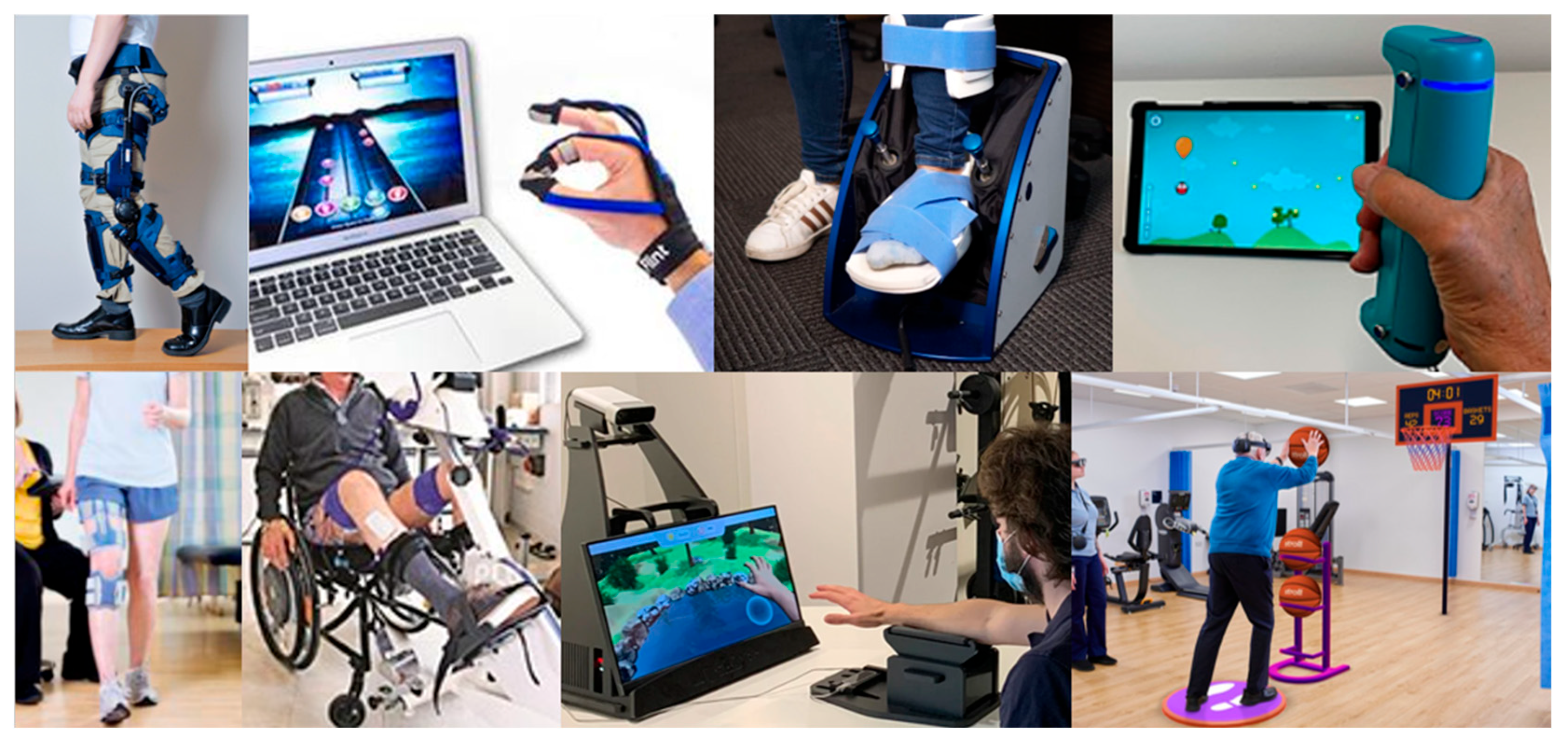
    FDA-Listed Interactive Devices for Home Movement Rehabilitation After Stroke: A Mixed-Methods Study of Availability, User Needs, Information Gaps, and an Accompanying Dataset

     Does your competent? doctor know of all 57 of these and determined the best ones for your recovery? Oh NO, knows nothing and has done nothing! WOW! You picked a winner!


    Here you are :

    Key FDA Lists for Interactive Devices [1]

    FDA-Listed Interactive Devices for Home Movement Rehabilitation After Stroke: A Mixed-Methods Study of Availability, User Needs, Information Gaps, and an Accompanying Dataset

    by 1,*, 1, 1,2, 1, 1 and 1
    1
    Mechanical and Aerospace Engineering Department, University of California Irvine, Irvine, CA 92617, USA
    2
    Department of Orthopaedics & Rehabilitation, University of New Mexico, Albuquerque, NM 87106, USA
    *
    Author to whom correspondence should be addressed.
    Bioengineering 2026, 13(4), 387; https://doi.org/10.3390/bioengineering13040387
    Submission received: 18 December 2025 / Revised: 6 March 2026 / Accepted: 24 March 2026 / Published: 27 March 2026
    (This article belongs to the Special Issue Technological Advances in Neurorehabilitation)

    Abstract

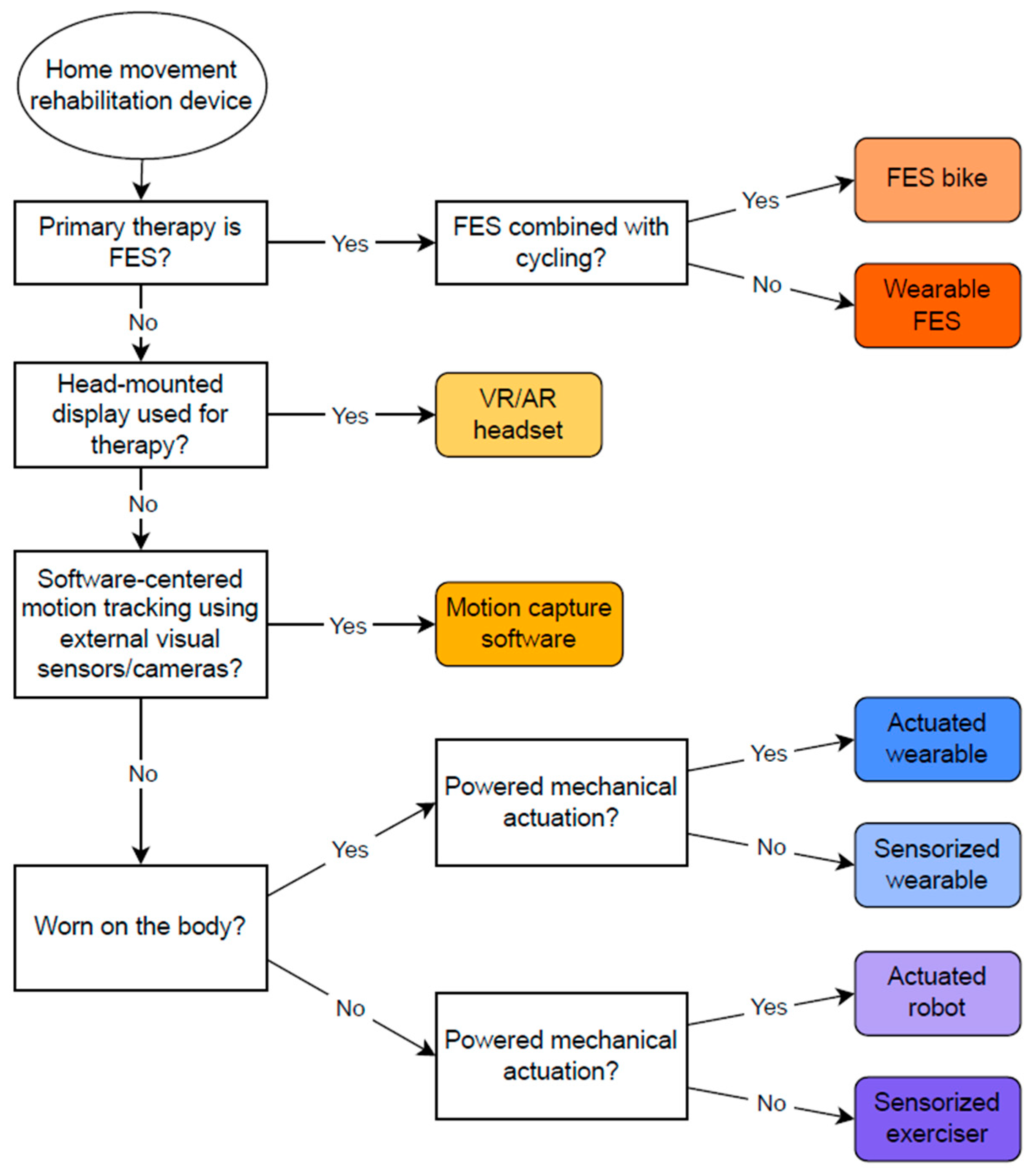
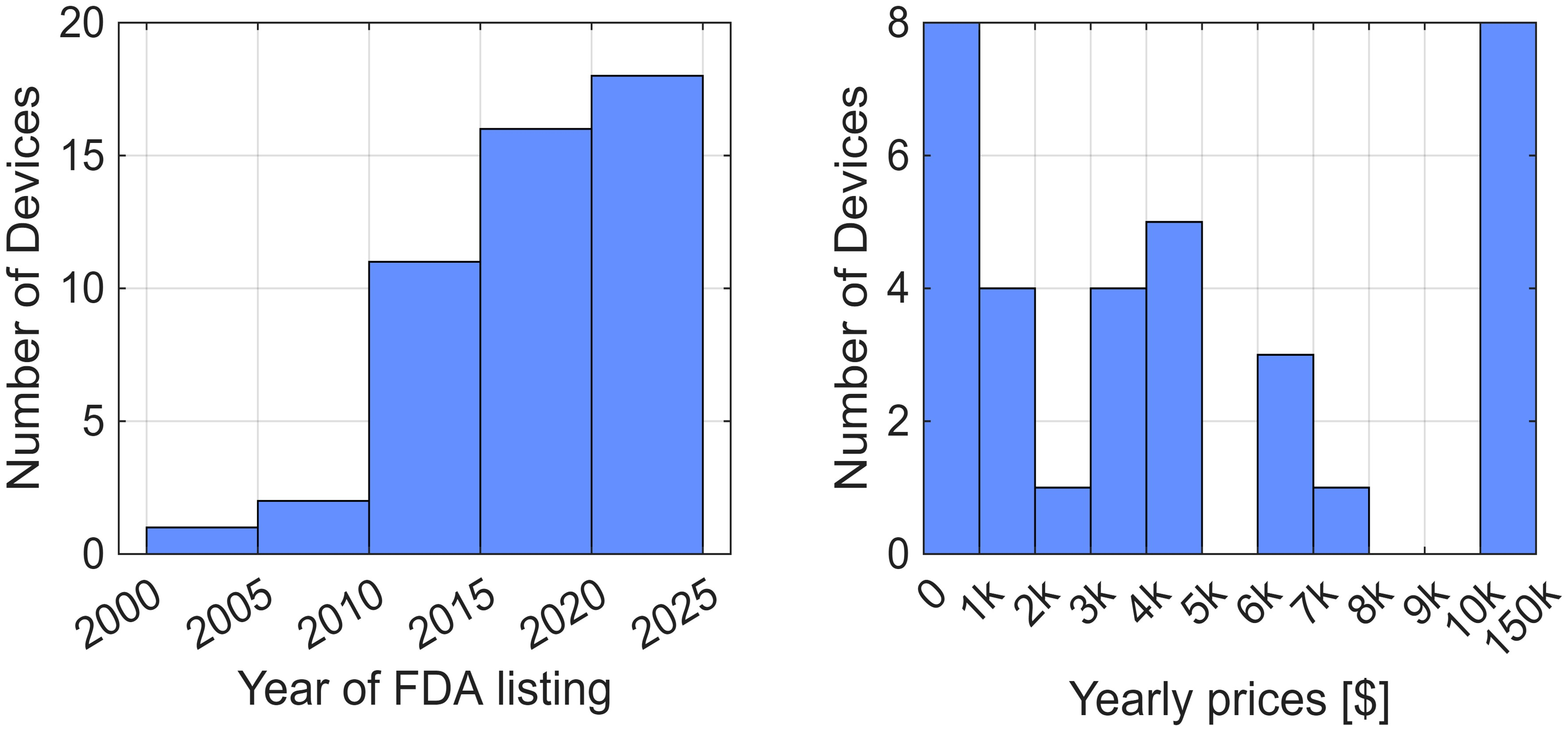
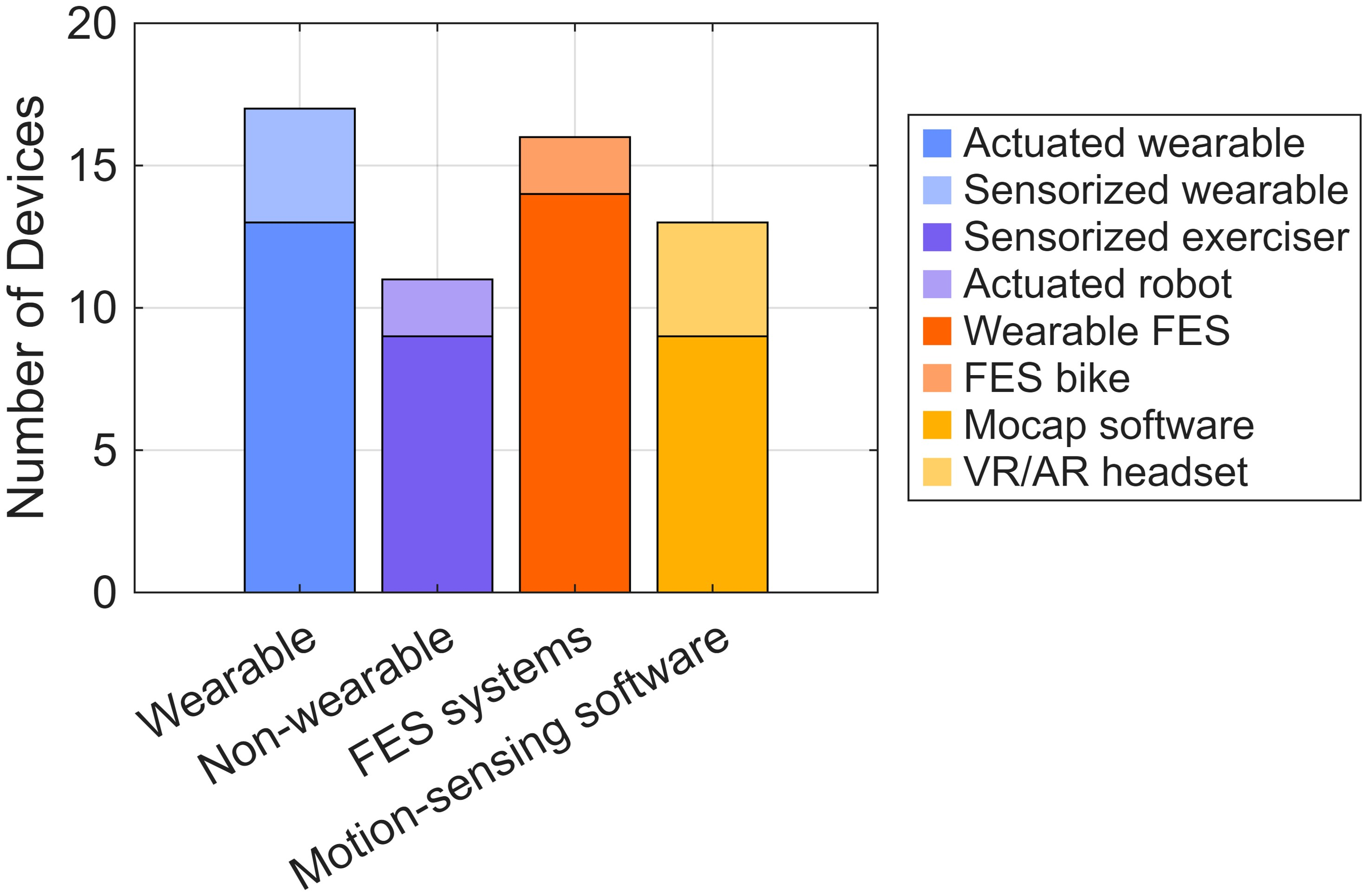
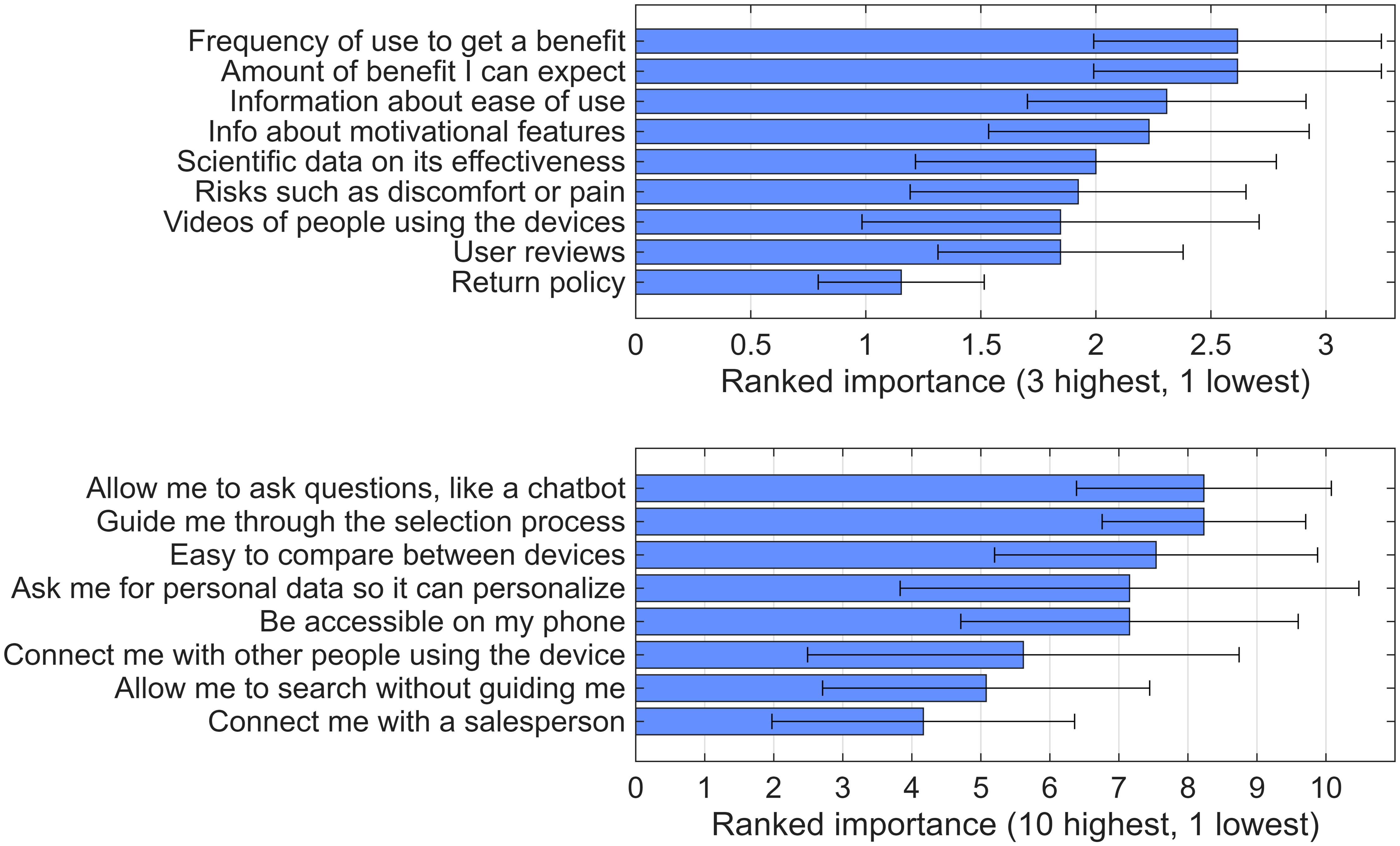
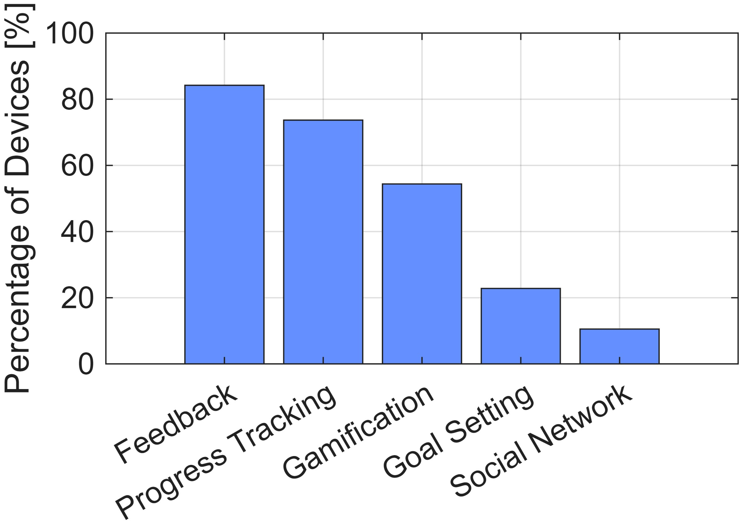
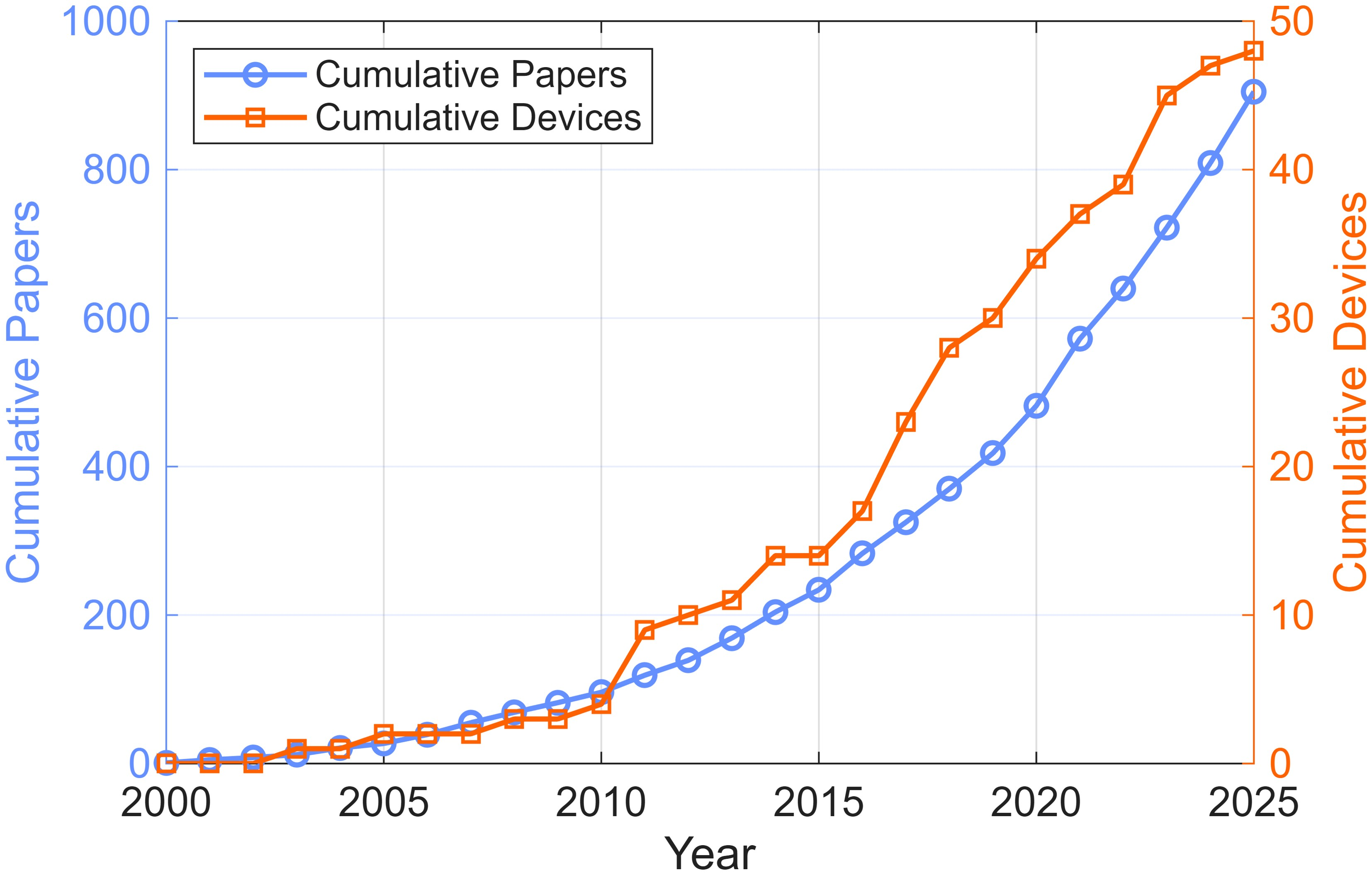
    Technologies for home movement rehabilitation after stroke are rapidly expanding. However, for consumers, the number and nature of available products are unclear, and the information provided by device manufacturers varies widely. To understand this landscape, we conducted a mixed-methods, descriptive study in which we used the U.S. Food and Drug Administration (FDA) database to identify interactive devices for stroke rehabilitation suitable for home use. We then surveyed 13 individuals with stroke to determine what information they most wanted about home-based rehabilitation devices and contacted manufacturers to obtain those details. Thirteen FDA codes were associated with stroke rehabilitation devices, encompassing 57 devices produced by 40 companies. Nearly half were categorized under two codes: QKC (interactive rehabilitation exercise devices) and GZI (neuromuscular stimulators). Among devices for which information was available, 71% were listed after 2015, and 23% cost under $1000. The top information priorities for individuals with stroke were required usage to achieve therapeutic benefit, expected benefit, ease of use, and motivational features. Despite repeated outreach, only 45% of companies responded to our queries; among those that did, details were vague and variable. These results confirm that a large and growing number of FDA-listed devices are now available for home-based post-stroke motor rehabilitation. We further identify a need to establish industry standards for reporting ease of use, motivational effectiveness, and dose–response characteristics to help the intended consumers select appropriate technologies. The curated dataset generated in this study is provided as a resource for future work and may support the development of accurate Artificial Intelligence-based interfaces for identifying and comparing rehabilitation devices.
    More at link.

    Friday, May 1, 2026

    New-Onset Constipation May Shape Stroke Recovery


    Your competent? doctor has been working on this problem for almost a decade, right?

    OH NO! Knows nothing AND does nothing!

    And your board of directors is so incompetent they can't recognize incompetence in their hospital!

     The incidence of constipation for stroke was 48%.  June 2017

    NO PROTOCOLS THAT WILL CURE IT.

     In my non-medical opinion, full physical recovery should lessen this problem immensely!

    New-Onset Constipation May Shape Stroke Recovery

    New-Onset Constipation After Stroke

    NEW-ONSET constipation after stroke was common and independently linked to poorer discharge outcomes in acute rehabilitation.

    Constipation may be an underrecognized complication in acute stroke care, with new data showing high rates of poststroke constipation and a measurable association with rehabilitation outcomes. In a cross sectional study of 600 patients with acute stroke, investigators examined the incidence, contributing factors, and discharge impact of constipation developing after stroke in patients with no previous history of the condition.

    Among all participants, 126 patients, or 21%, had a history of constipation. Poststroke constipation was identified in 278 patients, representing 46.3% of the cohort. New-onset constipation after stroke occurred in 184 patients, accounting for 38.8% of those without prior constipation.

    Risk Factors for New-Onset Constipation

    Several clinical and functional factors were associated with new-onset constipation after stroke. Hemorrhagic stroke, posterior circulation stroke, diabetes, use of osmotic diuretics, antacids, bedpan use, difficulty falling asleep, depression, and higher admission NIHSS scores were all identified as significant risk factors.

    The findings suggest that bowel dysfunction after stroke may reflect more than immobility or routine medication exposure. Sleep disruption and depression appeared to contribute to constipation risk, pointing to the need for broader assessment during the acute stage of stroke rehabilitation.

    Impact on Stroke Rehabilitation

    New-onset constipation was independently associated with poor discharge outcome after adjustment for confounders, with the strongest signal seen among patients with moderate stroke severity. This association reinforces the importance of early recognition, particularly in patients whose rehabilitation trajectory may be vulnerable to preventable complications.

    (WRONG, WRONG, WRONG! Survivors don't want it identified, you blithering idiots; they want it cured! And you're too stupid to deliver what is needed!)

    For clinicians, the results support routine screening for constipation risk in patients with stroke, including review of medication exposure, toileting method, neurologic severity, sleep quality, mood symptoms, and metabolic comorbidities. Identifying patients at risk may help optimize rehabilitation protocols and reduce barriers to recovery during the acute phase.

    (But you completely missed marijuana! Why? 

    Marijuana use linked with decreased constipation)

    While the study design does not establish causality, the high incidence of new-onset constipation after stroke and its association with discharge outcome highlight a clinically relevant target for early supportive care.

    Reference
    Lv Z et al. New-onset constipation at acute stage after stroke: incidence, risk factors, and impact on stroke rehabilitation. Frontiers in Neurology. 2026;17:1721157.

    Tuesday, April 7, 2026

    Linking eye movements, pupil responses, and brain networks in early cognitive decline

     Will your competent? doctor put this into a testing protocol, so if problems are found, that EXACT DEMENTIA PREVENTION PROTOCOL WILL BE USED?

    Do you prefer your doctor, hospital and board of director's incompetence NOT KNOWING? OR NOT DOING? Your choice; let them be incompetent or demand action!

    OH NO! your doctor KNOWS NOTHING AND DOES NOTHING! 


    Alzheimer's Research & Therapy
     We are providing an unedited version of this manuscript to give early access to its findings. 
    Before final publication, the manuscript will undergo further editing. Please note there may be errors present which affect the content, and all legal disclaimers apply.

    Abstract

    Background

    Early detection of Alzheimer’s disease (AD) requires biomarkers sensitive to pre-neurodegenerative dysfunction. Task-evoked ocular responses index arousal‑based gain and the stability of executive timing, but their relationship to brain structure, especially within AD‑signature cortex; regions that thin early in AD, remains poorly characterized. We tested whether ocular metrics map onto cortical thickness and subcortical volumes, and whether brain–ocular coupling differs between cognitively normal (CN) adults and those with mild cognitive impairment (MCI).

    Methods

    Participants with MCI (n = 212) and CN controls (n = 516) completed an interleaved prosaccade–antisaccade task; binocular pupil diameter and eye movements were time‑locked to cue and target onsets to derive pupil amplitude/variability and saccade metrics. Region‑wise linear mixed‑effects models quantified brain–ocular coupling to cortical thickness and subcortical volumes and tested group (CN vs. MCI) differences in coupling slopes.

    Results

    Diagnosis‑independent analyses showed that saccade latency variability (Latency SD) and pupil amplitude/variability exhibited robust region‑dependent coupling across cortical and subcortical volumes, whereas mean saccadic latency and pupil timing measures displayed no reliable spatial pattern.

    Diagnostic effects were modest and spatially selective but directionally opposite across modalities. For saccades, CN displayed positive thickness–variability slopes in frontal, cingulate, and insular cortices whereas MCI showed negative or near‑zero slopes. In the AD‑signature cortex, this inversionn CN(+)/MCI(−/0) was localized specifically to the supramarginal gyrus (Δβ≈–0.026). Subcortical volumes showed no significant diagnostic differences. For pupils, MCI showed more positive pupil–thickness coupling than CN within global cortex, most prominently in temporal, parietal, and insular regions, and within AD‑signature cortex this selective increase localized to medial temporal cortex (Δβ = 0.042) and to supramarginal gyrus (Δβ = 0.030); subcortical diagnostic differences were not significant after correction.

    Conclusions

    Task‑evoked ocular signals yield two complementary, region‑specific readouts of early Alzheimer‑relevant dysfunction. Pupil amplitude and variability reflect the strength of phasic, arousal‑linked responses arising from coordinated brainstem control hubs and show selective increases in MCI within medial temporal and temporoparietal regions that are vulnerable early in Alzheimer’s disease. Saccadic latency variability indexes loss of timing stability, revealing a thickness–stability inversion—positive in controls, absent or negative in MCI—that localizes to supramarginal gyrus within attention and executive‑control networks. Effects are modest and spatially circumscribed, positioning ocular measures as scalable, adjunct biomarkers of prodromal disease.