Changing stroke rehab and research worldwide now.Time is Brain! trillions and trillions of neurons that DIE each day because there are NO effective hyperacute therapies besides tPA(only 12% effective). I have 523 posts on hyperacute therapy, enough for researchers to spend decades proving them out. These are my personal ideas and blog on stroke rehabilitation and stroke research. Do not attempt any of these without checking with your medical provider. Unless you join me in agitating, when you need these therapies they won't be there.

What this blog is for:

My blog is not to help survivors recover, it is to have the 10 million yearly stroke survivors light fires underneath their doctors, stroke hospitals and stroke researchers to get stroke solved. 100% recovery. The stroke medical world is completely failing at that goal, they don't even have it as a goal. Shortly after getting out of the hospital and getting NO information on the process or protocols of stroke rehabilitation and recovery I started searching on the internet and found that no other survivor received useful information. This is an attempt to cover all stroke rehabilitation information that should be readily available to survivors so they can talk with informed knowledge to their medical staff. It lays out what needs to be done to get stroke survivors closer to 100% recovery. It's quite disgusting that this information is not available from every stroke association and doctors group.

Showing posts with label MRI. Show all posts

Wednesday, August 5, 2026

AI Maps Regional Brain Age and Alzheimer’s Risk

 You'll want your competent? doctor to run this on you so your poor aging areas can be CORRECTED BY EXACT PROTOCOLS!

AI Maps Regional Brain Age and Alzheimer’s Risk

Summary: Researchers trained a deep neural network on magnetic resonance imaging (MRI) scans from nearly 15,000 cognitively healthy individuals aged 19 to 100. Moving beyond traditional single-number brain age metrics, the model generates high-resolution 3D maps displaying local brain age acceleration. Applied to participants with mild cognitive impairment and Alzheimer’s disease, the AI identified localized premature aging concentrated in the hippocampus, amygdala, and frontal-temporal regions, establishing a strong correlation between localized structural degeneration and cognitive test performance.

Key Facts

  • Voxel-Level Spatial Resolution: Replaces single-number global “brain age” estimates with high-resolution 3D maps calculating regional aging at the level of individual voxels across the entire brain volume.
  • Baseline Asymmetry and Regional Dynamics: In healthy populations, the frontal and temporal lobes consistently appear biologically older than occipital and parietal regions, while the right hemisphere exhibits slightly more advanced structural aging than the left regardless of hand dominance.
  • Targeted Neurodegenerative Acceleration: Individuals with mild cognitive impairment and Alzheimer’s disease showed pronounced regional age acceleration concentrated in the hippocampus, amygdala, and deep memory pathways long before global changes manifest.
  • Cognitive Assessment Correlation: Accelerated local brain age directly mirrored lower scores on standardized cognitive assessments, with the tightest structure-function coupling occurring in advanced Alzheimer’s disease cases.
  • Prognostic Precision Care Potential: Provides a computational framework to track regional drug efficacy in clinical trials and identify early-stage dementia risk prior to overt clinical symptoms.

Source: USC

USC researchers have developed an approach that uses artificial intelligence to generate detailed maps that highlight differences in how distinct parts of the brain age.

The new model also sheds light on how patterns of brain changes correlate with changes in cognitive function across the lifespan, according to a new USC study published in the journal Proceedings of the National Academy of Sciences.

The researchers, led by Associate Professor Andrei Irimia of the USC Leonard Davis School of Gerontology, used magnetic resonance imaging from nearly 15,000 cognitively healthy individuals to train a deep learning AI model.

The data provided a baseline against which the model could measure local brain age, or how old specific regions of the brain appear. When the AI model was then used to analyze MRI images from people with mild cognitive impairment and Alzheimer’s disease, it revealed distinct patterns of accelerated aging in brain regions known to be affected early in neurodegeneration.

While most studies of brain age measure this phenomenon using a single number, the new model provides a much richer picture of typical aging and neurodegeneration. Rather than assigning a single “brain age” to an individual, the approach generates a detailed map showing how old different parts of the brain appear relative to what is typical for someone of the same chronological age.

“Not all brain regions age at the same rate,” Irimia said. “Some areas appear to be more resilient, while others are more vulnerable to aging and disease. By measuring local brain aging, we can identify where the brain is aging faster than expected and how those changes relate to cognitive function.”

Brain age as a biomarker

The research builds on previous efforts to estimate “brain age,” an emerging neuroimaging biomarker that compares a person’s brain structure to patterns seen in healthy people across the lifespan. Traditional methods typically reduce the brain to a single age estimate, which can obscure important regional differences.

The new approach instead measures local brain age at the voxel level — the three-dimensional units that make up an MRI scan — producing a much more detailed picture of structural aging throughout the brain.

“This more nuanced understanding of how the brain ages could pave the way for earlier identification of dementia, a better understanding of what factors affect risk and new ideas for treatment approaches,” Irimia said.

To develop the model, the researchers trained a deep-learning neural network using MRI scans from 14,748 cognitively normal adults ages 19 to 100 drawn from six large public datasets, including the UK Biobank, the Human Connectome Project and the Alzheimer’s Disease Neuroimaging Initiative.

They then tested the model using MRI scans from more than 1,900 additional participants in the Alzheimer’s Disease Neuroimaging Initiative, including cognitively normal adults, people with mild cognitive impairment and people with Alzheimer’s disease.

Across healthy adults, the model consistently found that the frontal and temporal lobes — regions involved in decision-making, memory and other higher cognitive functions — appeared biologically older than the parietal and occipital regions, which are involved in spatial awareness and sensory processing functions. The researchers also found that the brain’s right hemisphere tended to show slightly more advanced aging than the left, a pattern that persisted regardless of whether participants were right- or left-handed.

As cognitive impairment progressed, the differences became even more pronounced. Compared with cognitively normal adults, participants with mild cognitive impairment or Alzheimer’s disease showed significantly older local brain ages in structures that are among the first affected by Alzheimer’s pathology, including the hippocampus, amygdala and several deep brain regions involved in memory and cognitive processing.

The researchers also found that older local brain age was associated with poorer performance on cognitive assessments, strengthening the link between structural brain changes and real-world function. The strongest relationships appeared in people with Alzheimer’s disease, suggesting that regional brain aging may become increasingly informative as neurodegeneration advances.

What’s ahead

Because the model produces anatomically detailed maps, it could eventually help scientists better understand why some people experience faster decline in specific cognitive abilities than others. The approach may also prove useful for tracking disease progression or evaluating whether experimental therapies are slowing degeneration in targeted brain regions.

Although the findings are promising, Irimia emphasized that the method remains a research tool. The model was trained primarily on research-quality MRI data and will require additional validation using more diverse clinical datasets before it can be adopted in routine patient care.

The study also relied largely on cross-sectional data, meaning that future longitudinal studies will be needed to determine whether local brain aging can reliably predict who will progress from healthy aging to mild cognitive impairment or Alzheimer’s disease.

Still, the researchers believe that moving beyond a single measure of brain age represents an important advance for neuroscience.

“Brain aging isn’t uniform,” Irimia said. “By understanding how individual regions age, as well as how those patterns differ from person to person, we’re moving toward a much more precise understanding of healthy aging and neurodegenerative disease. Ultimately, that could help us identify people at risk earlier and develop more personalized approaches to preserving brain health.”

About the study

Irimia’s co-authors include first author Nikhil N. Chaudhari, Owen M. Vega Huerta, Samayan Bhattacharya and Nahian F. Chowdhury, all of USC.

Monday, May 18, 2026

ISMRM: MRI-based AI predicts organ aging before disease signs emerge

 If you really want to get into the minutiae of your aging. I'm not worried about my aging, I'll just get there. 

ISMRM: MRI-based AI predicts organ aging before disease signs emerge

A deep-learning model combining MRI scans with blood markers, imaging-derived biomarkers, and lifestyle data successfully predicted organ-specific biological age, according to poster data presented May 14 at the International Society of Magnetic Resonance in Medicine (ISMRM) meeting.

The model also identified accelerated aging in participants who went on to develop Alzheimer's disease and myocardial infarction, wrote a team led by Veronika Ecker of the University Hospital of Tuebingen in Germany.

"Biological age has the potential to capture the combined effects of genetic, lifestyle, and health-related factors on both individual- and organ-specific aging, but remains difficult to quantify," Ecker and colleagues noted. "Integration of MRI with other health-related data may improve estimation and provide insights into early disease risks."

MRI offers detailed information on both structure and function of the human body, capturing patterns that reflect individual differences in the aging process, the group explained, writing that "these patterns allow estimation of biological age which may diverge from chronological age due to genetic, environmental, and lifestyle factors."

The investigators developed a deep-learning model that combined 3D MPRAGE brain MRI and 2D+t cardiac cine MRI results with imaging-derived biomarkers, blood-based measures, and lifestyle information and applied it to 70,000 UK Biobank participants between the ages of 44 and 83 in an effort to predict biological age of the brain and heart and to identify accelerated aging. Because no ground truth for biological age exists, the model was also trained on a subcohort of healthy individuals in which biological and chronological age were assumed to approximate one another, the group explained.

Overall, Ecker and colleagues reported that the model detected a mean brain age gap of 1.98 years and a heart age gap of 0.81 years in disease subgroups compared with healthy controls – a result which suggests that it captures pathological aging processes before clinical diagnosis, they noted.

Predicted age gaps (= predicted age - chronological age) of the brain and heart across chronological age for a healthy test set (blue, upper row) and in comparison with a diseased subcohort (orange, lower row). Diseased subgroups are defined per organ (brain: patients developing Alzheimer's disease; heart: patients developing myocardial infarction).Predicted age gaps (= predicted age - chronological age) of the brain and heart across chronological age for a healthy test set (blue, upper row) and in comparison with a diseased subcohort (orange, lower row). Diseased subgroups are defined per organ (brain: patients developing Alzheimer's disease; heart: patients developing myocardial infarction).Veronika Ecker and ISMRM

"Integrating … complementary data sources can strengthen the robustness and interpretability of biological age prediction," the team wrote, concluding that "the model revealed consistent age-related embeddings, outperformed single-modality approaches, and captured accelerated aging in participants at higher disease risk."

Tuesday, July 1, 2025

Brain Scan Predicts Dementia Risk and Aging Speed in Midlife

 I'm sure our researchers could use this to prove the lost 5 cognitive years from your stroke but won't since there is NO leadership in stroke and NO protocol to recover those lost years!

Brain Scan Predicts Dementia Risk and Aging Speed in Midlife

Summary: A new tool developed by researchers can estimate how fast someone is aging by analyzing a single MRI brain scan, predicting chronic disease risk and dementia years before symptoms appear. Unlike traditional “aging clocks,” this model was trained using longitudinal data from the same individuals, eliminating generational biases.

The brain-based aging rate was strongly linked with poorer cognitive performance, shrinking memory regions, and increased dementia risk. People who aged faster were also more likely to develop chronic diseases and had a higher risk of early death.

Key Facts:

  • Early Detection: A single MRI can predict aging speed and dementia risk before symptoms arise.
  • Body-Brain Link: Faster brain aging is tied to overall health decline and higher mortality.
  • Global Applicability: The tool was accurate across diverse ethnic and socioeconomic populations.

Source: Duke University

Any high school reunion is a sharp reminder that some people age more gracefully than others. Some enter their older years still physically spry and mentally sharp. Others start feeling frail or forgetful much earlier in life than expected.

“The way we age as we get older is quite distinct from how many times we’ve traveled around the sun,” said Ahmad Hariri, professor of psychology and neuroscience at Duke University.

This shows a woman and a brain scan.
But in the meantime, the team hopes the tool will help researchers with access to brain MRI data measure aging rates in ways that aging clocks based on other biomarkers, such as blood tests, can’t. Credit: Neuroscience News

Now, scientists at Duke, Harvard and the University of Otago in New Zealand have developed a freely available tool that can tell how fast someone is aging, and while they’re still reasonably healthy — by looking at a snapshot of their brain.

From a single MRI brain scan, the tool can estimate your risk in midlife for chronic diseases that typically emerge decades later. That information could help motivate lifestyle and dietary changes that improve health.

In older people, the tool can predict whether someone will develop dementia or other age-related diseases years before symptoms appear, when they might have a better shot at slowing the course of disease.

“What’s really cool about this is that we’ve captured how fast people are aging using data collected in midlife,” Hariri said. “And it’s helping us predict diagnosis of dementia among people who are much older.”

The results were published July 1 in the journal Nature Aging.

Finding ways to slow age-related decline is key to helping people live healthier, longer lives. But first “we need to figure out how we can monitor aging in an accurate way,” Hariri said.

Several algorithms have been developed to measure how well a person is aging. But most of these “aging clocks” rely on data collected from people of different ages at a single point in time, rather than following the same individuals as they grow older, Hariri said.

“Things that look like faster aging may simply be because of differences in exposure” to things such as leaded gasoline or cigarette smoke that are specific to their generation, Hariri said.

The challenge, he added, is to come up with a measure of how fast the process is unfolding that isn’t confounded by environmental or historical factors unrelated to aging.

To do that, the researchers drew on data gathered from some 1,037 people who have been studied since birth as part of the Dunedin Study, named after the New Zealand city where they were born between 1972 and 1973.

Every few years, Dunedin Study researchers looked for changes in the participants’ blood pressure, body mass index, glucose and cholesterol levels, lung and kidney function and other measures — even gum recession and tooth decay.

They used the overall pattern of change across these health markers over nearly 20 years to generate a score for how fast each person was aging.

The new tool, named DunedinPACNI, was trained to estimate this rate of aging score using only information from a single brain MRI scan that was collected from 860 Dunedin Study participants when they were 45 years old.

Next the researchers used it to analyze brain scans in other datasets from people in the U.K., the U.S., Canada and Latin America.

Faster aging and higher dementia risk

Across data sets, they found that people who were aging faster by this measure performed worse on cognitive tests and showed faster shrinkage in the hippocampus, a brain region crucial for memory.

More soberingly, they were also more likely to experience cognitive decline in later years.

In one analysis, the researchers examined brain scans from 624 individuals ranging in age from 52 to 89 from a North American study of risk for Alzheimer’s disease.

Those who the tool deemed to be aging the fastest when they joined the study were 60% more likely to develop dementia in the years that followed. They also started to have memory and thinking problems sooner than those who were aging slower.

When the team first saw the results, “our jaws just dropped to the floor,” Hariri said.

Links between body and brain

The researchers also found that people whose DunedinPACNI scores indicated they were aging faster were more likely to suffer declining health overall, not just in their brain function.

People with faster aging scores were more frail and more likely to experience age-related health problems such as heart attacks, lung disease or strokes.

The fastest agers were 18% more likely to be diagnosed with a chronic disease within the next several years compared with people with average aging rates.

Even more alarming, they were also 40% more likely to die within that timeframe than those who were aging more slowly, the researchers found.

“The link between aging of the brain and body are pretty compelling,” Hariri said.

The correlations between aging speed and dementia were just as strong in other demographic and socioeconomic groups than the ones the model was trained on, including a sample of people from Latin America, as well as United Kingdom participants who were low-income or non-White.

“It seems to be capturing something that is reflected in all brains,” Hariri said.

The work is important because people worldwide are living longer. In the coming decades, the number of people over age 65 is expected to double, reaching nearly one fourth of the world’s population by 2050.

“But because we live longer lives, more people are unfortunately going to experience chronic age-related diseases, including dementia,” Hariri said.

Dementia’s economic burden is already huge. Research suggests that the global cost of Alzheimer’s care, for example, will grow from $1.33 trillion in 2020 to $9.12 trillion in 2050 — comparable or greater than the costs of diseases like lung disease or diabetes that affect more people.

Effective treatments for Alzheimer’s have proven elusive. Most approved drugs can help manage symptoms but fail to stop or reverse the disease.

One possible explanation for why drugs haven’t worked so far is they were started too late, when the Alzheimer’s proteins that build up in and around nerve cells have already done too much damage.

“Drugs can’t resurrect a dying brain,” Hariri said.

But in the future, the new tool could make it possible to identify people who may be on the way to Alzheimer’s sooner, and evaluate interventions to stop it — before brain damage becomes extensive, and without waiting decades for follow-up.

In addition to predicting our risk of dementia over time, the new clock will also help scientists better understand why people with certain risk factors, such as poor sleep or mental health conditions, age differently, said first author Ethan Whitman, who is working toward a Ph.D. in clinical psychology with Hariri and study co-authors Terrie Moffitt and Avshalom Caspi, also professors of psychology and neuroscience at Duke.

More research is needed to advance DunedinPACNI from a research tool to something that has practical applications in healthcare, Whitman added.

But in the meantime, the team hopes the tool will help researchers with access to brain MRI data measure aging rates in ways that aging clocks based on other biomarkers, such as blood tests, can’t.

“We really think of it as hopefully being a key new tool in forecasting and predicting risk for diseases, especially Alzheimer’s and related dementias, and also perhaps gaining a better foothold on progression of disease,” Hariri said.

Funding: The authors have filed a patent application for the work. This research was supported by the U.S. National Institute on Aging (R01AG049789, R01AG032282, R01AG073207), the UK Medical Research Council (MR/X021149/1), and the New Zealand Health Research Council (Programme Grant 16-604).

About this neuroimaging and brain aging research news

Author: Robin Smith
Source: Duke University
Contact: Robin Smith – Duke University
Image: The image is credited to Neuroscience News

Original Research: Open access.
DunedinPACNI Estimates the Longitudinal Pace of Aging From a Single Brain Image to Track Health and Disease” by Ahmad Hariri et al. Nature Aging

Thursday, May 22, 2025

New AI approach helps detect silent atrial fibrillation in stroke victims

 A major failure here is assuming that your hospital has done an MRI on you and has the AI ability.

New AI approach helps detect silent atrial fibrillation in stroke victims

Detecting atrial fibrillation (AF) from brain scans using AI could support future stroke care, according to a recent study published in the Karger journal Cerebrovascular Diseases.

A new study recently published in the journal Cerebrovascular Diseases shows that artificial intelligence (AI) may help physicians detect a common, but often hidden, cause of stroke by analyzing brain scans. The technology could make stroke care faster, more accurate, and more personalized.

The condition in focus is atrial fibrillation (AF) - a type of irregular heartbeat that increases stroke risk by five times. Because AF may not initially present symptoms, it often goes undiagnosed until a stroke has already occurred. Traditional detection methods, such as prolonged heart monitoring, can be expensive, invasive, and time-consuming.

This new research from the Melbourne Brain Centre and the University of Melbourne takes a different approach. By training a machine learning model on MRI images from patients who have already had strokes, the team taught the algorithm to recognize patterns linked to AF.

The researchers found that their AI model had "reasonable classification power" in telling apart strokes caused by AF from those caused by blocked arteries. In testing, the model achieved a strong performance score (AUC 0.81), suggesting that AI could become a valuable tool in helping doctors identify patients who might need further heart testing or treatment.

As the study notes, "machine learning is gaining greater traction for clinical decision-making and may help facilitate the detection of undiagnosed AF when applied to magnetic resonance imaging." Because MRIs are already a routine part of stroke care, this method doesn't require extra scans or procedures for patients - making it a low-cost, non-invasive way to support more targeted care.

The authors of the study emphasize the need for larger follow-up studies, but the potential is promising: Earlier detection of AF could lead to more timely treatment and fewer strokes.

"Early detection of atrial fibrillation (AF) is important to offer patients the best chance of preventing a serious cardioembolic stroke. However, many patients first present with an acute ischemic stroke for which the underlying cause of AF is silent because it is asymptomatic and intermittent," says Craig Anderson, Editor-in-Chief of the journal Cerebrovascular Diseases. "The work by Sharobeam et al. presents a novel approach to use AI-based algorithm to inform the diagnosis of AF according to the pattern of cerebral ischemia on MRI."

Source:
Journal reference:

Sharobeam, A., et al. (2025). Detecting atrial fibrillation by artificial intelligence enabled neuroimaging examination. Cerebrovascular Diseases. doi.org/10.1159/000543042.

Wednesday, April 16, 2025

Prognostic Value of Adding Magnetic Resonance Imaging to Computed Tomography in Acute Ischemic Stroke

 What stupidity, an additional scan does not lead to better recovery! You don't understand cause and effect at all!

Prognostic Value of Adding Magnetic Resonance Imaging to Computed Tomography in Acute Ischemic Stroke

First published: 15 April 2025

Funding: The authors received no specific funding for this work.

Kaixiang Chen and Jiafeng Ni contributed equally to this study.

ABSTRACT

Objective

To assess if magnetic resonance imaging (MRI) provides additional benefits over computed tomography (CT) in patients with acute ischemic stroke (AIS).

Methods

We retrospectively reviewed adult AIS patients who underwent an initial CT scan and received intravenous thrombolysis using rt-PA, dividing them into two groups: MRI plus CT and CT alone. Propensity-score matching (PSM) analysis was employed to reduce confounding biases.

Results

After PSM, two matched groups (168 pairs, n = 336 patients) were generated. There were no significant differences in the modified Rankin Scale (mRS) scores of 0–2 or 0–1 at 3 months between the two groups (both p > 0.05). Patients in the MRI plus CT group had significantly lower incidence rates of 7-day mortality (3.0% vs. 8.9%, p = 0.04), 30-day mortality (11.3% vs. 21.4%, p = 0.02), and symptomatic intracranial hemorrhage (SICH, 11.9% vs. 23.2%, p = 0.01). Multivariate logistic regression showed that the MRI plus CT-based regimen significantly reduced the risks of 7-day (OR = 0.02, 95% CI: 0.01–0.18; p < 0.01) and 30-day mortality (OR = 0.03, 95% CI: 0.01–0.13; p < 0.01), as well as SICH (OR = 0.27, 95% CI: 0.09–0.76; p = 0.01).

Conclusion

The addition of MRI to CT enhances prognostic value in AIS patients, as it is associated with significantly reduced risks of mortality and SICH.

Wednesday, June 26, 2024

Barriers to integrating portable Magnetic Resonance Imaging systems in emergency medical service ambulances for stroke care

 Why would you need something so slow as an MRI when you already have available these fast options?

TIME IS BRAIN and MRIs take a long time. Depending on the size of the area being scanned and how many images are taken, the whole procedure will take 15 to 90 minutes. 1.9 million neurons die per minute, so why are you letting that many die?

Hats off to Helmet of Hope - stroke diagnosis in 30 seconds; February 2017 

Smart Brain-Wave Cap Recognises Stroke Before the Patient Reaches the Hospital

 October 2023

And then this to rule out a bleeder.

New Device Quickly Assesses Brain Bleeding in Head Injuries - 5-10 minutes April 2017

The latest here:

Barriers to integrating portable Magnetic Resonance Imaging systems in emergency medical service ambulances for stroke care

Received 18 Mar 2024, Accepted 06 Jun 2024, Published online: 25 Jun 2024
 

   Abstract

This study examines the barriers to integrating portable Magnetic Resonance Imaging (MRI) systems into ambulance services to enable effective triaging of patients to the appropriate hospitals for timely stroke care and potentially reduce door-to-needle time for thrombolytic administration. The study employs a qualitative methodology using a digital twin of the patient handling process developed and demonstrated through semi-structured interviews with 18 participants, including 11 paramedics from an Emergency Medical Services system and seven neurologists from a tertiary stroke care centre. The interview transcripts were thematically analysed to determine the barriers based on the Systems Engineering Initiative for Patient Safety framework. Key barriers include the need for MRI operation skills, procedural complexities in patient handling, space constraints, and the need for training and policy development. Potential solutions are suggested to mitigate these barriers. The findings can facilitate implementing MRI systems in ambulances to expedite stroke treatment.

PRACTITONER SUMMARY

This study investigates the challenges of integrating portable MRI systems into ambulances for faster stroke care. It identifies key barriers such as operational skills, procedural complexities, space constraints, and policy development needs, and offers a few solutions to improve emergency stroke treatment.

Could Deep Learning Offer Quicker Acute Stroke Detection on Brain MRI Without the Need for T2WI Sequences?

But what are you using this MRI for if you've already done this fast stroke diagnosis?

TIME IS BRAIN and MRIs take a long time. Depending on the size of the area being scanned and how many images are taken, the whole procedure will take 15 to 90 minutes. 1.9 million neurons die per minute, so why are you letting that many die?

Hats off to Helmet of Hope - stroke diagnosis in 30 seconds; February 2017 

Smart Brain-Wave Cap Recognises Stroke Before the Patient Reaches the Hospital

 October 2023

And then this to rule out a bleeder.

New Device Quickly Assesses Brain Bleeding in Head Injuries - 5-10 minutes April 2017

The latest here:

Could Deep Learning Offer Quicker Acute Stroke Detection on Brain MRI Without the Need for T2WI Sequences?

Noting an average processing time of 24 seconds for deep learning detection of acute ischemic stroke on brain MRI, the authors of a new study said deep learning assessment of DWI and FLAIR sequences had equivalent sensitivity and AUROC to T2WI MRI.

Emerging research involving over 900 patients suggests the use of deep learning on brain magnetic resonance imaging (MRI) scans may obviate the need for T2WI MRI sequences in diagnosing acute ischemic stroke.

For the retrospective study, recently published in Academic Radiology, researchers evaluated the capability of a deep learning research application (Neuro Triage Application, version 1.2.5, Siemens Healthineers) in detecting acute ischemic stroke (AIS) in a cohort of 947 people (mean sage of 64) who had diffusion-weighted imaging (DWI) and fluid-attenuated inversion recovery sequences with brain MRI.

At an average processing time of 24 seconds, the deep learning application offered a 90 percent sensitivity rate, and 89 percent specificity rate and a 95 percent area under the receiver operating characteristic curve (AUROC) for diagnosing AIS on brain MRI, according to the study authors.


In a new study, researchers found that a deep learning application for brain MRI offered a 90 percent sensitivity rate and a 95 percent area AUROC for diagnosing acute ischemic stroke at an average processing time of 24 seconds. (AI-generated image courtesy of Adobe Stock.)

In a subgroup of 71 people who had additional T2WI sequences, the researchers found equivalent sensitivity (88 percent) and AUROC rates (88 percent) between the deep learning application (with no T2WI sequences) and the use of T2WI sequences. They also noted comparable specificity (81 percent for deep learning vs. 84 percent for T2WI) and accuracy rates (85 percent for deep learning vs. 83 percent for T2WI).

However, the study authors pointed out that use of T2WI sequences resulted in a 48-second processing time in comparison to 34 seconds with the deep learning application.

“ … The addition of T2WI to DWI and FLAIR does not provide any advantage when (the deep learning application) is employed to detect AIS. Given that the golden hour for stroke intervention is within 180 to 270 min, our findings suggest that T2WI can be omitted to shorten the detection time for AIS because any potential time delays caused by T2WI cannot be overlooked or regarded as minor,” wrote lead study author Jimin Kim, M.D., who is affiliated with the Department of Radiology at Eunpyeong St. Mary’s Hospital and the Catholic University of Korea College of Medicine in Seoul, Korea, and colleagues.

Assessing the performance of the deep learning application for differentiating between lacunar and non-lacunar AIS in a sub-analysis of 239 participants from the cohort, the researchers noted equivalent specificity (89 percent) as well as comparable accuracy rates (89 and 90 percent respectively) and processing time (22 seconds and 23 seconds respectively).

Three Key Takeaways

1. Deep learning efficiency. The use of a deep learning application significantly improves the efficiency of diagnosing acute ischemic stroke (AIS) on brain MRI, offering a high sensitivity (90 percent) and specificity (89 percent) with an average processing time of 24 seconds, which is faster than the traditional T2WI sequences.

2. Comparable diagnostic accuracy. The deep learning application shows comparable sensitivity, specificity, and accuracy rates to traditional T2WI sequences, suggesting that T2WI can be omitted without compromising diagnostic accuracy for AIS.

3. Time-savings in acute care settings. Omitting T2WI sequences in favor of the deep learning application may shorten the detection time for AIS, which could be crucial given the critical time window for stroke intervention. This is particularly important because time delays caused by T2WI may negatively impact patient outcomes.

However, the study authors also found that for non-lacunar AIS, the deep learning application had higher sensitivity (92 percent vs. 85 percent for lacunar AIS) and AUROC rates (96 percent vs. 92 percent). They noted prior research had revealed a slightly declining performance of the deep learning algorithm with respect to decreasing infarct size.

“This is likely attributed to the challenges that emerged when the (the deep learning application) attempted to detect very minor abnormalities. … However, the difference was marginal, so further evaluation is needed to confirm the significance of this difference,” added Kim and colleagues.

(Editor’s note: For related content, see “Can Deep Learning Automate Amyloid Positivity Assessment on Brain PET Imaging?,” “FDA Clears New Version of AI Segmentation Software for Brain MRI” and “Can AI Enhance MRI Detection of Amyloid-Related Abnormalities in Patients with Alzheimer’s Disease?”)

Beyond the inherent limitations of a single-center retrospective study, the authors acknowledged the small cohort (71 patients) utilized to assess the impact of T2WI MRI. They also noted that broader extrapolation of the study findings may be limited as all MRI scans were obtained with 3T devices from a single vendor.

Thursday, February 29, 2024

Can Deep Learning MRI Have an Impact in Suspected Stroke Cases?

 But aren't even these MRIs way too slow?

 TIME IS BRAIN; or don't you know that? 

Why not do these fast ones?

Hats off to Helmet of Hope - stroke diagnosis in 30 seconds; February 2017 

Smart Brain-Wave Cap Recognises Stroke Before the Patient Reaches the Hospital

 October 2023

And then this to rule out a bleeder.

New Device Quickly Assesses Brain Bleeding in Head Injuries - 5-10 minutes April 2017

Can Deep Learning MRI Have an Impact in Suspected Stroke Cases?

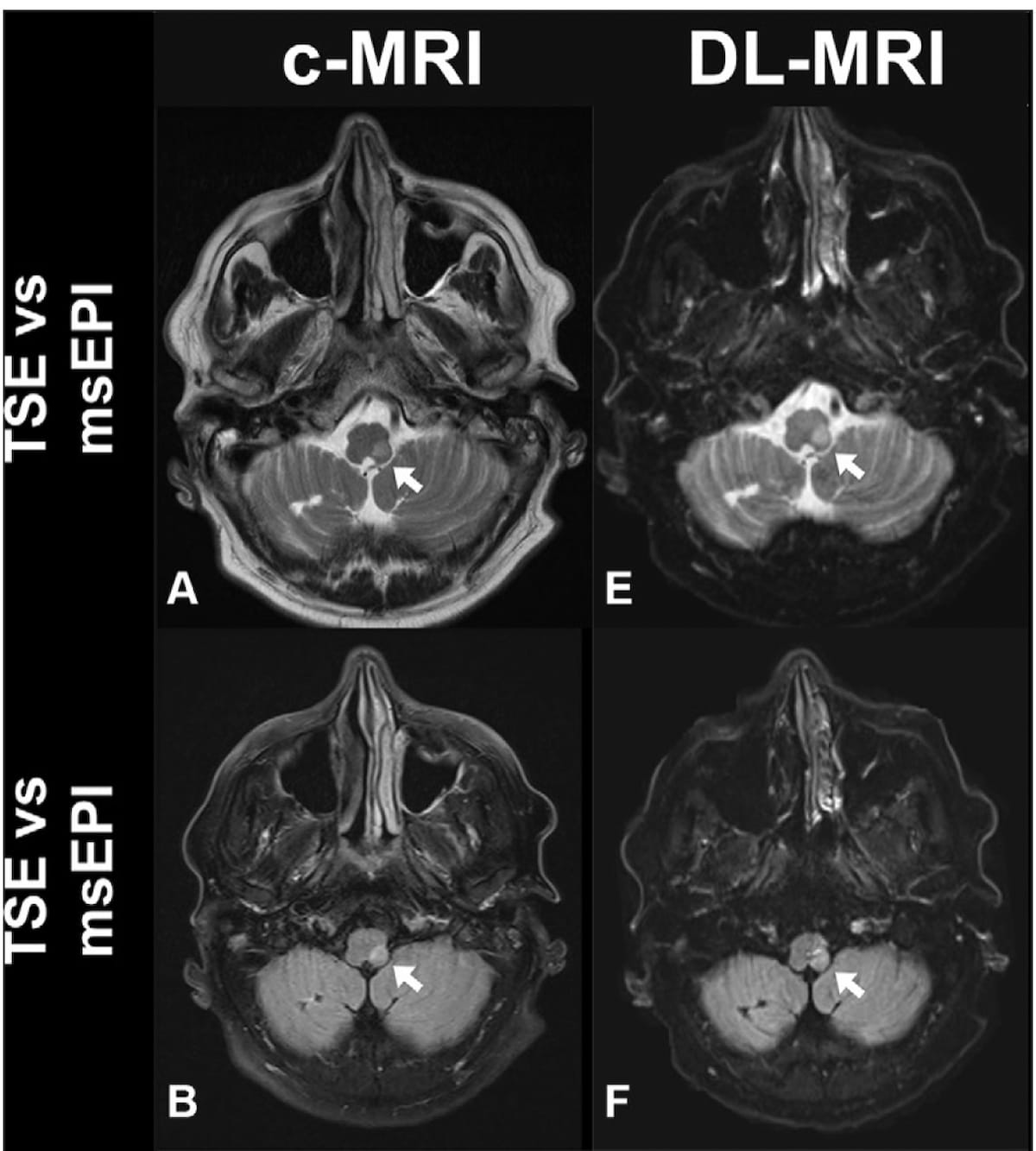
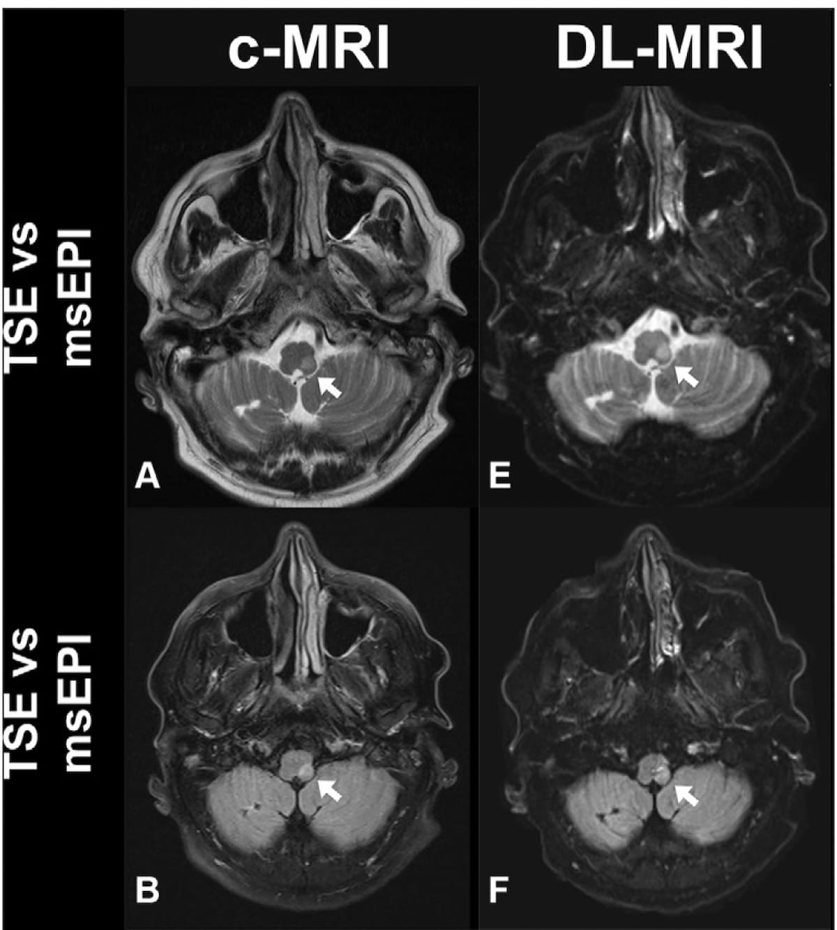
For patients with suspected acute stroke, researchers noted higher image quality for deep learning-accelerated MRI, which can be completed over 11 minutes sooner than similar sequences for conventional MRI.

Emerging research suggests that deep learning (DL) accelerated magnetic resonance imaging (MRI) offers similar signal properties for acute ischemic stroke, provides higher image quality, and reduces imaging time by more than 75 percent in comparison to conventional MRI.

For the prospective study, recently reported in Radiology, researchers reviewed data from 211 study participants (mean age of 65) with suspected acute stroke who had conventional MRI and DL accelerated MRI, which incorporated multi-shot echo-plantar imaging. The MRI sequences included T1-weighted, T2-weighted, diffusion-weighted imaging (DWI) and T2 fluid-attenuated inversion recovery (FLAIR) sequences, according to the study.

The study authors found that DL accelerated MRI and conventional MRI were interchangeable for the detection of acute ischemic lesions. While conventional MRI had slightly higher interrater agreement for acute ischemic lesion diagnosis in the left anterior circulation (94 percent vs. 93 percent) and right anterior circulation (94 percent vs. 91 percent), DL accelerated MRI had slightly higher interrater agreement in the posterior circulation (94 percent vs. 93 percent) and for relevant secondary findings (80 percent vs. 77 percent).

Can Deep Learning MRI Have an Impact In Suspected Stroke Cases?

Here one can see a comparison of conventional MRI and deep learning accelerated MRI that reveal acute infarction in a 75-year-old man. Researchers noted excellent image quality with deep learning accelerated MRI in 77.1 percent of readings in comparison to 61.9 percent of readings with conventional MRI. (Images courtesy of Radiology.)

Three reviewing radiologists also noted higher image quality with DL accelerated MRI. Out of 633 readings with DL accelerated MRI, the reviewing radiologists noted excellent image quality for 488 readings (77.1 percent) in comparison to 61.9 percent (392 of 633 readings) for conventional MRI.

The researchers also noted significant reductions in scanning time for DL accelerated MRI. The T2-weighted transverse plane sequence had an average acquisition time of 2 minutes, 39 seconds with conventional MRI vs. 24 seconds with DL accelerated MRI. T2 FLAIR sequences were completed in three minutes, 20 seconds on average with conventional MRI in comparison to one minute 22 seconds with DL accelerated MRI. Overall, the study authors noted a significant reduction in image acquisition time with DL accelerated MRI (14 minutes 18 seconds) in contrast to conventional MRI (3 minutes 4 seconds) for the detection of acute ischemic infarction.

Three Key Takeaways

  1. Similar signal properties for acute ischemic stroke detection. The study suggests that deep learning (DL) accelerated magnetic resonance imaging (MRI) provides similar signal properties for detecting acute ischemic stroke when compared to conventional MRI. This indicates that DL accelerated MRI can be a reliable alternative for acute ischemic lesion diagnosis.
  2. Higher image quality with DL accelerated MRI. Three reviewing radiologists noted higher image quality with DL accelerated MRI. The study found that DL accelerated MRI had excellent image quality for 77.1 percent of readings, compared to 61.9 percent for conventional MRI. This suggests that DL acceleration not only maintains but may even enhance image quality in certain cases.
  3. Significant reduction in imaging time. DL accelerated MRI demonstrated a substantial reduction in imaging time with over 75 percent reduction compared to conventional MRI. The study highlights specific sequences, such as T2-weighted transverse plane and T2 FLAIR, showing significantly shorter acquisition times with DL accelerated MRI. The potential to achieve faster imaging is crucial for improving efficiency in clinical settings, making it a valuable consideration for patients with suspected stroke.


“The deployment of DL-accelerated MRI reconstructions is becoming successively prevalent in MRI applications due to its potential to substantially reduce examination times while maintaining diagnostic image quality. Given the increasing demand for medical examinations and increasing financial constraints placed on health care systems, implementing this technique may be of great value,” wrote lead study author Sebastian Altmann, M.D., who is affiliated with the Department of Neuroradiology at the University Medical Center Mainz in Mainz, Germany, and colleagues.

While acknowledging that computed tomography (CT) is more often utilized in cases of suspected acute ischemic stroke due to availability and cost-effectiveness, the researchers emphasized stronger sensitivity of MRI for diagnosing early and small ischemic lesions.

“This may be of increasing relevance as randomized clinical trials have shown that patients with acute ischemic stroke of unknown onset and without a clear window of time could benefit from intravenous thrombolysis selected at DWI and (FLAIR) imaging,” pointed out Altmann and colleagues.

(Editor’s note: For related content, see “Study Says MRI Offers Most Cost-Effective Imaging for Dizziness Presentations in the ER,” “Practical Insights on CT and MRI Neuroimaging and Reporting for Stroke Patients” and “Can Abbreviated MRI Have an Impact in Neuroimaging?”)

Beyond the inherent limitations of a single-center study, the authors noted possible bias with manual assessment of DWI and ADC values for acute ischemic infarctions. The researchers did not assess other pathologic abnormalities aside from acute ischemic stroke and acknowledged that they did not acquire contrast-enhanced MRI views.


Thursday, December 21, 2023

Magnetic resonance image-based brain age as a discriminator of dementia conversion in patients with amyloid-negative amnestic mild cognitive impairment

I can't imagine getting an MRI paid for by insurance for this. 

Magnetic resonance image-based brain age as a discriminator of dementia conversion in patients with amyloid-negative amnestic mild cognitive impairment

Abstract

Patients with amyloid-negative amnestic mild cognitive impairment (MCI) have a conversion rate of approximately 10% to dementia within 2 years. We aimed to investigate whether brain age is an important factor in predicting conversion to dementia in patients with amyloid-negative amnestic MCI. We conducted a retrospective cohort study of patients with amyloid-negative amnestic MCI. All participants underwent detailed neuropsychological evaluation, brain magnetic resonance imaging (MRI), and [18F]-florbetaben positron emission tomography. Brain age was determined by the volumetric assessment of 12 distinct brain regions using an automatic segmentation software. During the follow-up period, 38% of the patients converted from amnestic MCI to dementia. Further, 73% of patients had a brain age greater than their actual chronological age. When defining ‘survival' as the non-conversion of MCI to dementia, these groups differed significantly in survival probability (p = 0.036). The low-educated female group with a brain age greater than their actual age had the lowest survival rate among all groups. Our findings suggest that the MRI-based brain age used in this study can contribute to predicting conversion to dementia in patients with amyloid-negative amnestic MCI.

Wednesday, November 8, 2023

Detection of Intracerebral Hemorrhage Using Low-Field, Portable Magnetic Resonance Imaging in Patients With Stroke

Well with NOTHING on how followup will get you 100% recovered; THIS IS TOTALLY FUCKING USELESS. Will you please solve stroke?

Detection of Intracerebral Hemorrhage Using Low-Field, Portable Magnetic Resonance Imaging in Patients With Stroke


Originally publishedhttps://doi.org/10.1161/STROKEAHA.123.043146Stroke. 2023;54:2832–2841

Abstract

BACKGROUND:

Neuroimaging is essential for detecting spontaneous, nontraumatic intracerebral hemorrhage (ICH). Recent data suggest ICH can be characterized using low-field magnetic resonance imaging (MRI). Our primary objective was to investigate the sensitivity and specificity of ICH on a 0.064T portable MRI (pMRI) scanner using a methodology that provided clinical information to inform rater interpretations. As a secondary aim, we investigated whether the incorporation of a deep learning (DL) reconstruction algorithm affected ICH detection.

METHODS:

The pMRI device was deployed at Yale New Haven Hospital to examine patients presenting with stroke symptoms from October 26, 2020 to February 21, 2022. Three raters independently evaluated pMRI examinations. Raters were provided the images alongside the patient’s clinical information to simulate real-world context of use. Ground truth was the closest conventional computed tomography or 1.5/3T MRI. Sensitivity and specificity results were grouped by DL and non-DL software to investigate the effects of software advances.

RESULTS:

A total of 189 exams (38 ICH, 89 acute ischemic stroke, 8 subarachnoid hemorrhage, 3 primary intraventricular hemorrhage, 51 no intracranial abnormality) were evaluated. Exams were correctly classified as positive or negative for ICH in 185 of 189 cases (97.9% overall accuracy). ICH was correctly detected in 35 of 38 cases (92.1% sensitivity). Ischemic stroke and no intracranial abnormality cases were correctly identified as blood-negative in 139 of 140 cases (99.3% specificity). Non-DL scans had a sensitivity and specificity for ICH of 77.8% and 97.1%, respectively. DL scans had a sensitivity and specificity for ICH of 96.6% and 99.3%, respectively.

CONCLUSIONS:

These results demonstrate improvements in ICH detection accuracy on pMRI that may be attributed to the integration of clinical information in rater review and the incorporation of a DL-based algorithm. The use of pMRI holds promise in providing diagnostic neuroimaging for patients with ICH.

Friday, February 4, 2022

Multimodal MRI reveals brain areas that can still ‘see’ after a stroke

WHOM is going to put this in a usable format so recovery can be applied to stroke survivors? We need to know the specific name of the leader and researcher so we can monitor progress. And you really think your stroke hospital subscribes to Physics World and requires the doctors to read it?

Multimodal MRI reveals brain areas that can still ‘see’ after a stroke

03 Feb 2022 Poulami Somanya Ganguly 
Stroke lesions
Anatomical imaging: Stroke lesions in the brain of one of the study participants. (Courtesy: CC BY 4.0/Front. Neurosci. 10.3389/fnins.2021.737215)

Scientists in the UK have used magnetic resonance imaging (MRI) to map the brain’s responses to visual stimuli after a stroke. In stroke survivors with vision loss, brain imaging revealed responsive areas that were inaccessible to current vision tests. The researchers’ findings could help clinicians better understand vision loss in stroke, and design tailored rehabilitation programmes for survivors. The researchers, from the University of Nottingham, describe their work in Frontiers in Neuroscience.

Every year, an estimated 100,000 people in the UK have a stroke. Globally, the number of strokes each year has risen by 70% since 1990, and stroke remains one of the leading causes of death and disability worldwide.

About a third of all stroke survivors experience visual field loss, in which a quarter or one half of their total visual field is impacted. Currently, there are no universally accepted rehabilitation strategies for vision loss in stroke, and the success of existing programmes varies significantly across survivors.

Visual field loss after stroke is usually diagnosed using perimetry, a technique that uses bright lights of differing size and brightness to test a patient’s visual response. However, perimetry only provides a coarse map of residual visual function and cannot pinpoint pathways in the brain that no longer process visual information.

To reveal more detail, the researchers used different MRI techniques – functional, anatomical and diffusion-weighted MRI – to chart affected visual pathways in the brain in four stroke survivors.

Mapping the the visual field

By measuring responses in the brain to visual stimuli, they were able to map the visual field in stroke survivors in much finer detail compared with perimetry. When they overlaid their visual field maps with perimetry maps, they were able to see spots in the visual field that still elicited a response from the brain.

Denis Schluppeck

“By examining different types of brain scans we can actually see areas of ‘residual vision’ – places where the eyes and brain can still process images, even if this doesn’t reach awareness,” explains Denis Schluppeck, senior author of the study. “Using MRI to pinpoint these areas of functional vision, clinicians could work with the stroke survivor and train them to recover some function in that particular spot.”

Rehabilitation strategies for vision loss in stroke include strengthening existing or alternative visual pathways in the brain. Combining brain imaging and optometric tests could provide a cost-effective way to understand which brain areas to focus on. The researchers note that their MRI protocol takes just one hour to perform.

“I think what was most challenging about the study was recruiting stroke survivors,” says first author Anthony Beh. “As with any clinical group, there are extra considerations to make when working closely with them, such as mobility challenges, past medical history and other stroke-related impairments. It was also important to get in touch with local stroke communities to visit and speak more about the project.”

Anthony Beh

Going forward, the researchers plan to use what they have learned to understand other forms of visual loss. “We hope to use a similar approach in children and younger individuals who have cortical visual impairment – visual loss that is not caused by damage to eyes or early parts of the visual pathway – to understand that set of disorders,” Schluppeck explains.