Changing stroke rehab and research worldwide now.Time is Brain! trillions and trillions of neurons that DIE each day because there are NO effective hyperacute therapies besides tPA(only 12% effective). I have 523 posts on hyperacute therapy, enough for researchers to spend decades proving them out. These are my personal ideas and blog on stroke rehabilitation and stroke research. Do not attempt any of these without checking with your medical provider. Unless you join me in agitating, when you need these therapies they won't be there.

What this blog is for:

My blog is not to help survivors recover, it is to have the 10 million yearly stroke survivors light fires underneath their doctors, stroke hospitals and stroke researchers to get stroke solved. 100% recovery. The stroke medical world is completely failing at that goal, they don't even have it as a goal. Shortly after getting out of the hospital and getting NO information on the process or protocols of stroke rehabilitation and recovery I started searching on the internet and found that no other survivor received useful information. This is an attempt to cover all stroke rehabilitation information that should be readily available to survivors so they can talk with informed knowledge to their medical staff. It lays out what needs to be done to get stroke survivors closer to 100% recovery. It's quite disgusting that this information is not available from every stroke association and doctors group.

Showing posts with label muscle movement. Show all posts

Tuesday, September 22, 2020

Individual Muscle Control using an Exoskeleton Robot for Muscle Function Testing

This is incredibly important, only 20 years old. If you can identify an individual muscle that is not working properly then you can go to muscle building sites and get EXACT exercises to work on those muscles.  This is your responsibility because your doctors and therapists have zero clues on how to get you recovered. My definition of recovery is 100%, your doctor and therapists will use the tyranny of low expectations to try to make you believe that their work to get you home and somewhat functioning is a success. If you didn't get to 100% recovery that failure is completely the fault of your doctor and therapists, they have had decades to get research going to solve all the problems in stroke. But DID NOTHING.

 Individual Muscle Control using an Exoskeleton Robot for Muscle Function Testing

2000, IEEE Transactions on Neural Systems and Rehabilitation Engineering
 Jun Ueda, Ding Ming, Vijaya Krishnamoorthy, Minoru Shinohara, and Tsukasa Ogasawara

 Abstract


 Healthy individuals modulate muscle activation pat- terns according to their intended movement and external environment. Persons with neurological disorders (e.g., stroke and spinal  cord injury), however, have problems in movement control due primarily to their inability to modulate their muscle activation pattern in an appropriate manner. A functionality test at the level of individual muscles that investigates the activity of a muscle of interest on various motor tasks may enable muscle-level force grading. To date there is no extant work that focuses on the application of exoskeleton robots to induce specific muscle activation in a systematic manner. This paper proposes a new method, named “individual muscle-force control” using a wearable robot (an exoskeleton robot, or a power-assisting device) to obtain a wider variety of muscle activity data than standard motor tasks, e.g., pushing a handle by hand. A computational algorithm systematically computes control commands to a wearable robot so that a desired muscle activation pattern for target muscle forces is induced. It also computes an adequate amount and direction of  a force that a subject needs to exert against a handle by his/her hand. This individual muscle control method enables users (e.g., therapists) to efficiently conduct neuromuscular function tests on target muscles by arbitrarily inducing muscle activation patterns.This paper presents a basic concept, mathematical formulation, and  solution of the individual muscle-force control and its implementation to a muscle control system with an exoskeleton-type robot for upper extremity. Simulation and experimental results in healthy individuals justify the use of an exoskeleton robot for future muscle function testing in terms of the variety of muscle activity data.
 

Tuesday, June 23, 2015

Stanford researchers provide insights into how human neurons control muscle movement

Your doctor should be able to use this to change and fine tune your stroke protocols.
http://scopeblog.stanford.edu/2015/06/23/stanford-researchers-provides-insights-into-how-human-neurons-control-muscle-movement/
Rina Shaikh-Lesko on June 23rd, 2015 No Comments

Stanford researchers provide insights into how human neurons control muscle movement

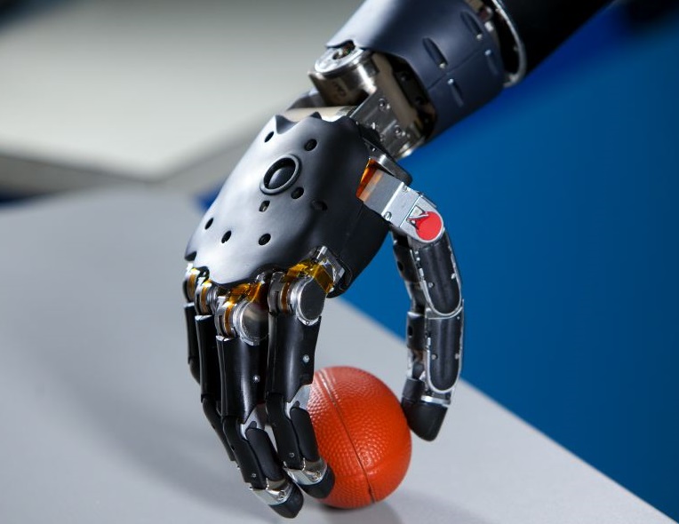
Brain-Controlled_Prosthetic_Arm_2A few years ago, a team led by Stanford researcher Krishna Shenoy, PhD, published a paper that proposed a new theory for how neurons in the brain controlled the movement of muscles: Rather than sending out signals with parceled bits of information about the direction and size of movement, Shenoy’s team found that groups of neurons fired in rhythmic patterns to get muscles to act.

That research, done in 2012, was in animals. Now, Shenoy and Stanford neurosurgeon Jamie Henderson, MD, have followed up on that work to demonstrate that human neurons function in the same way, in what the researchers call a dynamical system. The work is described in a paper published in the scientific journal eLife today. In our news release on the study, the lead author, postdoctoral scholar Chethan Pandarinath, PhD, said of the work:

    The earlier research with animals showed that many of the firing patterns that seem so confusing when we look at individual neurons become clear when we look at large groups of neurons together as a dynamical system.

The researchers implanted electrode arrays into the brains of two patients with amyotrophic lateral sclerosis (ALS), a neurodegenerative condition also known as Lou Gehrig’s disease. The new study provides further support for the initial findings and also lays the groundwork for advanced prosthetics like robotic arms that can be controlled by a person’s thoughts. The team is planning on working on computer algorithms that translate neural signals into electrical impulses that control prosthetic limbs.

Previously: Researchers find neurons fire rhythmically to create movement, Krishna Shenoy discusses the future of neural prosthetics at TEDxStanford, How does the brain plan movement? Stanford grad students explain in a video and Stanford researchers uncover the neural process behind reaction time

Stanford researchers provide insights into how human neurons control muscle movement
Brain-Controlled_Prosthetic_Arm_2A few years ago, a team led by Stanford researcher Krishna Shenoy, PhD, published a paper that proposed a new theory for how neurons in the brain controlled the movement of muscles: Rather than sending out signals with parceled bits of information about the direction and size of movement, Shenoy’s team found that groups of neurons fired in rhythmic patterns to get muscles to act.
That research, done in 2012, was in animals. Now, Shenoy and Stanford neurosurgeon Jamie Henderson, MD, have followed up on that work to demonstrate that human neurons function in the same way, in what the researchers call a dynamical system. The work is described in a paper published in the scientific journal eLife today. In our news release on the study, the lead author, postdoctoral scholar Chethan Pandarinath, PhD, said of the work:
The earlier research with animals showed that many of the firing patterns that seem so confusing when we look at individual neurons become clear when we look at large groups of neurons together as a dynamical system.
The researchers implanted electrode arrays into the brains of two patients with amyotrophic lateral sclerosis (ALS), a neurodegenerative condition also known as Lou Gehrig’s disease. The new study provides further support for the initial findings and also lays the groundwork for advanced prosthetics like robotic arms that can be controlled by a person’s thoughts. The team is planning on working on computer algorithms that translate neural signals into electrical impulses that control prosthetic limbs.
Previously: Researchers find neurons fire rhythmically to create movement, Krishna Shenoy discusses the future of neural prosthetics at TEDxStanford, How does the brain plan movement? Stanford grad students explain in a video and Stanford researchers uncover the neural process behind reaction time
- See more at: http://scopeblog.stanford.edu/2015/06/23/stanford-researchers-provides-insights-into-how-human-neurons-control-muscle-movement/#sthash.sUFVKzOj.dpuf

Monday, October 21, 2013

Muscle synergy space: learning model to create an optimal muscle synergy

Since we tend to have a lot of incorrect muscle synergies, your doctor can use this to tell us how to correct them to get to 100% recovery.
http://www.frontiersin.org/Journal/10.3389/fncom.2013.00136/full?utm_source=newsletter&utm_medium=email&utm_campaign=Neuroscience-w43-2013
Muscle redundancy allows the central nervous system (CNS) to choose a suitable combination of muscles from a number of options. This flexibility in muscle combinations allows for efficient behaviors to be generated in daily life. The computational mechanism of choosing muscle combinations, however, remains a long-standing challenge. One effective method of choosing muscle combinations is to create a set containing the muscle combinations of only efficient behaviors, and then to choose combinations from that set. The notion of muscle synergy, which was introduced to divide muscle activations into a lower-dimensional synergy space and time-dependent variables, is a suitable tool relevant to the discussion of this issue.
The synergy space defines the suitable combinations of muscles, and time-dependent variables vary in lower-dimensional space to control behaviors. In this study, we investigated the mechanism the CNS may use to define the appropriate region and size of the synergy space when performing skilled behavior. Two indices were introduced in this study, one is the synergy stability index (SSI) that indicates the region of the synergy space, the other is the synergy coordination index (SCI) that indicates the size of the synergy space.
The results on automatic posture response experiments show that SSI and SCI are positively correlated with the balance skill of the participants, and they are tunable by behavior training. These results suggest that the CNS has the ability to create optimal sets of efficient behaviors by optimizing the size of the synergy space at the appropriate region through interacting with the environment.

More for your doctor at the link.

Saturday, June 29, 2013

3-D Body Suit Put to Use in Healthcare Research

And using this our therapists could objectively determine all the muscles and joints that need correcting.

3-D Body Suit Put to Use in Healthcare Research


The suit, called MVN BIOMECH from Xsens out of Enschede, The Netherlands, is a 3-D human kinematic, camera-less measurement system with integrated small tracking sensors placed on the joints. Each sensor on the suit consists of three components: an accelerometer, magnetometers and a gyroscope. All together it gives information on each of the joints, the body segments between the joints and the 3-D movements. The technology sends the information a computer using a wireless signal which is then reconstructed into movements on the screen.

More at link.
 3-D Body Suit Put to Use in Healthcare Research

Tuesday, October 2, 2012

Movement Kinematics During a Drinking Task Are Associated With the Activity Capacity Level After Stroke

And the first thing I thought of was drinking alcohol(specifically beer). Sign me up for those elbow bending exercises.
 But objective would be great, make sure your therapists objectively analyze your movement problems. How else are they going to be able to document improvements?
  http://nnr.sagepub.com/content/26/9/1106.abstract?etoc

Abstract

Background. Kinematic analysis is a powerful method for an objective assessment of movements and is increasingly used as an outcome measure after stroke. Little is known about how the actual movement performance measured with kinematics is related to the common traditional assessment scales. The aim of this study was to determine the relationships between movement kinematics from a drinking task and the impairment or activity limitation level after stroke. Methods. Kinematic analysis of movement performance in a drinking task was used to measure movement time, smoothness, and angular velocity of elbow and trunk displacement (TD) in 30 individuals with stroke. Sensorimotor impairment was assessed with the Fugl-Meyer Assessment (FMA), activity capacity limitation with the Action Research Arm Test (ARAT), and self-perceived activity difficulties with the ABILHAND questionnaire. Results. Backward multiple regression revealed that the movement smoothness (similarly to movement time) and TD together explain 67% of the total variance in ARAT. Both variables uniquely contributed 37% and 11%, respectively. The TD alone explained 20% of the variance in the FMA, and movement smoothness explained 6% of the variance in the ABILHAND. Conclusions. The kinematic movement performance measures obtained during a drinking task are more strongly associated with activity capacity than with impairment. The movement smoothness and time, possibly together with compensatory movement of the trunk, are valid measures of activity capacity and can be considered as key variables in the evaluation of upper-extremity function after stroke. This increased knowledge is of great value for better interpretation and application of kinematic data in clinical studies.

Friday, September 28, 2012

Optimizing muscle power after stroke: a cross-sectional study

Ask your therapist and doctor to use this in your therapy protocol.
http://www.jneuroengrehab.com/content/9/1/67/abstract

Abstract (provisional)

Background

Stroke remains a leading cause of disability worldwide and results in muscle performance deficits and limitations in activity performance. Rehabilitation aims to address muscle dysfunction in an effort to improve activity and participation. While both muscle strength and power have an impact on activity performance, power has recently been acknowledged as contributing significantly to activity performance in this population. Therefore, rehabilitation efforts should include training of muscle power. However, little is known about what training parameters optimize muscle power performance in people with stroke. The purpose of this study was to investigate lower limb muscle power performance at differing loads in people with and without stroke.

Methods

A cross-sectional study design investigated muscle power performance in 58 hemiplegic and age matched control participants. Lower limb muscle power was measured using a modified leg press machine at 30, 50 and 70% of one repetition maximum (1-RM) strength.

Results

There were significant differences in peak power between involved and uninvolved limbs of stroke participants and between uninvolved and control limbs. Peak power was greatest when pushing against a load of 30% of 1RM for involved, uninvolved and control limbs. Involved limb peak power irrespective of load (Mean:220 [PLUS-MINUS SIGN] SD:134 W) was significantly lower (p < 0.05) than the uninvolved limb (Mean:466 [PLUS-MINUS SIGN] SD:220 W). Both the involved and uninvolved limbs generated significantly lower peak power (p < 0.05) than the control limb (Mean:708 [PLUS-MINUS SIGN] SD:289 W).

Conclusions

Significant power deficits were seen in both the involved and uninvolved limbs after stroke. Maximal muscle power was produced when pushing against lighter loads. Further intervention studies are needed to determine whether training of both limbs at lighter loads (and higher velocities) are preferable to improve both power and activity performance after stroke.

The complete article is available as a provisional PDF. The fully formatted PDF and HTML versions are in production.

Sunday, September 2, 2012

Muscle action and explanation for stroke rehab

I'm sure your therapists throw a lot of muscle names at you. This site can explain what they are referring to and for those of the do-it-yourself plan  you can use it for your mental imagery and action observation.
http://www.getbodysmart.com/
My first lookup was the latissimus dorsi to find out which muscle is causing me so much grief in trying to raise my arm due to spasticity. And as Steven Wolf says, 'Stroke patients need to rely more on their own problem solving to regain mobility',  this would be a good start for your problem solving.

Tuesday, June 26, 2012

What apps should your therapist have on their iPad to show you what they are doing for you?

I have to refer you to Mike Reinolds' blog for this one.
I wish I could see the VisibleBody one. Only 19.99, your therapist can afford this one  after billing one of your sessions. I may have to get the 29.99 one for my PC.
http://www.mikereinold.com/2012/06/best-ipad-apps-for-physical-therapy.html

Wednesday, April 18, 2012

Muscle Activation During Gait

Only 22 seconds
Visualisation of muscle activation during gait using an anatomically-based model of the lower limbs. Concentric contraction is indicated in yellow, isometric contraction in orange and eccentric contraction in purple respectively. (Research in musculoskeletal modelling at the Auckland Bioengineering Institute)
Something every PT should be able to show you. Ask them the difference in concentric, isometric and eccentric. And then ask for specific exercises to stop spasticity and improve your gait.
 http://www.youtube.com/watch?NR=1&feature=endscreen&v=GV6CAZiv5Zo

Thursday, January 5, 2012

To Speed People Up, Human Leg Muscle Slows Down

Researchers should be able to use something similar to determine exactly what is going on in impaired muscles. We could then use that knowledge to create therapies to address the problem areas.
http://www.healthcanal.com/bones-muscles/25175-Speed-People-Human-Leg-Muscle-Slows-Down.html

Other than Olympic race walkers, people generally find it more comfortable to run than walk when they start moving at around 2 meters per second – about 4.5 miles per hour.

North Carolina State University biomedical engineers Dr. Gregory Sawicki and Dr. Dominic Farris have discovered why: At 2 meters per second, running makes better use of an important calf muscle than walking, and therefore is a much more efficient use of the muscle’s – and the body’s – energy.

Published online this week in Proceedings of the National Academy of Sciences, the results stem from a first-of-its-kind study combining ultrasound imaging, high-speed motion-capture techniques and a force-measuring treadmill to examine a key calf muscle and how it behaves when people walk and run.

The study used ultrasound imaging in a unique way: A small ultrasound probe fastened to the back of the leg showed in real time the adjustments made by the muscle as study subjects walked and ran at various speeds.

The high-speed images revealed that the medial gastrocnemius muscle, a major calf muscle that attaches to the Achilles tendon, can be likened to a “clutch” that engages early in the stride, holding one end of the tendon while the body’s energy is transferred to stretch it. Later, the Achilles – the long, elastic tendon that runs down the back of the lower leg – springs into action by releasing the stored energy in a rapid recoil to help move you.

The study showed that the muscle “speeds up,” or changes its length more and more rapidly as people walk faster and faster, but in doing so provides less and less power. Working harder and providing less power means less overall muscle efficiency.

When people break into a run at about 2 meters per second, however, the study showed that the muscle “slows down,” or changes its length more slowly, providing more power while working less rigorously, thereby increasing its efficiency.

“The ultrasound imaging technique allows you to separate out the movement of the muscles in the lower leg and has not been used before in this context,” Farris says.

The finding sheds light on why speed walking is generally confined to the Olympics: muscles must work too inefficiently to speed walk, so the body turns to running in order to increase efficiency and comfort, and to conserve energy.

“The muscle can’t catch up to the speed of the gait as you walk faster and faster,” Sawicki says. “But when you shift the gait and transition from a walk to a run, that same muscle becomes almost static and doesn’t seem to change its behavior very much as you run faster and faster, although we didn’t test the muscle at sprinting rates.”

The research could help inform the best ways of building assistive or prosthetic devices for humans, or help strength and conditioning professionals assist people who have had spinal-cord injury or a stroke, Sawicki and Farris say.

The researchers are part of NC State’s Human PoWeR (Physiology of Wearable Robotics) Lab, directed by Sawicki. The joint Department of Biomedical Engineering is part of NC State’s College of Engineering and the University of North Carolina-Chapel Hill’s School of Medicine.

Thursday, December 15, 2011

Position-sensing technologies for movement analysis in stroke rehabilitation

Anything for our therapists to be able to identify movements and the specific muscles that need correction.

Link

Position-sensing technologies for movement analysis in stroke rehabilitation


Abstract

Research has focused on improvement of the quality of life of stroke patients. Gait detection, kinematics and kinetics analysis, home-based rehabilitation and telerehabilitation are the areas where there has been increasing research interest. The paper reviews position-sensing technologies and their application for human movement tracking and stroke rehabilitation. The review suggests that it is feasible to build a home-based telerehabilitation system for sensing and tracking the motion of stroke patients.

Keywords Position sensing - Motion tracking - Human movement - Rehabilitation - Inertial sensors

Wednesday, December 14, 2011

Effects of carbohydrate and caffeine co-ingestion on a reliable simulated soccer-specific protocol

For those who need an excuse for their caffeine habit.

Effects of carbohydrate and caffeine co-ingestion on a reliable simulated soccer-specific protocol


Abstract

The aim of this study was to evaluate the effect of co-ingesting carbohydrate and caffeine (CHO+CAF) in comparison to carbohydrate (CHO) and placebo (PLA), during a reliable soccer-specific test (Currell et al, 2009). 8 university-standard soccer players ingested a PLA, a 6.4% CHO or 6.4% CHO and 160 mg CAF (CHO+CAF) solution on three occasions, in a double-blind randomised cross-over design, with each trial separated by 7 days. The protocol was 90 min in duration, made up of ten 6 min exercise blocks, each followed by soccer-specific skills tests (agility, dribbling, heading and kicking accuracy). Dependant variables (Agility, dribbling, heading, kicking accuracy, glucose, lactate, HR and RPE) were analysed using one-way repeated measures ANOVA. Significant difference (p< 0.05) was found between CHO+CAF, CHO and PLA for each of the soccer-specific skill tests. Significant improvement (p= 0.02) was observed in agility time in CHO versus PLA trials, although no significant difference (p> 0.05) was reported for dribbling, heading and kicking accuracy. Blood glucose and lactate were elevated (p< 0.05) with CHO+CAF supplementation over PLA, but there was no difference (p> 0.05) compared to CHO. Blood glucose increased (p= 0.01) in the CHO trial compared to PLA, with no difference (p> 0.05) between CHO+CAF and CHO. No significant difference (p> 0.05) was reported for HR and RPE values across all trial conditions. Skill performance during simulated soccer activity improved with CHO+CAF supplementation in comparison to both CHO and PLA. CHO+CAF co-ingestion had no ergogenic benefit over CHO in the maintenance and availability of blood glucose however, CHO+CAF co-ingestion did allow players to sustain a higher work intensity as opposed to CHO and PLA beverages as shown by elevated blood lactate levels.

Wednesday, December 7, 2011

How The Brain Corrects Involuntary Bodily Movement

Another piece on information on the brain. One more item I will need to recreate.
http://www.medicalnewstoday.com/releases/238707.php
Researchers have identified the area of the brain that controls our ability to correct our movement after we've been hit or bumped - a finding that may have implications for understanding why subjects with stroke often have severe difficulties moving.

The fact that humans rapidly correct for any disturbance in motion demonstrates the brain understands the physics of the limb - scientists just didn't know what part of the brain supported this feedback response - until now.

Several pathways and regions of the central nervous system could contribute to our response to external knocks to the body, but researchers only recently discovered that the pathway through the primary motor cortex provides this knowledge of the physics of the limb.

"To say this process is complex is an understatement," says Stephen Scott, a neuroscience professor and motor behavior specialist in the Department of Biomedical and Molecular Sciences. "Voluntary movement is really, really hard in terms of the math involved. When I walk around, the equations of my motion are like a small book. The best physicists can't solve these complicated equations, but your brain can do it incredibly quickly."

The corrective movement pathway works by limiting and correcting the domino effect of involuntary bodily movement caused by an external blow. For example, a blow to the shoulder that causes the whole arm to swing about may require the brain to quickly turn on muscles in the shoulder, bicep, forearm and hand in order to regain control of the limb. Likewise, a football player who collides with an opponent during a game has to respond quickly to correct the movement and remain upright.

Strokes that take place in the primary motor cortex may cause varying levels of damage to this corrective movement pathway. This varying damage may explain why some stroke patients are able to improve their movement skills in rehabilitation and why some patients remain uncoordinated and unsteady.

Dr. Scott now wants to apply these findings to stroke patients by examining the damage these patients have to their sensory pathways and how this damage relates to movement problems. He believes that these findings may support an increased focus on first-stage sensory rehabilitation to help rebuild pathways that transmit sensory information to the brain before treatment moves to a focus on motor skills.