Use the labels in the right column to find what you want. Or you can go thru them one by one, there are only 33,991 posts. Searching is done in the search box in upper left corner. I blog on anything to do with stroke. DO NOT DO ANYTHING SUGGESTED HERE AS I AM NOT MEDICALLY TRAINED, YOUR DOCTOR IS, LISTEN TO THEM. BUT I BET THEY DON'T KNOW HOW TO GET YOU 100% RECOVERED. I DON'T EITHER BUT HAVE PLENTY OF QUESTIONS FOR YOUR DOCTOR TO ANSWER.
Changing stroke rehab and research worldwide now.Time is Brain!trillions and trillions of neuronsthatDIEeach day because there areNOeffective hyperacute therapies besides tPA(only 12% effective). I have 523 posts on hyperacute therapy, enough for researchers to spend decades proving them out. These are my personal ideas and blog on stroke rehabilitation and stroke research. Do not attempt any of these without checking with your medical provider. Unless you join me in agitating, when you need these therapies they won't be there.
What this blog is for:
My blog is not to help survivors recover, it is to have the 10 million yearly stroke survivors light fires underneath their doctors, stroke hospitals and stroke researchers to get stroke solved. 100% recovery. The stroke medical world is completely failing at that goal, they don't even have it as a goal. Shortly after getting out of the hospital and getting NO information on the process or protocols of stroke rehabilitation and recovery I started searching on the internet and found that no other survivor received useful information. This is an attempt to cover all stroke rehabilitation information that should be readily available to survivors so they can talk with informed knowledge to their medical staff. It lays out what needs to be done to get stroke survivors closer to 100% recovery. It's quite disgusting that this information is not available from every stroke association and doctors group.
Do you
prefer your doctor, hospital and board of director's incompetence NOT
KNOWING? OR NOT DOING? Your choice; let them be incompetent or demand
action!
We’re sharing this article early to provide faster access to peer-reviewed, accepted research. It is citable and carries a permanent DOI. This version is subject to further edits and will be replaced automatically by the final Version of Record. All legal disclaimers apply.
Abstract
Background
Tranexamic acid (TXA) is commonly used in trauma care to decrease bleeding, but its effectiveness and safety in prehospital settings, notably in traumatic brain injury (TBI), remain controversial. This review aimed to systematically evaluate the evidence on prehospital tranexamic acid (TXA) use in trauma and traumatic brain injury (TBI), with quantitative meta-analysis restricted to randomized comparative evidence and non-randomized evidence summarized narratively.
Methods
We conducted a systematic review with quantitative meta-analysis restricted to independent randomized comparative studies evaluating prehospital TXA versus placebo, usual care, or no TXA. Observational studies, secondary analyses, and survey studies were summarized separately and were not pooled with randomized trials in the primary efficacy or safety meta-analysis. outcomes included mortality (28/30-day, in-hospital), thromboembolic events, seizures, and neurological consequences. Risk of bias was evaluated using Cochrane risk of bias tools for randomized controlled trials (RoB2) and non-randomized studies by Risk Of Bias In Non-randomized Studies of Interventions (ROBINS-I).
Results
Twenty-one reports met the broad review criteria. The RCT-only analysis of 28- or 30-day mortality included three independent parent randomized trials: PATCH-Trauma/Gruen et al. 2023, STAAMP/Guyette et al. 2021, and ROC-TXA/Rowell et al. 2020. Prehospital TXA was associated with lower 28- or 30-day mortality (RR 0.81, 95% CI 0.68–0.95). The TBI mortality result was derived solely from ROC-TXA/Rowell et al. 2020 and was therefore a single-study estimate rather than a pooled estimate (RR 0.87, 95% CI 0.64–1.20). In-hospital mortality was reported only by STAAMP/Guyette et al. 2021 and was also a single-study estimate (RR 0.87, 95% CI 0.57–1.33). No statistically significant increase in thromboembolic events or seizures was demonstrated. However, the DVT and PE point estimates were above the null, and their wide confidence intervals did not exclude a clinically relevant increase in thromboembolic risk.
Conclusion
Prehospital TXA may reduce 28- or 30-day mortality in selected broader trauma populations, particularly among patients at risk of hemorrhage. No statistically significant increase in thromboembolic events or seizures was demonstrated; however, clinically relevant thromboembolic harm cannot be excluded because the DVT and PE point estimates were above the null, and the confidence intervals were wide.
How long will it take for your competent? doctor/hospital to get human testing going for stroke related neurodegeneration? NEVER? So your stroke medical 'professionals' are useless?
Do you
prefer your doctor, hospital and board of director's incompetence NOT
KNOWING? OR NOT DOING? Your choice; let them be incompetent or demand
action!
Mohamed Arif1*, V. Gayathri2, Radhika Ravindran3,P. Kalaivani2, R. Siva2, Shonam Tamrakar2
1Gidaa Life Sciences Private Limited, 287, 9th Cross, Bapuji Layout Near Chandra Layout, Vijayanagara, Bangalore, Karnataka 560040, India.
2Centre for Toxicology and Developmental Research, Sri Ramachandra Institute of Higher Education and Research (Deemed to be University), No. 1,
Ramachandra Nagar, Porur, Chennai 600116, Tamil Nadu, India.
3Tissue Engineering and Biomaterials Lab, Department of Biotechnology, IIT Madras, India.
*Corresponding Author: Mohamed Arif, Gidaa Life Sciences Private Limited, 287, 9th Cross, Bapuji Layout Near Chandra Layout, Vijayanagara,
Bangalore, Karnataka 560040, India.
DOI: https://doi.org/10.58624/SVOANE.2026.07.030
Received: July 31, 2026
Published: August 18, 2026
Citation: Arif M, Gayathri V, Ravindran R, Kalaivani P, Siva R, Tamrakar S. A Novel Curcumin-Based Formulation Offers Protection Against
Neurodegeneration in Rat Models of AlCl3-Induced Alzheimer’s Disease. SVOA Neurology 2026, 7:4, 220-238. doi.org/10.58624/SVOANE.2026.07.030
Abstract
The exact mechanisms underlying Alzheimer’s disease (AD) pathogenesis are not fully understood, and
effective disease-modifying treatments remain lacking despite decades of AD research. Therefore,
alternative therapeutic approaches that may target multiple mechanisms of action underlying AD and have a
better safety profile than synthetic drugs, such as phytotherapy, are being explored. We aimed to assess the
neuroprotective properties of a novel curcumin formulation fortified with andrographolides and piperine (MAG
XXI) in rat models of aluminium chloride–induced AD. Overall, 30 male Wistar rats were included and divided
into five groups (a healthy control group, a non-treated AD group, and three AD groups treated with donepezil
or 200/400 mg/kg body weight of MAG XXI). The Morris water maze, passive avoidance, and elevated plus
maze tests were performed on the rats. Tissue samples from the cortex and hippocampus of the rats were
then subjected to biochemical evaluation of neuronal, oxidative stress, and inflammatory markers. Compared
to non-treated rats, donepezil-treated rats and high-dose (400 mg/kg body weight) MAG XXI–treated rats
showed a mild but significant improvement in the Morris water maze and elevated plus maze test findings
and a marked and significant improvement in the passive avoidance task results. Furthermore, oxidative
stress markers, inflammatory markers, and neuronal markers improved overall in the donepezil-treated group
and in both MAG XXI–treated groups. Notably, the improvement in the oxidative stress markers was more
marked with MAG XXI (both doses) than with donepezil. Histopathological examination revealed lower
incidence rates of neurofibrillary tangles, gliosis, and neuronal degeneration in the high-dose MAG XXI and
donepezil groups. Notably, cresyl staining revealed minimal-to-mild cell dispersion in the donepezil group,
whereas normal neuronal cells with well-lineated cell bodies and Nissl substance were observed in the high
dose MAG XXI group. No adverse events were noted in the MAG XXI groups. MAG XXI could be a promising
alternative for AD treatment because it appears to exhibit neuroprotective properties, as demonstrated by its
ability to alleviate oxidative stress and neuroinflammation. However, further clinical trials involving humans
are necessary to corroborate the present study’s results
Do you
prefer your doctor, hospital and board of director's incompetence NOT
KNOWING? OR NOT DOING? Your choice; let them be incompetent or demand
action!
Researchers have pioneered a way to create miniaturized copies of a person's carotid artery that could help doctors predict and manage the patient's stroke risk, a new study suggests.
In the research, published in July in the journalCell Biomaterials, scientists used these "arteries-on-a-chip" to monitor how real blood flows through a patient's carotid arteries, which carry blood to the brain, face and neck. This could enable doctors to identify not just how and what type of clots form in that specific patient, but also determine which medications would be most effective in dealing with the blockage.
While the technique is currently a proof-of-concept, someday, it could help medical practitioners to better tailor treatments for each patient.
"When the inner lining of an artery is damaged, material underneath the cells, including collagen, becomes exposed to the blood," Zhao said. A bloodborne protein called von Willebrand factor (VWF) grabs hold of platelets from the flowing blood. Those platelets then stick together and recruit more platelets, building a clot, he said.
But forming a clot is only half the story; it's what this mass does next that determines the risk to the patient.
In some cases, the growing ball of clotted blood will stay firmly rooted to the artery wall, slightly impeding blood flow but not posing any immediate danger. However, if tiny fragments break off this static clot, they can travel toward the brain, where they risk blocking smaller vessels and thus causing a stroke.
"Clinically, we are very good at imaging how narrow an artery is," Zhao said, "but narrowing alone does not tell us exactly how a clot will behave."
This is where artery-on-a-chip models come in, he said. Rather than relying purely on medical scans, the team's model recreates the exact shape and structure of an individual patient's blood vessels using 3D printing. To create the models, they first use a patient’s existing CT scans to 3D print a plastic replica of their carotid artery, including any narrowing caused by atherosclerosis. Next, the inside of this plastic structure is coated with collagen, and then cells that line the carotid artery get layered on top. Blood is passed through the replica artery, mimicking the speed and pressure of blood flow in the body.
Do you
prefer your doctor, hospital and board of director's incompetence NOT
KNOWING? OR NOT DOING? Your choice; let them be incompetent or demand
action!
As Alzheimer’s disease and other forms of dementia continue to rise, many people are looking for ways to protect their brain health as they age. One neurophysiologist believes one physical exercise may stand above the rest when it comes to the brain.
Nicola argued that the exercise engages nearly the entire body. “If done correctly, the deadlift can use almost every muscle in your body,” she said, listing muscles including the erector spinae (deep back muscles extending from the pelvis to the base of the skull), glutes, quadriceps, serratus anterior (a muscle on the side of the chest), and calves. Recruiting so many muscle groups at once, the movement would also be comparable to a barbell squat, she said.
She also suggested that lifting heavier weights places greater demands on the brain. “I want both men and women lifting heavy because…think of your brain as real estate,” Nicola said. “There’s real estate in your brain reserved for lifting heavy.”
According to Nicola, moving heavier loads requires more neural resources, resulting in a greater “neural drive” that she believes benefits the brain.
The discussion comes amid growing concern over dementia worldwide. Alzheimer’s disease is the most common cause of dementia, a condition characterized by progressive declines in memory, thinking, and everyday functioning. Alzheimer’s disease is responsible for around 60 to 80 percent of dementia cases, according to the Alzheimer’s Association.
While experts continue to search for treatments and preventive strategies, physical activity is increasingly recognized as one of the most important modifiable lifestyle factors associated with healthy aging and reduced dementia risk.
Physicians who spoke to Newsweek noted that while the deadlift is beneficial for brain health, no single exercise has been proven to prevent Alzheimer’s disease.
Neurophysiologist Reveals the 1 Exercise They’d Never Skip for Brain Health
Nuances on Deadlifts and Benefits for the Brain
Dr. Joanna Fong-Isariyawongse, a triple board-certified neurologist, sleep specialist, and epileptologist, told Newsweek that there’s “some nuance” to the idea that one exercise is best or that heavier lifting is always better.
Research suggests different forms of exercise support different aspects of brain health, she explained. Resistance training appears particularly helpful for overall cognition; aerobic activities, such as brisk walking, benefit memory, while balance-focused exercises, such as tai chi, have performed well in recent studies involving older adults, she noted.
“So, rather than one single exercise, a combination tends to offer the broadest protection,” Fong-Isariyawongse said.
Dr. Sergio Guiteau, a board-certified family medicine and sports medicine physician, told Newsweek that there is currently no evidence directly linking heavier weightlifting to greater neuroprotection or lower dementia rates.
“What we do know is that resistance training, as well as any activity or exercise that challenges balance, coordination, and learning, will decrease dementia risk more than just trying to maximize weight on a deadlift,” Guiteau said.
Dr. Parth Bhavsar, a board-certified family medicine physician and founder of TeleDirectMD, emphasized that brain protection depends on much more than lifting heavier weights. He told Newsweek that factors including consistency, technique, cardiovascular fitness, sleep quality, blood pressure control, insulin sensitivity, and avoiding injuries all contribute to long-term brain health.
Bhavsar noted: “If a person can perform deadlifts safely, it is one of the best strength exercises. However, the best brain-protective exercise is an exercise that you will be able to perform safely, progressively, and consistently,” and “heavy lifting can be a part of it.”
Deadlift Alternatives That Can Be Just as Effective
The experts stressed that people do not need to perform heavy deadlifts to gain potential brain benefits.
“The encouraging part is that none of this requires deadlifting heavy,” Fong-Isariyawongse said. Machine weights, resistance bands, bodyweight exercises, walking, and tai chi all appear to provide meaningful brain-health benefits while carrying a lower risk of injury.
Bhavsar listed numerous alternatives, including trap-bar deadlifts, kettlebell deadlifts from an elevated surface, Romanian deadlifts with light dumbbells, sit-to-stand exercises, goblet squats, step-ups, leg presses, farmer carries, resistance-band rows, and sled pushes or pulls.
Dr. Mariam Zakhary, a physician specializing in physical medicine, rehabilitation, and sports medicine from the Ikon Recovery Center, told Newsweek: “Deadlifts are a great compound exercise due to their ability to recruit multiple large muscle groups and the fact that they challenge coordination, balance, force generation, and motor function.”
However, she added that people can obtain similar functional benefits through sit-to-stand movements, chair squats, step-ups, resistance-band exercises, low-weight hip hinges, and supervised machine-based strength training.
Guiteau likewise highlighted squats, step-ups, and resistance-band exercises as adaptable options, particularly for people with mobility limitations or prior injuries.
The Most Important Factor for Brain Health
The experts largely agreed on one key point when it comes to exercise and brain health—consistency matters more than any single exercise.
Bhavsar noted that physical inactivity is identified as a modifiable dementia risk factor in the 2024 Lancet commission report on dementia prevention. He also pointed to recommendations from the United States Centers for Disease Control and Prevention (CDC) and World Health Organization (WHO) encouraging regular physical activity to help reduce the risk of cognitive decline.
Zakhary agreed that “consistency is key,” noting that “the most beneficial form of exercise will be the one that safely challenges both your cardiovascular system and your musculoskeletal system and allows you to sustain the exercise over time.”
Fong-Isariyawongse added that physical activity is only part of the equation. Learning a language, playing an instrument, or mastering another new skill can also produce measurable changes in brain connectivity and volume.
Taken together, the experts suggest that while the deadlift may be an excellent strength exercise for many people, the strongest evidence supports a broader approach, rather than any single movement alone.
Do you have a tip on a health or science story that Newsweek should be covering? Do you have a question about the brain? Let us know via life@newsweek.com.
Repeating the reporting on this since repetition may be the only way to get thru the thick heads in your stroke hospitals! Print this out and slap your doctor with it, asking when s/he will initiate research in humans to prove this out!
Do you
prefer your doctor, hospital and board of director's incompetence NOT
KNOWING? OR NOT DOING? Your choice; let them be incompetent or demand
action!
The brain evidently can regenerate itself better than previously assumed after injuries or certain autoimmune diseases. Using a mouse model, researchers at the University of Zurich have demonstrated that special supporting and nourishing cells repopulate damaged areas of the brain by initially sending only newly formed cell nuclei there.
Glial cells are supporting and nourishing cells in the brain. Star-shaped glial cells called astrocytes are vital to the functioning of neurons. They supply the nerve cells with nutrients, help to regulate blood flow and keep brain tissue healthy. It had long been assumed that when astrocytes are lost – as happens, for instance, in brain injuries or autoimmune diseases such as rare neuromyelitis optica spectrum disorder, in which the body's own antibodies destroy these cells – the adult brain cannot fully replace them.
Regenerative astrocytes repair damaged tissue
A new study by co-lead authors Marina Herwerth and Matthias Wyss from the Institute of Pharmacology and Toxicology at the University of Zurich (UZH) has now overturned that assumption: their research team headed by Bruno Weber discovered a specialized group of "regenerative" astrocytes in the brains of living mice that step in on the perimeter of the damaged area of the brain to rebuild the cells.
The findings of our study reveal a previously unknown ability of the adult brain to repair itself. They point toward new ways of supporting recovery from ailments involving the loss of astrocytes."
Bruno Weber, University of Zurich
Only cell nuclei migrate
The researchers used two-photon microscopy to observe the brains of living mice in real time over a period of several weeks and mapped which genes switch on in which areas of the brain. This way they were able to identify the special astrocytes that take care of rebuilding injured tissue. But those cells don't just divide, they also perform a remarkable feat: "they send the newly formed nuclei of their daughter cells gliding across long distances to repopulate the damaged area of the brain and knit the astrocyte network back together," Weber explains.
Starting points for targeted regeneration
The discovery of how adult brain cell nuclei migrate through the long star-shaped extensions of astrocytes to injured tissue expands comprehension of how the brain organizes and regenerates itself after certain injuries. If those mechanisms can be selectively activated, that could help to more effectively repair damaged brain tissue, restore astrocyte networks and thus improve recovery after certain brain disorders. "We were able to identify numerous genes and signaling pathways that are temporarily activated during repair. They could serve as starting points in the future for influencing post-disease and -injury regeneration processes," Weber stresses.
Herwerth, M., et al. (2026). Focal astrocyte loss reveals nuclear translocation during lesion repopulation. Nature Neuroscience. DOI: 10.1038/s41593-026-02354-5. https://www.nature.com/articles/s41593-026-02354-5
Since your competent? hospital can't even afford music therapy they'll never get a tilt-table! Your hospital is into the NOT DOING ANYTHING for stroke patients that is newer than your latest doctor's medical degree! Don't expect anything new at all because INCOMPETENCE!
Your board of directors is so incompetent they all need to be fired!
We are providing an unedited version of this manuscript to give early access to its findings. Before final publication, the manuscript will undergo further editing. Please note there may be errors present which affect the content, and all legal disclaimers apply.
Abstract
Background
This study compared changes in physical function following tilt-table robot-assisted training between stroke patients with and without sarcopenia. The aim was to investigate whether sarcopenia influences functional recovery during rehabilitation.
Methods
A total of 74 stroke patients who received tilt-table robot-assisted training at Gwangju G Rehabilitation Hospital between July 2022 and June 2024 were included. Participants were classified into sarcopenia (SRG, n = 45) and non-sarcopenia (NSRG, n = 29) groups according to the 2019 Asian Working Group for Sarcopenia criteria. All participants underwent a 4-week tilt-table robot-assisted training program combined with the hospital's standard multidisciplinary convalescent rehabilitation program. Functional outcomes, including muscle strength (MMT), balance (BBS), gait (FAC), and activities of daily living (K-MBI), were assessed before and after the intervention. Within-group and between-group comparisons were performed using the Wilcoxon signed-rank test and Mann–Whitney U test, respectively. Multivariable linear regression analyses were performed to identify factors independently associated with post-intervention functional outcomes.
Results
Both groups showed significant improvements in muscle strength, balance, gait, and activities of daily living following the intervention (all p < .05). Participants without sarcopenia demonstrated significantly greater improvements in balance and activities of daily living than those with sarcopenia (both p < .05), whereas no between-group differences were observed for muscle strength or gait. Multivariable regression analysis demonstrated that sarcopenia remained independently associated with lower post-intervention K-MBI scores after adjustment for age, sex, baseline cognition, and baseline functional status.
Conclusions
Both groups demonstrated significant functional improvements during the intervention period. Although participants with sarcopenia showed relatively smaller improvements in balance and activities of daily living, they also achieved meaningful functional gains. These findings suggest that individualized rehabilitation strategies may be particularly important for stroke patients with sarcopenia. Further controlled prospective studies are needed to clarify the independent effects of tilt-table robot-assisted training.
Trial registration: This trial was approved by the Public Institutional Review Board of the Ministry of Health and Welfare (P01-202411-01-011) and registered in the Clinical Research Information Service of Korea (KCT0010038).
How long will it take for your competent? doctor/hospital to get human testing going for stroke? NEVER?
Do you
prefer your doctor, hospital and board of director's incompetence NOT
KNOWING? OR NOT DOING? Your choice; let them be incompetent or demand
action!
Your stroke medical 'professionals' have been creating protocols on astrocytes for over a decade, right!
Summary: Researchers discovered a previously unknown mechanism through which the adult mammalian brain repairs itself following focal injuries or autoimmune damage. Using two-photon microscopy and longitudinal gene mapping in living mouse models, researchers identified a specialized population of “regenerative” astrocytes capable of repopulating damaged brain regions.
Rather than relying solely on classical cell body division at the site of injury, these specialized astrocytes situated along the lesion perimeter send newly formed daughter cell nuclei gliding long distances through their star-shaped cellular extensions. These migrating cell nuclei repopulate the depleted lesion zone, re-establishing functional astrocyte networks.
This discovery overturns long-held assumptions regarding the limited regenerative capacity of adult glial networks, revealing molecular signaling pathways that could serve as therapeutic targets for traumatic brain injury and autoimmune conditions such as neuromyelitis optica spectrum disorder (NMOSD).
Key Facts
Overturning Dogma on Glial Regeneration: Demonstrates that the adult central nervous system possesses a previously unrecognized capability to replace lost astrocytes and restore damaged tissue architecture.
Mechanism of Long-Distance Nuclear Migration: Specialized regenerative astrocytes remain at the lesion boundary and send newly generated cell nuclei gliding across long astrocytic extensions into the depleted injury core.
Rebuilding Functional Glial Networks: Astrocytes perform vital homeostatic functions, including nutrient supply to neurons, blood flow regulation via end-feet, and extracellular ion balance, making their network reconstruction essential for neuronal survival.
Targeted Clinical Applications: Holds therapeutic relevance for neurotraumatic brain injuries and rare autoimmune conditions like neuromyelitis optica spectrum disorder (NMOSD), where autoantibodies selectively destroy astrocytes.
Molecular Targets for Therapeutics: The team identified specific genes and signaling pathways temporarily activated during nuclear migration, providing potential targets for pharmacological interventions to accelerate brain repair.
Source: University of Zurich
The brain evidently can regenerate itself better than previously assumed after injuries or certain autoimmune diseases. Using a mouse model, researchers at the University of Zurich have demonstrated that special supporting and nourishing cells repopulate damaged areas of the brain by initially sending only newly formed cell nuclei there.
Glial cells are supporting and nourishing cells in the brain. Star-shaped glial cells called astrocytes are vital to the functioning of neurons. They supply the nerve cells with nutrients, help to regulate blood flow and keep brain tissue healthy.
It had long been assumed that when astrocytes are lost – as happens, for instance, in brain injuries or autoimmune diseases such as rare neuromyelitis optica spectrum disorder, in which the body’s own antibodies destroy these cells – the adult brain cannot fully replace them.
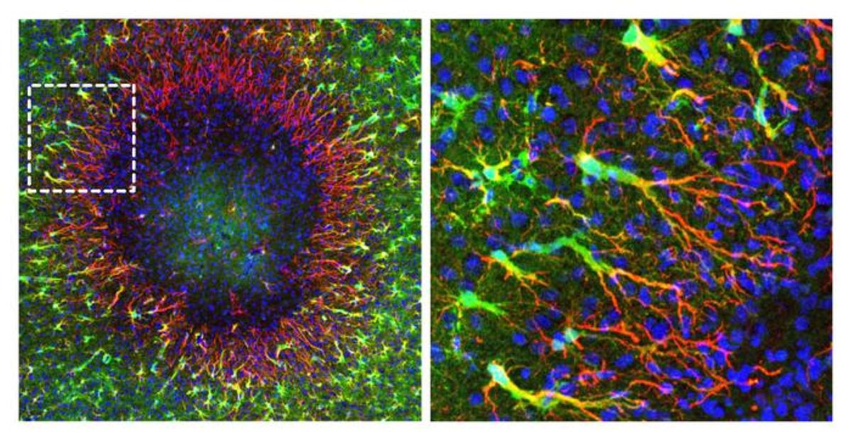
The image on the left shows a brain lesion (diameter: just under 0.5 mm). Around the perimeter of the lesion, the newly discovered “regenerative” astrocytes begin to seal the defect by forming long cellular extensions (shown in red). Newly formed cell nuclei (shown in blue) migrate along the cellular extensions toward the damaged area. Unaltered astrocytes (shown in green) surround the lesion area. The image on the right shows an enlargement of the marked area in the left image. Credit: Institute of Pharmacology and Toxicology, University of Zurich
Regenerative astrocytes repair damaged tissue
A new study by co-lead authors Marina Herwerth and Matthias Wyss from the Institute of Pharmacology and Toxicology at the University of Zurich (UZH) has now overturned that assumption: their research team headed by Bruno Weber discovered a specialized group of “regenerative” astrocytes in the brains of living mice that step in on the perimeter of the damaged area of the brain to rebuild the cells.
“The findings of our study reveal a previously unknown ability of the adult brain to repair itself. They point toward new ways of supporting recovery from ailments involving the loss of astrocytes,” Weber says.
Only cell nuclei migrate
The researchers used two-photon microscopy to observe the brains of living mice in real time over a period of several weeks and mapped which genes switch on in which areas of the brain. This way they were able to identify the special astrocytes that take care of rebuilding injured tissue. But those cells don’t just divide, they also perform a remarkable feat: “they send the newly formed nuclei of their daughter cells gliding across long distances to repopulate the damaged area of the brain and knit the astrocyte network back together,” Weber explains.
Starting points for targeted regeneration
The discovery of how adult brain cell nuclei migrate through the long star-shaped extensions of astrocytes to injured tissue expands comprehension of how the brain organizes and regenerates itself after certain injuries. If those mechanisms can be selectively activated, that could help to more effectively repair damaged brain tissue, restore astrocyte networks and thus improve recovery after certain brain disorders.
“We were able to identify numerous genes and signaling pathways that are temporarily activated during repair. They could serve as starting points in the future for influencing post-disease and -injury regeneration processes,” Weber stresses.
Key Questions Answered:
Q: How do “regenerative” astrocytes differ from standard cell division during tissue repair?
A: Instead of whole cells migrating or simply dividing locally, these specialized astrocytes remain at the perimeter of the damaged area. They divide and send the newly formed nuclei of their daughter cells gliding long distances through their extended cellular processes directly into the injured zone to rebuild the network.
Q: What conditions cause the loss of astrocytes in the adult brain?
A: Astrocytes are lost during traumatic brain injuries, strokes, and specific neuroinflammatory or autoimmune conditions, most notably Neuromyelitis Optica Spectrum Disorder (NMOSD), where the body’s immune system produces autoantibodies that target and destroy astrocytes.
Q: How was this nuclear migration observed in real time?
A: Researchers at the University of Zurich used in vivo two-photon microscopy in living mouse models over several weeks. This allowed them to track living cells, observe nuclear movement through astrocytic branches, and map corresponding gene expression changes as the tissue repaired itself.
Editorial Notes:
This article was edited by a Neuroscience News editor.
Journal paper reviewed in full.
Additional context added by our staff.
About this neuroscience research news
Author: Kurt Bodenmueller Source: University of Zurich Contact: Kurt Bodenmueller – University of Zurich Image: The image is credited to Institute of Pharmacology and Toxicology, University of ZurichOriginal Research: Open access.
“Focal astrocyte loss reveals nuclear translocation during lesion repopulation” by Marina Herwerth, Matthias T. Wyss, Nicola B. Schmid, Anna Lasne, Jacqueline Condrau, Luca Ravotto, José María Mateos Melero, Andres Kaech, Gustav Bredell, Carolina Thomas, Rachel Kim, Petra Kukanja, Vladyslav L. Korobeynyk, Christine Stadelmann, Thomas Misgeld, Jeffrey L. Bennett, Sebastian Jessberger, Aiman S. Saab, Shane A. Liddelow & Bruno Weber. Nature Neuroscience DOI:10.1038/s41593-026-02354-5
A competent? doctor and hospital WOULD INSURE HUMAN TESTING OCCURS! Do you have competence among in your stroke medical 'professionals'?
Do you prefer your doctor, hospital and board of director's incompetence NOT KNOWING? OR NOT DOING? Your choice; let them be incompetent or demand action!
Summary: Researchers
discovered a previously unknown mechanism through which the adult
mammalian brain repairs itself following focal injuries or autoimmune
damage. Using two-photon microscopy and longitudinal gene mapping in
living mouse models, researchers identified a specialized population of
“regenerative” astrocytes capable of repopulating damaged brain regions.
Rather
than relying solely on classical cell body division at the site of
injury, these specialized astrocytes situated along the lesion perimeter
send newly formed daughter cell nuclei gliding long distances through
their star-shaped cellular extensions. These migrating cell nuclei
repopulate the depleted lesion zone, re-establishing functional
astrocyte networks.
This discovery
overturns long-held assumptions regarding the limited regenerative
capacity of adult glial networks, revealing molecular signaling pathways
that could serve as therapeutic targets for traumatic brain injury and
autoimmune conditions such as neuromyelitis optica spectrum disorder
(NMOSD).
Key Facts
Overturning Dogma on Glial Regeneration:
Demonstrates that the adult central nervous system possesses a
previously unrecognized capability to replace lost astrocytes and
restore damaged tissue architecture.
Mechanism of Long-Distance Nuclear Migration:
Specialized regenerative astrocytes remain at the lesion boundary and
send newly generated cell nuclei gliding across long astrocytic
extensions into the depleted injury core.
Rebuilding Functional Glial Networks:
Astrocytes perform vital homeostatic functions, including nutrient
supply to neurons, blood flow regulation via end-feet, and extracellular
ion balance, making their network reconstruction essential for neuronal
survival.
Targeted Clinical Applications:
Holds therapeutic relevance for neurotraumatic brain injuries and rare
autoimmune conditions like neuromyelitis optica spectrum disorder
(NMOSD), where autoantibodies selectively destroy astrocytes.
Molecular Targets for Therapeutics:
The team identified specific genes and signaling pathways temporarily
activated during nuclear migration, providing potential targets for
pharmacological interventions to accelerate brain repair.
Source: University of Zurich
The
brain evidently can regenerate itself better than previously assumed
after injuries or certain autoimmune diseases. Using a mouse model,
researchers at the University of Zurich have demonstrated that special
supporting and nourishing cells repopulate damaged areas of the brain by
initially sending only newly formed cell nuclei there.
Glial
cells are supporting and nourishing cells in the brain. Star-shaped
glial cells called astrocytes are vital to the functioning of neurons.
They supply the nerve cells with nutrients, help to regulate blood flow
and keep brain tissue healthy.
It had
long been assumed that when astrocytes are lost – as happens, for
instance, in brain injuries or autoimmune diseases such as rare
neuromyelitis optica spectrum disorder, in which the body’s own
antibodies destroy these cells – the adult brain cannot fully replace
them.
Regenerative astrocytes repair damaged tissue
A
new study by co-lead authors Marina Herwerth and Matthias Wyss from the
Institute of Pharmacology and Toxicology at the University of Zurich
(UZH) has now overturned that assumption: their research team headed by
Bruno Weber discovered a specialized group of “regenerative” astrocytes
in the brains of living mice that step in on the perimeter of the
damaged area of the brain to rebuild the cells.
“The findings of our study reveal a previously unknown ability of the
adult brain to repair itself. They point toward new ways of supporting
recovery from ailments involving the loss of astrocytes,” Weber says.
Only cell nuclei migrate
The
researchers used two-photon microscopy to observe the brains of living
mice in real time over a period of several weeks and mapped which genes
switch on in which areas of the brain. This way they were able to
identify the special astrocytes that take care of rebuilding injured
tissue. But those cells don’t just divide, they also perform a
remarkable feat: “they send the newly formed nuclei of their daughter
cells gliding across long distances to repopulate the damaged area of
the brain and knit the astrocyte network back together,” Weber explains.
Starting points for targeted regeneration
The
discovery of how adult brain cell nuclei migrate through the long
star-shaped extensions of astrocytes to injured tissue expands
comprehension of how the brain organizes and regenerates itself after
certain injuries. If those mechanisms can be selectively activated, that
could help to more effectively repair damaged brain tissue, restore
astrocyte networks and thus improve recovery after certain brain
disorders.
“We were able to identify
numerous genes and signaling pathways that are temporarily activated
during repair. They could serve as starting points in the future for
influencing post-disease and -injury regeneration processes,” Weber
stresses.
Key Questions Answered:
Q: How do “regenerative” astrocytes differ from standard cell division during tissue repair?
A:
Instead of whole cells migrating or simply dividing locally, these
specialized astrocytes remain at the perimeter of the damaged area. They
divide and send the newly formed nuclei of their daughter cells gliding
long distances through their extended cellular processes directly into
the injured zone to rebuild the network.
Q: What conditions cause the loss of astrocytes in the adult brain?
A:
Astrocytes are lost during traumatic brain injuries, strokes, and
specific neuroinflammatory or autoimmune conditions, most notably
Neuromyelitis Optica Spectrum Disorder (NMOSD), where the body’s immune
system produces autoantibodies that target and destroy astrocytes.
Q: How was this nuclear migration observed in real time?
A:
Researchers at the University of Zurich used in vivo two-photon
microscopy in living mouse models over several weeks. This allowed them
to track living cells, observe nuclear movement through astrocytic
branches, and map corresponding gene expression changes as the tissue
repaired itself.
Editorial Notes:
This article was edited by a Neuroscience News editor.
Journal paper reviewed in full.
Additional context added by our staff.
About this neuroscience research news
Author: Kurt Bodenmueller Source: University of Zurich Contact: Kurt Bodenmueller – University of Zurich Image: The image is credited to Institute of Pharmacology and Toxicology, University of Zurich
Original Research: Open access. “Focal astrocyte loss reveals nuclear translocation during lesion repopulation”
by Marina Herwerth, Matthias T. Wyss, Nicola B. Schmid, Anna Lasne,
Jacqueline Condrau, Luca Ravotto, José María Mateos Melero, Andres
Kaech, Gustav Bredell, Carolina Thomas, Rachel Kim, Petra Kukanja,
Vladyslav L. Korobeynyk, Christine Stadelmann, Thomas Misgeld, Jeffrey
L. Bennett, Sebastian Jessberger, Aiman S. Saab, Shane A. Liddelow &
Bruno Weber. Nature Neuroscience DOI:10.1038/s41593-026-02354-5