Changing stroke rehab and research worldwide now.Time is Brain! trillions and trillions of neurons that DIE each day because there are NO effective hyperacute therapies besides tPA(only 12% effective). I have 523 posts on hyperacute therapy, enough for researchers to spend decades proving them out. These are my personal ideas and blog on stroke rehabilitation and stroke research. Do not attempt any of these without checking with your medical provider. Unless you join me in agitating, when you need these therapies they won't be there.

What this blog is for:

My blog is not to help survivors recover, it is to have the 10 million yearly stroke survivors light fires underneath their doctors, stroke hospitals and stroke researchers to get stroke solved. 100% recovery. The stroke medical world is completely failing at that goal, they don't even have it as a goal. Shortly after getting out of the hospital and getting NO information on the process or protocols of stroke rehabilitation and recovery I started searching on the internet and found that no other survivor received useful information. This is an attempt to cover all stroke rehabilitation information that should be readily available to survivors so they can talk with informed knowledge to their medical staff. It lays out what needs to be done to get stroke survivors closer to 100% recovery. It's quite disgusting that this information is not available from every stroke association and doctors group.

Showing posts with label innovation. Show all posts

Thursday, October 23, 2025

INNOVATIONS IN STROKE REHABILITATION

I see no innovation, just rehashed crapola guidelines. 

I'd say this is totally fucking useless.

We may as well go back to blood letting as a stroke prescription as discussed in the 1843 book, 'An Essay on the Nature and Treatment of Apoplexy'.

 INNOVATIONS IN STROKE REHABILITATION

K. Shtereva, P. Dobrev, N. Bekir*, S. Valeva, K. Mollova Department of Health Care, Medical College, Trakia University, Stara Zagora, Bulgaria 

 ABSTRACT 

 Stroke rehabilitation requires a personalized and multidisciplinary approach. Stroke remains a leading cause of disability, and innovative technologies such as robotics, virtual reality, and telerehabilitation show significant potential for improving patients' motor and cognitive functions. Robotic systems provide precise, repetitive movements, while virtual reality enhances motivation through engaging and realistic environments. The combination of technologies creates a synergistic effect and leads to higher efficiency. Telerehabilitation and brain-computer interface (BCI) robots expand access to therapy and allow recovery even in severe cases. 
Objective: This article aims to analyze the role and effectiveness of these innovations in the context of modern post-stroke rehabilitation. 
Materials and Methods: A review of recent literature was conducted, including scientific publications related to the rehabilitation of ischemic or hemorrhagic stroke and the use of innovative technologies. Conclusion: Robotic systems, virtual reality, telerehabilitation, and BCI robots complement traditional therapies and offer new approaches to overcoming long-term disability. Clinical evidence shows significant benefits from their use, especially in the early phases after stroke. 

Wednesday, June 4, 2025

Stroke survivors get new tools to lead more independent lives with help from an innovation lab

 At least they are directly working with survivors.

Stroke survivors get new tools to lead more independent lives with help from an innovation lab   

The lab brings together patients, therapists, engineers and designers to come up with products and services to improve patients’ recovery process.

Stroke survivors get new tools to lead more independent lives with help from an innovation lab
Designer Orion Dai helps a stroke survivor to put on a wearable robotic glove that he created for rehabilitation purposes
Read a summary of this article on FAST.

SINGAPORE: Stroke survivors in Singapore are co-developing solutions with researchers and designers through an innovation lab, which is aiming to help them lead independent and functional lives again.

The lab was launched last month by Stroke Support Station (S3), a community-based charity focusing on stroke rehabilitation and wellness.

It is currently trialling projects like a wearable robotic device to help with stroke rehabilitation at home and a gym that will provide special equipment for stroke patients to maintain fitness while reducing the risk of secondary strokes.

Stroke is one of the leading causes of adult disability in Singapore. The number of cases has gone up by 58 per cent to nearly 10,000 in 2021, as compared to about 6,000 a decade ago, according to the Ministry of Health.                 

Saturday, August 5, 2023

Innovations in Stroke Recovery and Rehabilitation

They suggest nothing about recovery, just biomarkers which are just a way to predict failure to recover. I'd say this is totally fucking useless.

We may as well go back to blood letting as a stroke prescription as discussed in the 1843 book, 'An Essay On The Nature and Treatment of Apoplexy'. I see no innovation, just rehashed crapola guidelines.

Ask your doctor what is the basis for their treatment of your stroke disabilities and why they moved away from bloodletting.  My doctor knew nothing and did nothing as proven by the writing of three E.T.(Evaluate and treat) prescriptions.


Innovations in Stroke Recovery and Rehabilitation

Published:August 02, 2023DOI:https://doi.org/10.1016/j.pmr.2023.07.003
Figure thumbnail fx1
Joan Stilling, MD, MS, FRCPC, Editor
Despite improvements in acute stroke management, many stroke survivors remain with substantial disability. Efforts to more effectively harness the brain’s plasticity and capacity for recovery have been gradually providing a more robust array of evidence to help guide practitioners. At the same time, new treatments, such as cryoneurolysis, are being incorporated into the management of long-term sequelae of stroke. In this special issue focusing on stroke rehabilitation, contributors review our current understanding of mechanisms involved in motor recovery and motor learning following stroke. Techniques to impact and augment motor control and motor learning are discussed. Biomarkers of functional outcomes following stroke are presented with regards to prognostication of both upper and lower limb recovery, with the expectation that these predictive models will ultimately be used to guide individualized treatment.
To read this article in full you will need to make a payment

Sunday, May 8, 2022

Innovative approaches to the rehabilitation of upper extremity hemiparesis using virtual environments

Has your stroke hospital been innovative enough in 12.5 years to  bring this in? Or are they still in the dark ages when bloodletting was a stroke intervention?

We may as well go back to blood letting as a stroke prescription as discussed in the 1843 book, 'An Essay On The Nature and Treatment of Apoplexy'.

Ask your doctor what is the basis for their treatment of your stroke disabilities and why they moved away from bloodletting.  My doctor knew nothing and did nothing as proven by the writing of three E.T.(Evaluate and treat) prescriptions.


Innovative approaches to the rehabilitation of upper extremity hemiparesis using virtual environments

Author manuscript; available in PMC 2017 Oct 19.
Published in final edited form as:
 

Abstract

Aim

Upper-extremity interventions for hemiparesis are a challenging aspect of stroke rehabilitation. Purpose of this paper is to report the feasibility of using virtual environments (VEs) in combination with robotics to assist recovery of hand-arm function and to present preliminary data demonstrating the potential of using sensory manipulations in VE to drive activation in targeted neural regions.

Methods

We trained 8 subjects for 8 three hour sessions using a library of complex VE’s integrated with robots, comparing training arm and hand separately to training arm and hand together. Instrumented gloves and hand exoskeleton were used for hand tracking and haptic effects. Haptic Master robotic arm was used for arm tracking and generating three-dimensional haptic VEs. To investigate the use of manipulations in VE to drive neural activations, we created a “virtual mirror” that subjects used while performing a unimanual task. Cortical activation was measured with functional MRI (fMRI) and transcranial magnetic stimulation.

Results

Both groups showed improvement in kinematics and measures of real-world function. The group trained using their arm and hand together showed greater improvement. In a stroke subject, fMRI data suggested virtual mirror feedback could activate the sensorimotor cortex contralateral to the reflected hand (ipsilateral to the moving hand) thus recruiting the lesioned hemisphere.

Conclusion

Gaming simulations interfaced with robotic devices provide a training medium that can modify movement patterns. In addition to showing that our VE therapies can optimize behavioral performance, we show preliminary evidence to support the potential of using specific sensory manipulations to selectively recruit targeted neural circuits.

Keywords: Paresis, Robotics, Recovery of function, Arm, therapy, Stroke

Improvement in upper extremity post-stroke function has been recalcitrant to current therapeutic interventions with only 5% of all patients regaining full use of their upper extremity following intensive therapy. Stroke rehabilitation has focused on the facilitation of isolated movements through spasticity reduction, passive guidance and graded movement assistance. Current emphasis is on repetitive task practice and utilizing principles of motor learning such as regulating practice schedules and augmenting feedback. It is believed that these motor learning principles parallel the practice principles purported to effect neuroplasticity. Evidence that plasticity is “use-dependent” and intensive massed and repeated practice may be necessary to modify neural organization.

Virtual reality (VR) technology may be an appropriate means to provide these plasticity-mediated therapies using motor learning principles. The first generation of these computerized systems provided motivating environments in which tasks could be practiced repetitively. Training schedules, specificity and frequency of visual and auditory feedback could be objectively monitored and quantified. Currently treatment interventions are being developed to take advantage of technological advances such as the improvement in robotic design, the development of haptic interfaces, and the integration of these devices with virtual environments. Studies have shown that robotically-facilitated repetitive movement training might be an effective stimulus for normalizing upper extremity motor control in persons with moderate to severe impairments who have difficulties in performing unassisted movements. Several authors have integrated VE with adaptive robotic systems to train the hemiparetic upper extremity.

Although considerable progress has been made in developing these devices, the clinical evidence demonstrating effectiveness of these systems has not yet reached the highest levels of evidence found in systematic reviews and randomized controlled studies. Many of the studies supporting the use of interactive robotics and virtual environments interfaced with movement tracking and sensing glove systems consist of case studies, small feasibility studies, or studies without control groups. Unfortunately, in fact, the assimilation of technology into rehabilitation has not been as fast or as extensive as in other branches of medicine.

If repetition and skill learning are important for motor learning and recovery of function, what do these technologies add over and above real-world task practice? This is an important question. What can training within an interactive virtual environment, either with or without robotic devices contribute to skill learning and improved motor control? Do gaming environments, augmented visual, auditory and haptic feedback provide added value to the learning process? Are these technological interventions able to model and incorporate accepted rehabilitation practices such as physical assistance and graded progression of tasks to adjust the kinematics of the movement during training? We believe that the combination of virtual environments and robotics can be effectively used both as a training tool and and as a test of hypotheses regarding the benefits of various rehabilitation approaches. These capabilities are afforded through the quantitative evaluation tools inherent to this technology.

We present an example of such a study that tests two different approaches to hand rehabilitation. The prevailing paradigm for upper extremity rehabilitation describes the need to develop proximal control and mobility of the shoulder prior to initiating training on the hand. This has been the accepted rehabilitation method for many years. An increasing number of human and animal studies have reported that movement practice increases the area and density of motor cortex correlated with that movement, with the possibility that this expansion of motor territory influences representations occupying adjacent territory. It is not clear whether this expansion of cortical representations occurs through sharing of cortical tissue among representations or through competition for cortical territory. These findings prompt us to reconsider the rehabilitation strategy that encourages early shoulder activation post-stroke. In general, there is better return of upper arm function post-stroke than of the hand. Does early motor activity of the upper arm and shoulder hinder recovery of hand function because of cortical competition facilitated through intensive motor activity? Several small studies exploring the concept of providing additional hand training during conventional therapy or training the hand while the upper arm is deafferented and deefferented through regional anesthesia have shown positive changes in hand function.

The purpose of this paper is to report preliminary findings on the feasibility of using special designed virtual environments in combination with robotics to assist recovery of hand-arm function post-stroke. The first section presents preliminary findings that address the above mentioned competition hypothesis. Two groups of post-stroke patients were trained: one group used virtual reality training simulations to train the hand and arm together as a functional unit and one group trained hand and arm separately. We hypothesized that balancing and integrating the training of both proximal and distal components of the upper extremity will minimize over-representation of the upper arm.

In the second section, we report preliminary data that demonstrate the potential of sensory manipulations in VE to selectively drive activation in targeted neural regions. For this, we used a task, mirror visual feedback (MVF), which in smaller-scale studies has shown promise in aiding recovery of hand-arm function after stroke., We simulated the MVF effect in VE by creating a “virtual mirror” as subjects performed a unimanual task. We simultaneously measured cortical activation with functional magnetic resonance imaging (fMRI) and, in a separate session, with transcranial magnetic stimulation (TMS). We hypothesized that the virtual mirror effect would be associated with activation of the motor cortex in the hemisphere ipsilateral to the moving hand (i.e. contralateral to the mirror-reflected hand).

More at link.


Thursday, December 16, 2021

Can innovation play a role in stroke care and diagnosis?

Innovation is not the problem. The problem is that there is NO stroke leadership executing a stroke strategy to deliver 100% recovery. There are thousands of pieces of research out there that if just followed up and solved would vastly lessen the stroke burden.  Execution is the problem.

Can innovation play a role in stroke care and diagnosis?

by


Thursday, May 21, 2020

Tiny implanted sensors monitor brain injuries, then dissolve away

You'll have to ask your doctor and stroke hospital if this ever made it to human testing. Because with just a few modifications with nanosensors we could listen in on individual neuron signals and figure out EXACTLY what happens during neuroplasticity.  Assuming of course that there is anyone in stroke research that has two neurons to rub together for a spark of innovation.


Tiny implanted sensors monitor brain injuries, then dissolve away


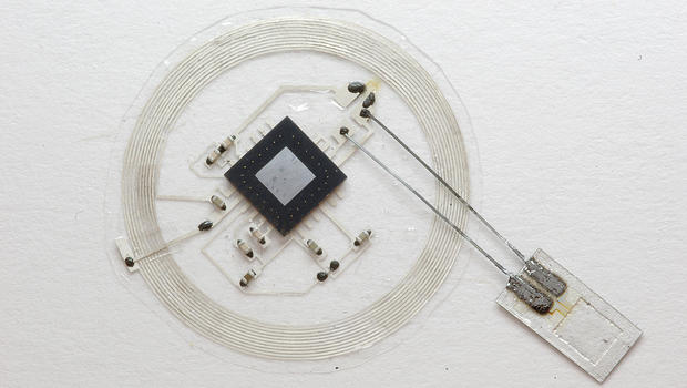
A tiny implantable brain sensor could someday monitor conditions within the skull before dissolving away. Currently being tested in rats, this technology tracks temperature and pressure levels, potentially offering a new option for how brain injuries are studied and treated. By melting away when no longer useful, these micro-sized sensors remove the risks associated with current technology used to monitor brain injuries, according to a University of Illinois press release.
The study, led by John A. Rogers, professor of material sciences and engineering at the University of Illinois at Urbana-Champaign, and Wilson Ray, professor of neurological surgery at the Washington University School of Medicine in St. Louis, is published in the journal Nature.
"This is a new class of electronic biomedical implants," Rogers said in the release. "These kinds of systems have potential across a range of clinical practices, where therapeutic or monitoring devices are implanted or ingested, perform a sophisticated function, and then resorb harmlessly into the body after their function is no longer necessary."
Rogers stressed that the technology now used for monitoring after traumatic brain injuries or brain surgery can be dangerous for patients. Systems involve bulky wires that restrict patients' movements, and invasive implants may lead to brain hemorrhages, allergic reactions, and infections.
By contrast, the tiny dissolvable silicon devices that Rogers' team developed are smaller than a grain of rice. They are naturally biodegradable -- built up on incredibly thin silicon sheets -- and dissolve away after a few weeks of monitoring brain activity. They melt harmlessly into the human body's own fluids, the researchers say.
These devices are sensitive to pressure levels in the intracranial fluid that surrounds the brain, and have a temperature sensor synced to a wireless postage stamp-sized transmitter that is placed on top of the skull.
sensor-transmitter.jpg
The small sensor connects to an embeddable wireless transmitter that lies on top of the skull. Courtesy of John A. Rogers
"If you simply could throw out all the conventional hardware and replace it with very tiny, fully implantable sensors capable of the same function, constructed out of bioresorbable materials in a way that also eliminates or greatly miniaturizes the wires, then you could remove a lot of the risk and achieve better patient outcomes," Rogers added. "We were able to demonstrate all of these key features in animal models, with a measurement precision that's just as good as that of conventional devices."
The lab tests with rats were to see how compatible these tiny devices would be with a living organism's body.
So, what's the next phase? The team hopes to move toward human trials soon.
"The ultimate strategy is to have a device that you can place in the brain -- or in other organs in the body -- that is entirely implanted, intimately connected with the organ you want to monitor and can transmit signals wirelessly to provide information on the health of that organ, allowing doctors to intervene if necessary to prevent bigger problems," Rory Murphy, a neurosurgeon at Washington University who was a co-author of the paper.


Friday, February 14, 2020

The Art of Lucid Dreaming: Over 60 Powerful Practices to Help You Wake Up in Your Dreams

If your doctor hasn't already trained you in lucid dreaming you need to find someone with an innovative idea streak in them. Lucid dreaming would seem to combine both action observation and motor imagery. So extremely likely very good for your rehab.

The Art of Lucid Dreaming: Over 60 Powerful Practices to Help You Wake Up in Your Dreams

Saturday, March 16, 2019

Development of a battery-free ultrasonically powered functional electrical stimulator for movement restoration after paralyzing spinal cord injury

Should be able to be used in stroke rehab if ANYONE in stroke has any innovative functioning brain cells at all. But that will never occur. Stroke brains are fossilized and don't work, which is why they are in the stroke field. 

Development of a battery-free ultrasonically powered functional electrical stimulator for movement restoration after paralyzing spinal cord injury 

  • ,
  • ,
  • ,
  • ,
  • ,
  • ,
  • ,
  • and
  • Email author
Contributed equally
Journal of NeuroEngineering and Rehabilitation201916:36
  • Received: 16 November 2018
  • Accepted: 22 February 2019
  • Published:

Abstract

Background

Functional electrical stimulation (FES) is used to restore movements in paretic limbs after severe paralyses resulting from neurological injuries such as spinal cord injury (SCI). Most chronic FES systems utilize an implantable electrical stimulator to deliver a small electric current to the targeted muscle or nerve to stimulate muscle contractions. These implanted stimulators are generally bulky, mainly due to the size of the batteries. Furthermore, these battery-powered stimulators are required to be explanted every few years for battery replacement which may result in surgical failures or infections. Hence, a wireless power transfer technique is desirable to power these implantable stimulators.

Methods

Conventional wireless power transduction faces significant challenges for safe and efficient energy transfer through the skin and deep into the body. Inductive and electromagnetic power transduction is generally used for very short distances and may also interfere with other medical measurements such as X-ray and MRI. To address these issues, we have developed a wireless, ultrasonically powered, implantable piezoelectric stimulator. The stimulator is encapsulated with biocompatible materials.

Results

The stimulator is capable of harvesting a maximum of 5.95 mW electric power at an 8-mm depth under the skin from an ultrasound beam with about 380 mW/cm2 of acoustic intensity. The stimulator was implanted in several paraplegic rats with SCI. Our implanted stimulator successfully induced several hindlimb muscle contractions and restored leg movement.

Conclusions

A battery-free miniature (10 mm diameter × 4 mm thickness) implantable stimulator, developed in the current study is capable of directly stimulating paretic muscles through external ultrasound signals. The required cost to develop the stimulator is relatively low as all the components are off the shelf.

Wednesday, February 20, 2019

Locomotion and cadence detection using a single trunk-fixed accelerometer: validity for children with cerebral palsy in daily life-like conditions

Our stroke researchers should be able to use this to objectively determine stroke gait problems. Then with that objective data we could map rehab protocols that fix those specific problems. But that won't occur, we don't have two neurons to rub together to create a spark of innovative thought in stroke. 

Locomotion and cadence detection using a single trunk-fixed accelerometer: validity for children with cerebral palsy in daily life-like conditions

  • Email author,
  • ,
  • ,
  • ,
  • and
Journal of NeuroEngineering and Rehabilitation201916:24
  • Received: 5 July 2018
  • Accepted: 25 January 2019
  • Published:

Background

Physical therapy interventions for ambulatory youth with cerebral palsy (CP) often focus on activity-based strategies to promote functional mobility and participation in physical activity. The use of activity monitors validated for this population could help to design effective personalized interventions by providing reliable outcome measures. The objective of this study was to devise a single-sensor based algorithm for locomotion and cadence detection, robust to atypical gait patterns of children with CP in the real-life like monitoring conditions.

Methods

Study included 15 children with CP, classified according to Gross Motor Function Classification System (GMFCS) between levels I and III, and 11 age-matched typically developing (TD). Six IMU devices were fixed on participant’s trunk (chest and low back/L5), thighs, and shanks. IMUs on trunk were independently used for development of algorithm, whereas the ensemble of devices on lower limbs were used as reference system. Data was collected according to a semi-structured protocol, and included typical daily-life activities performed indoor and outdoor.
The algorithm was based on detection of peaks associated to heel-strike events, identified from the norm of trunk acceleration signals, and included several processing stages such as peak enhancement and selection of the steps-related peaks using heuristic decision rules. Cadence was estimated using time- and frequency–domain approaches. Performance metrics were sensitivity, specificity, precision, error, intra-class correlation coefficient, and Bland-Altman analysis.

Results

According to GMFCS, CP children were classified as GMFCS I (n = 7), GMFCS II (n = 3) and GMFCS III (n = 5). Mean values of sensitivity, specificity and precision for locomotion detection ranged between 0.93–0.98, 0.92–0.97 and 0.86–0.98 for TD, CP-GMFCS I and CP-GMFCS II-III groups, respectively.
Mean values of absolute error for cadence estimation (steps/min) were similar for both methods, and ranged between 0.51–0.88, 1.18–1.33 and 1.94–2.3 for TD, CP-GMFCS I and CP-GMFCS II-III groups, respectively. The standard deviation was higher in CP-GMFCS II-III group, the lower performances being explained by the high variability of atypical gait patterns.

Conclusions

The algorithm demonstrated good performance when applied to a wide range of gait patterns, from normal to the pathological gait of highly affected children with CP using walking aids.

Wednesday, January 2, 2019

20 billion nanoparticles talk to the brain using electricity

I'm sure with ANY innovative brains at all in the stroke world we could easily see many applications for stroke rehab. BCI? TMS? etc. etc. stem cell  placement? axon pathfinding? neurite outgrowth? magnetic delivery of tPA with much smaller boluses?

20 billion nanoparticles talk to the brain using electricity


Read more: Click here to read the original, longer version of this story.


ELECTRICITY is the brain’s language, and now we can speak to it without wires or implants. Nanoparticles can be used to stimulate regions of the brain electrically, opening up novel ways to treat brain diseases.
When magneto-electric nanoparticles are subjected to an external magnetic field, they produce an electric field. If such nanoparticles are placed next to neurons, this electric field should allow them to communicate.
To find out, Sakhrat Khizroev of Florida International University in Miami and his team inserted 20 billion of these nanoparticles into the brains of mice. They then switched on a magnetic field. An electroencephalogram showed that the brain region surrounded by nanoparticles lit up, stimulated by the electric field that had been generated (Future Medicine, doi.org/44b).
The more pets you meet as a baby, the lower your risk of allergie


Monday, December 24, 2018

A history of innovations in heart disease and stroke

What the hell, this is just trying to whitewash all the problems in stroke still needing solutions. If you believe this crap you would think having a stroke is not bad. 

A history of innovations in heart disease and stroke


For decades, medical schools and teaching hospitals have been at the forefront of developing new techniques and technologies to prevent, diagnose, and treat heart disease and stroke. Here are some of the more notable ones.
Innovations-photo-man-computer-stroke_992.jpg
Six hours. For years, that was the time window for removing a blood clot in a stroke victim. Go beyond that six-hour mark and the benefits decrease dramatically, physicians believed. But in January 2018, the American Heart Association and the American Stroke Association released new guidelines with a new time window: 16 to 24 hours, depending on the patient.
The decision was based on two studies led by Stanford University Medical Center and the University of Pittsburgh Medical Center (UPMC), with the participation of dozens of teaching hospitals. The idea, however, came from discussions that occurred almost 10 years earlier at UPMC.
“It’s been a labor of love,” says Tudor Jovin, MD, assistant professor of neurology and neurosurgery at the University of Pittsburgh School of Medicine, who was a principal investigator for one of the trials and currently serves as director of UPMC’s Stroke Institute. (UPMC had the largest number of patients in the trial.) “We encountered a lot of resistance because people didn't believe that these trials were feasible — that they would show what we thought they would show. There was a lot of skepticism.”
Jovin’s comments underscore why medical schools and teaching hospitals are known for medical innovation.  Not only do many of them offer the latest treatments and cutting-edge technology, they frequently house world-class research facilities. And they employ a range of experts, from experienced biomedical researchers and clinicians, to students, residents, and fellows, all of whom bring a unique perspective to the work.
“You get the innovative thoughts of new doctors with the experience and wisdom of those who have been doing research for a long time,” says Mona Bahouth, MD, assistant professor in the department of neurology at Johns Hopkins School of Medicine. And that, she says, results in challenges to the intellectual status quo.
Indeed, teaching hospitals have a history of innovations that have dramatically reduced mortality from heart disease and stroke. Between 1969 and 2015, deaths from heart disease declined 68%, due in part to research funded by the National Institutes of Health (NIH) and conducted at medical schools and teaching hospitals around the country. Here are some of those key contributions.

Heart disease

Since 1950, 80% of the Nobel Prize winners related to cardiovascular disease have been affiliated with American medical schools and teaching hospitals. Among the achievements: Biomedical researchers at academic medical centers helped show that treating even moderate hypertension reduced cardiac-related deaths. Research pioneered at medical schools and teaching hospitals also led to new standards of care for treating coronary artery disease and new medical and surgical treatments.
The work of one institution often leads to developments at another. Two medical professors at Columbia University won the Nobel Prize in 1958 for discoveries related to heart catherization. That research led the Cleveland Clinic and the University of Oregon to conduct pioneering work on coronary angiography in the 1950s and ‘60s, although it was “an accidental discovery,” says Joaquin Cigarroa, MD, head of the Oregon Health & Science University School of Medicine’s (OHSU) division of cardiovascular medicine. In 1958, F. Mason Sones Jr., a pediatric cardiologist at the Cleveland Clinic, was conducting a cardiac catherization. As his resident injected 50mm of dye into a patient’s aorta, the catheter moved slightly and some of the dye inadvertently entered a coronary artery.
“That began the era of consistent angiography,” says Cigarroa.
At OHSU, in 1964, Charlie Dotter became the first physician to perform an angioplasty on a peripheral artery. The patient was an 82-year-old woman suffering from blocked circulation in her leg. Physicians wanted to amputate, but she refused. A surgeon knew Dotter, who used a guide wire and Teflon catheters to dilate a superficial femoral artery stenosis.
“He demonstrated that you could actually push aside plaque in an artery,” says Cigarroa.
Between 1969 and 2015, deaths from heart disease declined 68%, due in part to research funded by the National Institutes of Health (NIH) and conducted at medical schools and teaching hospitals around the country.
Researchers working at medical schools and teaching hospitals have also helped develop everything from less-invasive angioplasty procedures to robotic surgery to surgeries that don’t require stopping the heart or using a heart-lung machine. Physicians at OHSU, Harvard, and Georgetown, among others, have developed and improved artificial heart valves. Procedures pioneered at teaching hospitals — including the Cleveland Clinic and Stanford — led to the development of Transcatheter Aortic Valve Replacement (TAVR), which allows physicians to treat aortic stenosis without open-heart surgery and often without long-term recovery. Middle school teacher Susan Strong, who underwent TAVR surgery at the University of Colorado Hospital in 2014, notes that she attended a full-day seminar one day after the procedure.
Researchers working at medical schools and teaching hospitals have also played a critical role in developing heart transplant surgeries and ventricular assist devices. The first such device was implanted by faculty members at the Baylor College of Medicine. At OHSU, researchers are currently working on an artificial heart designed for permanent use. (The only patented artificial heart is for temporary use.) The device would replace two ventricles with a single titanium tube. A rod in the tube moves back and forth to send blood to the lungs.
“It has gone through the engineering testing and we have done some short-term experiments,” says Cigarroa. “It shows promise, which is exciting.”

Stroke

The groundbreaking studies published in 2018 that increased the window for life-saving clot removal surgery are the latest in a long history of improvements to stroke treatment. How much has changed? Since 1969, the stroke mortality rate has declined by 71%(but what about 100% recovery?), due in large part to NIH-funded research on treatments and prevention conducted at medical schools and teaching hospitals.
In 1995, an NIH-funded clinical trial established the first FDA-approved treatment for ischemic stroke – the drug r-tPA(Which fails at full recovery 88% of thew time) (tissue plasminogen activator). Even more notable, clinical trials conducted in part at academic medical centers have established the importance of improved blood pressure control, decreased smoking rates and the use of statins in stroke prevention.
More recently, telestroke and teleneurology programs are making further inroads in reducing the death rate from stroke. Potential stroke victims need specialized expertise within hours of experiencing symptoms, but that can be difficult in rural areas. In the Stroke Belt — an 11-state region consisting of Mississippi, Tennessee, Louisiana, Kentucky, Georgia, North Carolina, Alabama, South Carolina, Arkansas, Indiana, and Virginia — stroke rates are 34% higher than in other parts of the country, the CDC reports. Because of that, many teaching hospitals provide access to experts through audio and video links.
After speaking with patients and reviewing medical records and imaging and lab results, physicians can determine if the stroke is ischemic or hemorrhagic, offer advice on whether to use the emergency stroke drug r-tPA, and recommend whether a patient should be sent for surgery. The many teaching hospitals that provide telestroke and teleneurology include Emory University Hospital, the Medical University of South Carolina Medical Center, Nebraska Medicine, the Ohio State Heath System, the Ronald Reagan UCLA Medical Center, and Yale New Haven Health. The University of Utah Health System provides telestroke services to more than 25 sites in Utah and elsewhere. And Penn State Health Milton S. Hershey Medical Center partners with regional hospitals for its telestroke program, called LionNet.
“At our core is a mission to advance gaps in knowledge that can impact patients. That is done at a basic science level, through clinical trials, through innovations in how we teach, and in partnership with communities. It is who we are.”
Joaquin Cigarroa, MD
Oregon Health & Science University School of Medicine
The Cleveland Clinic is reaching stroke patients through its Mobile Stroke Unit, an ambulance-like vehicle with staff, equipment, and medications for diagnosing and treating strokes. The vehicle also includes a lab to test blood samples, a portable CT scanner that can send images to the hospital, and a telemedicine link to the hospital’s neurologists. A 2017 study in Neurology found that patients received thrombolysis 38.5 minutes sooner(So what? How fast does it need to be delivered to get 100% recovery?) via the Cleveland Clinic’s mobile unit than they would with traditional stroke procedures. The University of Texas Health Science Center at Houston (UT Health) introduced the first Mobile Stroke Unit as part of a clinical trial in 2014; others with units include the University of Tennessee Health Science Center and Indiana University Health.
Medical schools and teaching hospitals have also been innovators when it comes to patient safety. In a recent study by researchers at UT Health, stroke patients treated at teaching hospitals were less likely to be readmitted than those who weren’t.
Other innovators are focused on recovery. Since 2015, the Stanford Stroke Recovery Program has focused on improving “gait, arm function, and cognition after stroke.” The laboratories have worked on new therapies, noninvasive brain stimulation, and medical devices. Among its more interesting trials: The program is testing the StrokeCoach, a rehab program that uses the Apple Watch to offer exercises and assess the progress of a weak arm following a stroke. It’s also testing devices to improve hand function and muscle weakness.
At Johns Hopkins, researchers are bringing together engineers, nurses, physicians, and other experts to create a treatment room that becomes “the treatment machine for a stroke patient,” says Bahouth. “Think of it as a bio room that measures and responds to patients’ needs. That will require a lot of innovative work between technologists, engineers, and informatics people, but I think that's where we're moving: To test a concept that creates a whole environment of healing for stroke patients.”
Innovative ideas like this are part of the DNA of teaching hospitals — and they will continue to save lives.
“At our core is a mission to advance gaps in knowledge that can impact patients,” says Cigarroa. “That is done at a basic science level, through clinical trials, through innovations in how we teach, and in partnership with communities. It is who we are.”

Monday, November 26, 2018

Inflammation in stroke: the role of cholinergic, purinergic and glutamatergic signaling

You'll have to hope your stroke hospital has the brains and innovative ability to create a protocol  to reduce the outcome of cerebral ischemia or at least followup with research. That makes a huge assumption that your stroke hospital is following stroke research at all. If not your board of directors needs to be fired. 

First Published May 4, 2018 Review Article
The inflammatory response is a major factor in stroke pathophysiology and contributes to secondary neuronal damage in both acute and chronic stages of the ischemic injury. Recent work in experimental cerebral ischemia has demonstrated the involvement of neurotransmitter signaling in the modulation of neuroinflammation. The present review discusses recent findings on the therapeutic potential and diagnostic perspectives of cholinergic, purinergic and glutamatergic receptors and transporters in experimental stroke. It provides evidence of the role of neurotransmission signaling as a promising inflammatory biomarker in stroke. Finally, recent molecular imaging studies using positron emission tomography of cholinergic receptors and glutamatergic transporters are outlined along with their potential as novel anti-inflammatory therapy to reduce the outcome of cerebral ischemia.

Thursday, October 25, 2018

Study reveals how the brain overcomes its own limitations

With just the TINIEST bit of forward thinking and innovation our stroke medical 'professionals' could figure out how to apply this to stroke rehab. But since we don't have two functioning neurons to rub together in all of the stroke medical world, stroke survivors will continue to be screwed.  I would suggest we just throw all the problems in stroke to MIT and the MIT Media Lab and have them solve them as part of their graduating requirements. But our fucking failures of stroke associations don't have enough brains to do that because they consider stroke treatable.

Study reveals how the brain overcomes its own limitations

Strategies to compensate for uncertainty help the brain succeed at difficult mental computations.


Imagine trying to write your name so that it can be read in a mirror. Your brain has all of the visual information you need, and you’re a pro at writing your own name. Still, this task is very difficult for most people. That’s because it requires the brain to perform a mental transformation that it’s not familiar with: using what it sees in the mirror to accurately guide your hand to write backward.
MIT neuroscientists have now discovered how the brain tries to compensate for its poor performance in tasks that require this kind of complicated transformation. As it also does in other types of situations where it has little confidence in its own judgments, the brain attempts to overcome its difficulties by relying on previous experiences.
“If you’re doing something that requires a harder mental transformation, and therefore creates more uncertainty and more variability, you rely on your prior beliefs and bias yourself toward what you know how to do well, in order to compensate for that variability,” says Mehrdad Jazayeri, the Robert A. Swanson Career Development Professor of Life Sciences, a member of MIT’s McGovern Institute for Brain Research, and the senior author of the study.
This strategy actually improves overall performance, the researchers report in their study, which appears in the Oct. 24 issue of the journal Nature Communications. Evan Remington, a McGovern Institute postdoc, is the paper’s lead author, and technical assistant Tiffany Parks is also an author on the paper.
Noisy computations
Neuroscientists have known for many decades that the brain does not faithfully reproduce exactly what the eyes see or what the ears hear. Instead, there is a great deal of “noise” — random fluctuations of electrical activity in the brain, which can come from uncertainty or ambiguity about what we are seeing or hearing. This uncertainty also comes into play in social interactions, as we try to interpret the motivations of other people, or when recalling memories of past events.
Previous research has revealed many strategies that help the brain to compensate for this uncertainty. Using a framework known as Bayesian integration, the brain combines multiple, potentially conflicting pieces of information and values them according to their reliability. For example, if given information by two sources, we’ll rely more on the one that we believe to be more credible.
In other cases, such as making movements when we’re uncertain exactly how to proceed, the brain will rely on an average of its past experiences. For example, when reaching for a light switch in a dark, unfamiliar room, we’ll move our hand toward a certain height and close to the doorframe, where past experience suggests a light switch might be located.
All of these strategies have been previously shown to work together to increase bias toward a particular outcome, which makes our overall performance better because it reduces variability, Jazayeri says.
Noise can also occur in the mental conversion of sensory information into a motor plan. In many cases, this is a straightforward task in which noise plays a minimal role — for example, reaching for a mug that you can see on your desk. However, for other tasks, such as the mirror-writing exercise, this conversion is much more complicated.
“Your performance will be variable, and it’s not because you don’t know where your hand is, and it’s not because you don’t know where the image is,” Jazayeri says. “It involves an entirely different form of uncertainty, which has to do with processing information. The act of performing mental transformations of information clearly induces variability.”
That type of mental conversion is what the researchers set out to explore in the new study. To do that, they asked subjects to perform three different tasks. For each one, they compared subjects’ performance in a version of the task where mapping sensory information to motor commands was easy, and a version where an extra mental transformation was required.
In one example, the researchers first asked participants to draw a line the same length as a line they were shown, which was always between 5 and 10 centimeters. In the more difficult version, they were asked to draw a line 1.5 times longer than the original line.
The results from this set of experiments, as well as the other two tasks, showed that in the version that required difficult mental transformations, people altered their performance using the same strategies that they use to overcome noise in sensory perception and other realms. For example, in the line-drawing task, in which the participants had to draw lines ranging from 7.5 to 15 centimeters, depending on the length of the original line, they tended to draw lines that were closer to the average length of all the lines they had previously drawn. This made their responses overall less variable and also more accurate.
“This regression to the mean is a very common strategy for making performance better when there is uncertainty,” Jazayeri says.
Noise reduction
The new findings led the researchers to hypothesize that when people get very good at a task that requires complex computation, the noise will become smaller and less detrimental to overall performance. That is, people will trust their computations more and stop relying on averages.
“As it gets easier, our prediction is the bias will go away, because that computation is no longer a noisy computation,” Jazayeri says. “You believe in the computation; you know the computation is working well.”
The researchers now plan to further study whether people’s biases decrease as they learn to perform a complicated task better. In the experiments they performed for the Nature Communications study, they found some preliminary evidence that trained musicians performed better in a task that involved producing time intervals of a specific duration.
The research was funded by the Alfred P. Sloan Foundation, the Esther A. and Joseph Klingenstein Fund, the Simons Foundation, the McKnight Endowment Fund for Neuroscience, and the McGovern Institute.