Changing stroke rehab and research worldwide now.Time is Brain! trillions and trillions of neurons that DIE each day because there are NO effective hyperacute therapies besides tPA(only 12% effective). I have 523 posts on hyperacute therapy, enough for researchers to spend decades proving them out. These are my personal ideas and blog on stroke rehabilitation and stroke research. Do not attempt any of these without checking with your medical provider. Unless you join me in agitating, when you need these therapies they won't be there.

What this blog is for:

My blog is not to help survivors recover, it is to have the 10 million yearly stroke survivors light fires underneath their doctors, stroke hospitals and stroke researchers to get stroke solved. 100% recovery. The stroke medical world is completely failing at that goal, they don't even have it as a goal. Shortly after getting out of the hospital and getting NO information on the process or protocols of stroke rehabilitation and recovery I started searching on the internet and found that no other survivor received useful information. This is an attempt to cover all stroke rehabilitation information that should be readily available to survivors so they can talk with informed knowledge to their medical staff. It lays out what needs to be done to get stroke survivors closer to 100% recovery. It's quite disgusting that this information is not available from every stroke association and doctors group.

Showing posts with label nano. Show all posts

Thursday, June 19, 2025

Smart Thrombosis Care: The Rise of Closed-Loop Diagnosis-to-Treatment Nano Systems

 There is much earlier research on nano stuff for recovery. HAS YOUR INCOMPETENT? DOCTOR DONE NOTHING WITH THIS? So, you DON'T have a functioning stroke doctor, do you? And complete incompetence(for over a decade!) in the stroke medical world which seems to have NO idea on how to solve stroke! They are all blithering idiots straight from the pages of Monty Python. Like this:

Monty Python's Flying Circus - Upper Class Twit of the Year (1971)
  • nanoparticles (83 posts to June 2012)
  • nanomissiles (1 post to June 2022)
  • nanoneedles (2 post to March 2015)
  • nanopatch (1 post to August 2024)
  • nanopeptide (1 post to October 2017)
  • nanophytomedicine (1 post to May 2021)
  • nanopropellers (1 post to May 2020)
  • nanoRobots (14 posts to March 2012)
  • nanorockets (1 post to December 2016)
  • nanorods (1 post to January 2016)
  • nanosensors (3 posts to November 2017)
  • nanospear (1 post to June 2018)
  • nanospheres (1 post to June 2025)
  • nanostructures (1 post to November 2019)
  • nanosubmarine (1 post to November 2015)
  • nanotechnology (9 posts to December 2014)
  • nanotherapeutic (5 posts to April 2015)
  • nanotubes(5 posts to February 2012)
  • Nanovesicles (2 posts to November 2021)
  • nanowires (23 posts to January 2012)
  • Nanozymes (2 posts to June 2024)
  • Smart Thrombosis Care: The Rise of Closed-Loop Diagnosis-to-Treatment Nano Systems

    Authors Wu JZhang YChen WHao TRan CZhou YShen YYou WWang T

    Received 27 March 2025

    Accepted for publication 10 June 2025

    Published 19 June 2025 Volume 2025:20 Pages 7851—7868

    DOI https://doi.org/10.2147/IJN.S530884

    Checked for plagiarism Yes

    Review by Single anonymous peer review

    Peer reviewer comments 2

    Editor who approved publication: Prof. Dr. RDK Misra


    Jiong Wu,1,* Yuanyuan Zhang,2,* Wu Chen,1,* Tianjiao Hao,1 Chuanjiang Ran,1 Yuanyuan Zhou,1 Yan Shen,1 Wei You,3 Tao Wang4

    1Department of Pharmaceutics, School of Pharmacy, China Pharmaceutical University, Nanjing, Jiangsu Province, 210009, People’s Republic of China; 2Department of Pharmacy, Jiangsu Cancer Hospital, Jiangsu Institute of Cancer Research, Nanjing Medical University Affiliated Cancer Hospital, Nanjing, People’s Republic of China; 3Department of Cardiology, Nanjing First Hospital, Nanjing Medical University, Nanjing, People’s Republic of China; 4Department of Clinical Laboratory, Second People’s Hospital of Taixing City, Taixing, Jiangsu Province, 225400, People’s Republic of China

    *These authors contributed equally to this work

    Correspondence: Wei You, Email youwei@njmu.edu.cn Tao Wang, Email 13775743588@163.com

    Abstract: Thrombosis continues to be a leading cause of morbidity and mortality worldwide, presenting complex pathophysiological challenges that complicate effective diagnosis and treatment. A holistic approach to thrombosis management, incorporating integrated diagnostic and therapeutic systems, is essential for improving patient outcomes. This review explores the emerging concept of closed-loop diagnosis-to-treatment nanosystems in thrombosis care, with a focus on integrating advanced technologies. Specifically, we examine the targeting of critical components involved in thrombosis, including platelets, coagulation factors, endothelial cells, the fibrinolytic system, and the immune system. Techniques such as platelet aggregation assays, coagulation function tests, biomarker detection, and nanotechnology-based therapies are discussed. Moreover, the application of these integrated systems is reviewed in both the acute and chronic phases of thrombosis, covering conditions such as acute coronary syndrome, acute pulmonary embolism, chronic deep vein thrombosis, and post-surgical thrombosis prevention. Finally, the review highlights potential future developments in integrated thrombosis care, with an emphasis on personalized treatment strategies and the role of emerging technologies in enhancing clinical outcomes. These insights underscore the transformative potential of closed-loop nano-systems in achieving more precise, timely, and effective thrombosis management.

    Keywords: thrombosis, nanotechnology, integrated management

    Graphical Abstract:
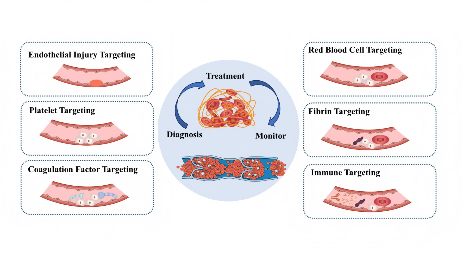
    Introduction

    Thrombosis-related diseases, including deep vein thrombosis (DVT), pulmonary embolism (PE), and coronary artery thrombosis, impose a substantial global health burden. These conditions contribute to high morbidity and mortality rates, with venous thromboembolism (VTE) alone affecting millions of individuals annually.1 In the United States and Europe, VTE accounts for an estimated 300,000 to 600,000 deaths per year, often due to complications such as PE. The economic burden is equally significant, as healthcare systems allocate substantial resources to hospitalization, long-term anticoagulation therapy, and the management of recurrent thrombotic events.2 Development of diagnosis and treatment of thrombosis is shown in Figure 1. Despite advancements in thrombosis management, major challenges persist in early diagnosis, risk stratification, and individualized treatment, underscoring the urgent need for more effective and integrated approaches.3

    Thursday, December 3, 2015

    Nanofibrous scaffolds supporting optimal central nervous system regeneration: an evidence-based review

    Your rehab doctor should be contacting these researchers immediately in order to create a stroke protocol for this. If not, fire them.
    https://www.dovepress.com/articles.php?article_id=24827
    Authors Kamudzandu M, Roach P, Fricker RA, Yang Y
    Received 26 July 2015
    Accepted for publication 9 October 2015
    Published 2 December 2015 Volume 2015:3 Pages 123—131
    DOI http://dx.doi.org/10.2147/JN.S70337
    Checked for plagiarism Yes
    Review by Single-blind
    Peer reviewers approved by Dr Saberi Hooshang
    Peer reviewer comments 3
    Editor who approved publication: Dr Hongyun Huang
    Munyaradzi Kamudzandu, Paul Roach, Rosemary A Fricker, Ying Yang

    Institute for Science and Technology in Medicine, School of Medicine, Keele University, Stoke-on-Trent, UK

    Abstract: Restoration of function following damage to the central nervous system (CNS) is severely restricted by several factors. These include the hindrance of axonal regeneration imposed by glial scars resulting from inflammatory response to damage, and limited axonal outgrowth toward target tissue. Strategies for promoting CNS functional regeneration include the use of nanotechnology. Due to their structural similarity, synthetic nanofibers could play an important role in regeneration of CNS neural tissue toward restoration of function following injury. Two-dimensional nanofibrous scaffolds have been used to provide contact guidance for developing brain and spinal cord neurites, particularly from neurons cultured in vitro. Three-dimensional nanofibrous scaffolds have been used, both in vitro and in vivo, for creating cell adhesion permissive milieu, in addition to contact guidance or structural bridges for axons, to control reconnection in brain and spinal cord injury models. It is postulated that nanofibrous scaffolds made from biodegradable and biocompatible materials can become powerful structural bridges for both guiding the outgrowth of neurites and rebuilding glial circuitry over the “lesion gaps” resulting from injury in the CNS.

    Download Article [PDF] 

    Monday, November 16, 2015

    Rice makes light-driven nanosubmarine

    Our researchers should be jumping for joy, This should allow exact delivery of drugs within the brain and put in sensors that tell us how neuroplasticity works and if neurogenesis/stem cells survive. So many uses and I bet no-one in stroke will even think of the possibilities.
    http://www.rdmag.com/news/2015/11/rice-makes-light-driven-nanosubmarine?
    Though they're not quite ready for boarding a lá "Fantastic Voyage," nanoscale submarines created at Rice University are proving themselves seaworthy.
    Each of the single-molecule, 244-atom submersibles built in the Rice lab of chemist James Tour has a motor powered by ultraviolet light. With each full revolution, the motor's tail-like propeller moves the sub forward 18 nanometers.
    And with the motors running at more than a million RPM, that translates into speed. Though the sub's top speed amounts to less than 1 inch per second, Tour said that's a breakneck pace on the molecular scale.
    "These are the fastest-moving molecules ever seen in solution," he said.
    Expressed in a different way, the researchers reported this month in the American Chemical Society journal Nano Letters that their light-driven nanosubmersibles show an "enhancement in diffusion" of 26 percent. That means the subs diffuse, or spread out, much faster than they already do due to Brownian motion, the random way particles spread in a solution.
    While they can't be steered yet, the study proves molecular motors are powerful enough to drive the sub-10-nanometer subs through solutions of moving molecules of about the same size.
    "This is akin to a person walking across a basketball court with 1,000 people throwing basketballs at him," Tour said.
    Tour's group has extensive experience with molecular machines. A decade ago, his lab introduced the world to nanocars, single-molecule cars with four wheels, axles and independent suspensions that could be "driven" across a surface.
    Tour said many scientists have created microscopic machines with motors over the years, but most have either used or generated toxic chemicals. He said a motor that was conceived in the last decade by a group in the Netherlands proved suitable for Rice's submersibles, which were produced in a 20-step chemical synthesis.
    "These motors are well-known and used for different things," said lead author and Rice graduate student Victor García-López. "But we were the first ones to propose they can be used to propel nanocars and now submersibles."
    The motors, which operate more like a bacteria's flagellum than a propeller, complete each revolution in four steps. When excited by light, the double bond that holds the rotor to the body becomes a single bond, allowing it to rotate a quarter step. As the motor seeks to return to a lower energy state, it jumps adjacent atoms for another quarter turn. The process repeats as long as the light is on.
    For comparison tests, the lab also made submersibles with no motors, slow motors and motors that paddle back and forth. All versions of the submersibles have pontoons that fluoresce red when excited by a laser, according to the researchers. (Yellow, sadly, was not an option.)
    "One of the challenges was arming the motors with the appropriate fluorophores for tracking without altering the fast rotation," García-López said.
    Once built, the team turned to Gufeng Wang at North Carolina State University to measure how well the nanosubs moved.
    "We had used scanning tunneling microscopy and fluorescence microscopy to watch our cars drive, but that wouldn't work for the submersibles," Tour said. "They would drift out of focus pretty quickly."
    The North Carolina team sandwiched a drop of diluted acetonitrile liquid containing a few nanosubs between two slides and used a custom confocal fluorescence microscope to hit it from opposite sides with both ultraviolet light (for the motor) and a red laser (for the pontoons).
    The microscope's laser defined a column of light in the solution within which tracking occurred, García-López said. "That way, the NC State team could guarantee it was analyzing only one molecule at a time," he said.
    Rice's researchers hope future nanosubs will be able to carry cargoes for medical and other purposes. "There's a path forward," García-López said. "This is the first step, and we've proven the concept. Now we need to explore opportunities and potential applications."
    Source: Rice University

    Monday, June 30, 2014

    2nd Annual International Translational Nanomedicine (ITNano) Conference has been set for July 25 - 27th

    Every decent stroke association and stroke hospital should be attending to figure out how to get drugs thru the blood brain barrier so when we finally get some useful neuoplasticity and neurogenesis drugs we will have a way to deliver them.
    http://www.northeastern.edu/itnano/
    Northeastern University in Boston July 25 to 27th, 2014.

    Thursday, May 2, 2013

    Nanoscopic Optical Imaging

    Our researchers should be able to use this to answer the question -
    Are neurons Good, Bad or Ugly? Telling us the answer how neuroplasticity works so we can create repeatable protocols. With that our therapists would actually have useable therapies to give us for recovery rather than compensation.
    http://zhuang.harvard.edu/storm.html

    Friday, December 21, 2012

    Building human body parts

    This looks quite disturbing but this should be one of the steps in helping rebuild our damaged brains. Use stem cells and the scaffolding provided and grow neurons externally, just to prove it can be done. Next step is to create the same nutrient rich location inside your brain and populate it with stem cells to create both neurons and blood vessels. This doesn't take a rocket scientist to figure this out. If we had a decent stroke association(One that I was leading) this would be being worked on. But alas, we have incompetent ones with no sense of helping survivors.
    Great pictures at the link.
    http://cnnphotos.blogs.cnn.com/2012/12/21/the-human-body-parts-store/?hpt=hp_c3
    Alex Seifalian’s lab at University College London is helping humans who lose body parts to repair their bodies the way a newt would if it lost its tail – by growing another.
    The researchers in his lab, which Seifalian calls “the human body parts store,” create the body parts with synthetic materials and a patient’s stem cells.
    The lab builds a scaffold of the needed body part with a porous nanocomposite material, developed and patented by the team, and then puts it in a bioreactor with some of the patient’s bone marrow. The patient’s cells cover the scaffold and fill its many holes so that it essentially becomes the patient’s own.
    After it is inserted into the patient, it’s absorbed by the body and replaced by new cells over time.
    The team has successfully developed a small artery bypass graft and an artificial trachea, or windpipe, both first-evers that are now at work inside patients.
    Seifalian’s lab, at UCL’s Department of Nanotechnology and Regenerative Medicine, recently took on a compassionate case of growing a nose for a 56-year-old man who had had his nose removed during cancer treatment. The man had a prosthetic plastic nose attached to glasses that he could wear, but he chose to not go out in public very often.
    Earlier in December, after the nose had been forming in a glass jar for about four weeks, the lab-grown nose was implanted under the man’s arm. The patient’s doctor will move it to his face after it further develops under his skin. For the first year, the nostrils will remain sealed to avoid infection.
    “You work in a lab all alone, don’t see the future of it,” Seifalian said. “What’s most exciting is that the things we make go to patients.”

    Saturday, October 20, 2012

    Halting Brain Injury

    A fascinating idea, lets get clinical trials started.
    http://www.medpagetoday.com/Nephrology/KidneyTransplantation/35451?utm_content=&utm_medium=email&utm_campaign=DailyHeadlines&utm_source=WC&xid=NL_DHE_2012-10-20&eun=g424561d0r&userid=424561&email=oc1dean@yahoo.com&mu_id=5523591
    The administration of nontoxic carbon particles stabilized the cerebral neurovasculature in a rodent model of traumatic brain injury, restoring critical blood flow and preventing the secondary damage caused by hypotension and free radical release, researchers reported.
    Damage to the neurovascular unit is a major predictor of outcome after brain injury, even in mild concussions, when release of reactive oxygen species such as superoxide interferes with resuscitative efforts. Previous efforts to counteract this damaging process using antioxidants have been unsuccessful for reasons including the need for the presence of additional molecules that contribute detoxification.
    But carbon particles have no need for these other detoxifying factors, so researchers led by Thomas A. Kent, MD, from Baylor University in Houston tested antioxidant carbon particles in rats following controlled cortical impact and induced hypotension.
    "Remarkably," they wrote in ACS Nano, the particle treatment not only restored cerebral blood flow, but also normalized the free radical profile in the animals, suggesting that this approach could provide a novel means of interrupting the cascade of events following brain injury and improving the neurologic outcome for patients.

    Friday, July 20, 2012

    Control of growth and inflammatory response of macrophages and foam cells with nanotopography

    This could explain why we are getting atherosclerosis.
    http://www.nanoscalereslett.com/content/pdf/1556-276X-7-394.pdf
    Background
    Recent fabrication of nanostructured materials with different surface properties has generated a great deal of interest for developing implant materials, i.e., cardiovascular, dental, orthopedic, percutaneous, subcutaneous, and auditory [1-5]. The interface between nanostructured materials and biological tissues is likely to vary dependent upon the surface properties of the nanomaterial. Understanding the degree of toxicity induced by the unique cellular interaction of nanostructured materials is a major concern before utilization in biomedical applications [6-8]. Therefore, fabricating biocompatible materials which are designed to perform specific functions within living organisms has become a key component for generating nanodevices for biomedical applications, including implants.  Macrophages play a critical role during innate and acquired immune responses through the phagocytosis of foreign material. During an immune response, macrophages are typically the first cell type to respond and will secrete proteins (cytokines and chemokines) in order to recruit more immune cells to the site of injury. Atherosclerosis is a pathological process that takes place in the major arteries and is the underlying cause of heart attacks, stroke, and peripheral artery disease. The earliest detectable lesions, called fatty streaks, contain macrophage foam cells that are derived from recruited monocytes. The formation of these foam cells correlates to inflammatory responses [9-11]. In particular, immune cells such as monocytes and macrophages play a key role in mediating host tissue response to implants in the foreign body reaction. One study demonstrated that the macrophage receptor with collagenous structure (MARCO) displayed limited expression in healthy cells but increased in expression around the synovial fluid following hip replacements [12]. This study indicated that the presence of a foreign body can generate an immune response, and the continued presence of the foreign body can potentially lead to macrophage buildup and production of foam cells.  Recent reports have shown that microscaled landscapes are able to direct shape and migration of cultured cells. When cultured on ridges and grooves of nanoscale dimensions, cells migrate more extensively to the ridges than into the grooves. Cell shape is aligned and extended in the direction of the groove [13]. Osteoblasts grown on a fibrous matrix composed of multiwalled carbon nanofibers (100 nm in diameter) exhibit increased proliferation compared to those on flat glass surfaces [14-16]. Nanodots larger than 100 nm in diameter induced an apoptosis-like morphology for NIH-3T3 fibroblast cells [17]. Breast epithelial cells proliferate and form multicellular spheroids on interwoven polyamide fibers fabricated using electrospinning polymer solution onto a glass slide [18]. A 3-D nanofibrillar surface covalently modified with tenascin-C-derived peptides enhances neuronal growth in vitro [19].  The cardiomyoblast H9c2 shows induced cell adhesion and cytoskeleton organization on nanodot arrays smaller than 50 nm [20].
    Recently, arrays of nanodots with defined diameter and depth have been fabricated using aluminum nanopores as a template during oxidation of tantalum thin films [21]. The pore size of aluminum oxide is controllable and uniformly distributed; the depth of dots depends on the
    voltage applied; thus, it can serve as a convenient mold to fabricate tantalum into a nanodot array of specific diameter and depth. The structure containing nanodots of uniform size could serve as a comparable nanolandscape to probe cellular response at the molecular level.
    Although many implant surface topographies are commercially available, there is generally a lack of detailed comparative histological studies at the nano-interface that document how these surfaces interact with living cells, in particular immune cells. In the present study, different sizes of nanodot arrays ranging from 10 to 200 nm were used to evaluate the growth
    and inflammatory response of macrophages and foam cells.


    Rest at the link, a total of 18 pages, baffle your doctor with questions from here.

    Saturday, June 30, 2012

    Concentration-dependent effects of fullerenol on cultured hippocampal neuron viability

    Better viability of new neurons is a good thing.
     http://www.dovepress.com/articles.php?article_id=10259
    Background: Recent studies have shown that the biological actions and toxicity of the water-soluble compound, polyhydroxyfullerene (fullerenol), are related to the concentrations present at a particular site of action. This study investigated the effects of different concentrations of fullerenol on cultured rat hippocampal neurons.
    Methods and results: Fullerenol at low concentrations significantly enhanced hippocampal neuron viability as tested by MTT assay and Hoechst 33342/propidium iodide double stain detection. At high concentrations, fullerenol induced apoptosis confirmed by Comet assay and assessment of caspase proteins.
    Conclusion: These findings suggest that fullerenol promotes cell death and protects against cell damage, depending on the concentration present. The concentration-dependent effects of fullerenol were mainly due to its influence on the reduction-oxidation pathway.

    Keywords: fullerenol, nanomaterial, neurotoxicity, neuroprotection, hippocampal neuron

    Sunday, April 15, 2012

    Nanoparticles home in on human tumors growing in mice’s brains, increase accuracy of surgical removal

    With an innovative stroke researcher we could use the same technology to deliver drugs that stop the cascade of neuronal death and deliver stem cells to the damaged area. :Rant-On: And then we might actually get closer to 100% recovery.:Rant-Off:
    http://scopeblog.stanford.edu/2012/04/nanoparticles-home-in-on-human-tumors-growing-in-mices-brains-increase-accuracy-of-surgical-removal/
    Like special-forces troops laser-tagging targets for a bomber pilot, tiny particles that home in on malignant brain tumors, such as glioblastoma, may someday enable neurosurgeons to remove these malignancies with unprecedented accuracy.
    As I wrote in my release about a new study just published in Nature Medicine by Stanford radiology chief Sam Gambhir, MD, PhD, and his colleagues:
    The prognosis for glioblastoma is bleak: the median survival time without treatment is three months. Surgical removal of such tumors — a virtual imperative whenever possible — prolongs the typical patient’s survival by less than a year. One big reason for this is that it is almost impossible for even the most skilled neurosurgeon to remove the entire tumor while sparing normal brain.
    It’s thought that microscopic remnants of the particularly rough-edged – and particularly nasty – brain tumors called glioblastomas may be largely responsible for this tumor’s ultimate recalcitrance to surgical removal.
    Gambhir and his teammates have engineered brain-tumor-seeking particles measuring less than five one-millionths of an inch in diameter – about one-sixtieth that of a human red blood cell. These particles’ gold centers are coated with materials that, in the study, made it possible to do three things: (1) preoperatively locate a human glioblastoma tumor growing in a mouse’s brain; (2) envision the tumor mass the as it’s being excised; and (3) importantly, guide the scalpel immediately afterward in the removal of myriad minuscule tentacles that snake outward from these tumors’ bulk to invade healthy surrounding tissue. Similarly, the nanoparticles highlighted isolated microscopic tumor-cell patches, called micrometastases, dotting otherwise healthy tissue. Normally, these would have been invisible to the surgeon’s naked eye, and thus might have lived to recur another day.
    Previously: Nanomedicine moves one step closer to reality

    Saturday, March 10, 2012

    Nanoparticle device co. that could reduce drug-making costs gets fresh capital

    Every researcher looking at drugs to cross the blood brain barrier should be rejoicing at this development. Too bad I'm not starting out in graduate school, fascinating possibilities for stroke research if only someone would take advantage of it.
    http://www.medcitynews.com/2012/03/nanoparticle-device-co-that-could-reduce-drug-making-costs-gets-fresh-capital/?utm_source=rss&utm_medium=rss&utm_campaign=nanoparticle-device-co-that-could-reduce-drug-making-costs-gets-fresh-capital
    A nanoparticle device company that produces devices that could reduce drug-making costs has secured follow-on funding from a Pennsylvania economic development agency.

    Ben Franklin Technology Partners of Northeast Pennsylvania invested $50,000 in the business in its latest funding round and has allocated a total of $400,000 to it since 2008.

    Xigo Nanotools is based in Danville, Pennsylvania and develops devices that pharmaceutical companies use for drug formulation development. The company plans to use the investment to make manufacturing preparations as it prepares to expand sales.

    Advertisement
    Its main instrument, the Acorn Area device, is a patented, shoebox-size device that rapidly measures the wetted surface area of nanoparticles using magnetic resonance imaging. The measurements tell pharmaceutical companies both the extent and the nature of the particle liquid interface.”Drugs generally have to be dissolved in order for them to provide a therapeutic benefit and the bioavailability of drugs is directly related to their wetted surface area,” said Sean Race, the co-founder of the company.

    The company was co-founded by Race and Dr. David Fairhurst in 2005, and it started selling its devices in 2010. In response to emailed questions, Race said: “Most drugs are manufactured, delivered or consumed as suspensions of particles in a liquid. … If you can re-engineer the active particle ingredient (API) particles to achieve the same therapeutic benefit at a lower concentration, then you save more money, and less drug is metabolized, so there is a secondary benefit to the consumer.”

    Nanoparticle properties are key predictors of material performance and there currently are no efficient methods to measure wetted nanoparticle surface area, according to a statement from Ben Franklin that describes the company. In addition to pharmaceuticals, the device also has applications for energy, electronics and ceramics.

    The company worked with Johnson & Johnson (NYSE:JNJ) to establish its first beta site.

    Race added: “In addition, we can provide information about the drug formulation such as physical stability, so we are involved in formulation development as well as process development and, in the long run, quality labs, although we don’t have any applications in pharma that well advanced.”

    Race said the company is seeking to raise $2 million and is in investment discussions with a few different venture groups.

    The use of nanoparticles has been a growing source of interest in medicine, particularly in the past decade, since nanoparticles could potentially be directed to specific, targeted cells such as cancer cells and be used to develop more targeted, tailored treatments for individuals. Some companies are developing gold nanoparticles to treat cancer, like CytImmune. Although there has been some criticism that the market forecast for nanoparticles has been overhyped at the expense of some potential risks, the outlook is still strong.

    Saturday, February 11, 2012

    Scale of the Universe 2012

    Cool site for seeing the relative sizes of objects, if you scroll far enough to the left you get down to nanometers where we are interested in using that to deliver drugs through the blood brain barrier.

    109 or .000000001 meters

    http://onemorelevel.com/game/scale_of_the_universe_2012

    Monday, January 9, 2012

    Nanoparticles Hold Promise as Potential Vehicle for Drug Delivery in Brain

    I was blogging about this numerous times last year.
    http://www.sciencedaily.com/releases/2012/01/120109132752.htm
    In the images of fruit flies, clusters of neurons are all lit up, forming a brightly glowing network of highways within the brain.


    It's exactly what University at Buffalo researcher Shermali Gunawardena was hoping to see: It meant that ORMOSIL, a novel class of nanoparticles, had successfully penetrated the insects' brains. And even after long-term exposure, the cells and the flies themselves remained unharmed.

    The particles, which are tagged with fluorescent proteins, hold promise as a potential vehicle for drug delivery.

    Each particle is a vessel, containing cavities that scientists could potentially fill with helpful chemical compounds or gene therapies to send to different parts of the human body. Gunawardena is particularly interested in using ORMOSIL -- organically modified silica -- to target problems within neurons that may be related to neurodegenerative disorders including Alzheimer's disease.

    The recent study on fruit flies is a step toward making this happen, demonstrating that long-term exposure to ORMOSIL, through breathing and feeding, did not injure the animals.

    The research appeared in the journal PLoS ONE on Jan. 3.

    "We saw that after feeding these nanoparticles in the fruit fly larvae, the ORMOSIL was going mainly into the guts and skin. But over time, in adult flies, you could see it in the brain. These results are really fascinating because these particles do not show any toxic effects on the whole organism or the neuronal cells," said Gunawardena, an assistant professor of biological sciences and a researcher in UB's Institute for Lasers, Photonics and Biophotonics.

    The ORMOSIL particles she is investigating are a unique variety crafted by a research group led by Paras N. Prasad, the UB institute's executive director. Each particle contains cavities that can hold drugs, which can be released when the particles are exposed to light.

    Besides Gunawardena and Prasad, co-authors on the study include Farda Barandeh, Phuong-Lan Nguyen, Rajiv Kumar, Gary J. Iacobucci, Michelle L. Kuznicki, Andrew Kosterman and Earl J. Bergey, all from UB.

    Gunawardena is an expert in axonal transport. This involves the movement of motor proteins along neurons' thread-like axon. These molecular motors, called kinesins and dyneins, carry "cargo" including vital proteins to and from the synapse and cell body of neurons.

    In this neuronal highway system, one problem that can occur is an axonal blockage, which resembles a traffic jam in neurons. Proteins aggregate in a clump along the axon.

    Researchers don't know whether these obstructions contribute to disorders such as Alzheimer's or Parkinson's diseases, which are characterized by unusual build-ups of proteins called amyloids and Lewy bodies.

    But the amyloid precursor protein involved in Alzheimer's disease has been shown to have a role in axonal transport, and if axonal obstructions do turn out to be an early indicator for neurodegeneration seen in Alzheimer's disease, eliminating blockages could help prevent or delay the onset of disease.

    That's where ORMOSIL comes in: Gunawardena hopes to use these nanoparticles to target drugs to protein jams along axons, breaking up the accumulations.

    Success, if possible, is still a long way off. But the potential benefit is great. Gunawardena calls the research a "high-risk, high-rewards" endeavor.

    The next step is for her team to see if they can find a way to force the ORMOSIL to latch onto motor proteins. (The nanoparticles, on their own, do not move along axons.)

    Friday, December 16, 2011

    Nanoparticles: second-hand bolt and cancer.

    Originally in Thai. If they can direct nanoparticles to cancer they can direct them to plaque and clots. Cool picture at the link.
    Original in Thai: 

    Nanoparticles: second-hand bolt and cancer.

    translated to English:
    http://translate.google.com/translate?sl=th&tl=en&js=n&prev=_t&hl=en&ie=UTF-8&layout=2&eotf=1&u=http%3A%2F%2Fwww.biomed.in.th%2Fnano_particles%2F

    Nanotech Treatment Shows Promise against Spinal Cord Injury in Mice

    It should also be able to be used to bridge gaps across dead brain areas. 

    Nanotech Treatment Shows Promise against Spinal Cord Injury in Mice


    For release: Monday, May 19, 2008

    Several experimental treatments for spinal cord injury involve a relatively simple idea: implant a "scaffold" at the site of the injury to support the regrowth of severed connections. Unfortunately, many kinds of scaffolds – from pieces of living nerve tissue to artificial polymers – have been tested in animal models of spinal cord injury, and they tend to produce disappointing results. Typically, damaged nerve cells will grow into a scaffold, but then fail to extend through it and beyond, where new connections could truly improve functional outcomes.

    In a study reported in the Journal of Neuroscience*, scientists describe a new kind of polymer scaffold, designed using nanotechnology. They show that the polymer stimulates axons – the nerve cell extensions that run up and down the spinal cord – to regrow all the way across a spinal injury. In mice, a single injection of the polymer given one day after a spinal cord injury led to functional improvements that would be "life-changing" in humans, said John Kessler, M.D., the study's senior investigator and the chairman of neurology at Northwestern University in Chicago.

    "This is not a magic bullet, but it's a promising advance," said Dr. Kessler, who is supported by the National Institute of Neurological Disorders and Stroke (NINDS).

    Long after an injury, the damaged part of the spinal cord remains an inhospitable place for axons. The injured spinal cord lacks the signals that guide axons during embryonic development, and it becomes filled with scar tissue formed by cells called glia. The new polymer, produced through a collaboration between Dr. Kessler and Samuel Stupp, Ph.D., a biomaterials engineer at Northwestern, is an attempt to solve those problems.

    The polymer consists of a carbon-based molecule that forms nanofibers (10,000 times finer than a human hair), and ultimately assembles into a nanogel, upon contact with bodily tissues or fluids. That means the polymer can be injected into the spinal cord in liquid form, which carries less risk of additional damage compared to implanting a pre-formed scaffold, Dr. Kessler said.

    The polymer also contains fragments of laminin – a protein that stimulates axon growth. Perhaps most importantly, once the gelatinous scaffold forms, it lasts for about two weeks and then disintegrates. This apparently gives axons the extra nudge they need to keep growing until they cross the lesion. Previously tested scaffolds "seem to create such a favorable environment that the axons don't want to leave," Dr. Kessler explained.

    Dr. Kessler has spent the better part of his career studying regenerative medicine, particularly the biology of the stem cells that make neurons and glia. He began to focus on spinal cord regeneration in 2001, when his teenage daughter Allison was in a skiing accident that left her paralyzed from the waist down. (Now in her early 20s, she is a Harvard alum and a graduate student at the London School of Economics and Political Science.)

    Drs. Kessler and Stupp originally developed the nanogel thinking it could serve as a delivery vehicle for stem cells and a niche where the cells – stimulated by laminin and shielded from negative cues in the spinal cord – would morph into neurons. But in an earlier study, they found that the nanogel not only stimulated cultured stem cells to turn into neurons, it also suppressed them from becoming astrocytes – the glia that produce scar tissue.

    "We reasoned that if the nanogel had the same effect inside the spinal cord, it would limit the amount of glial scarring," Dr. Kessler said.

    In their current study, Dr. Kessler and his colleagues injected the polymer into mice 24 hours after a spinal cord injury that caused hindlimb paralysis. By nine weeks, the treated mice were using their hindlimbs to take coordinated, weight-bearing steps.

    Inside the spinal cords of the mice, even those that were not treated, axons appeared to grow into the damaged area over several weeks. However, in the treated mice, axons were more likely to enter the lesion, and they also tended to penetrate into it more deeply. By 11 weeks, about 35 percent of descending axons (those that extend downward from the brain) had grown all the way across the lesion in treated mice, while none grew across the lesion in untreated mice. As expected, the treated mice also had less glial scarring than the untreated mice, which may have made it easier for the regrowing axons to leave the scaffold area and make new functional connections within the spinal cord.

    Additional data from animal models will be necessary before the nanogel can be tested in humans with spinal cord injury, Dr. Kessler said. The researchers are investigating the long-term effects of the treatment in mice, as well as its mode of action. They have evidence that the nanogel not only inhibits stem cells from becoming astrocytes, but also encourages them to become oligodendrocytes, the cells that form a protective myelin sheath around spinal axons.

    NINDS is a component of the National Institutes of Health (NIH) within the Department of Health and Human Services. The NIH — The Nation's Medical Research Agency — includes 27 Institutes and Centers and is the primary Federal agency for conducting and supporting basic, clinical, and translational medical research. It investigates the causes, treatments, and cures for both common and rare diseases. For more information about NIH and its programs, visit http://www.nih.gov.

    Tuesday, November 15, 2011

    Nano-drugs against stroke (health)

    Someone using my idea of nanoparticles delivering drugs thru the blood-brain barrier and fixing part of the cascade of death. Who else is researching this style of recovery?
    http://health-med-news.com/health/nano-drugs-against-stroke-health/

    For the first time, the effectiveness of nano-drug for the protection of the cells of the nervous system after a cerebral vascular accident (stroke), has been demonstrated. STROKE is the third cause of mortality in Italy and the second in the world. It is a study international, published in the journal Pnas, which demonstrates the usefulness of these nano-drugs. The study involved including the Institute of neurosciences at the National Research Council of Pisa (In – Cnr), the University of Florence and the University College London.

    “During a stroke, many damaged neurons activate a series of biochemical factors that cause the death of nerve cells in the term,” said Tommaso Pizzorusso, Coordinator of the research. “It is a protein, called Caspasi 3, which causes the”cell suicide”in particular.” This protein can be effectively inhibited by the use of small molecules of RNA, called silencing RNA (siRNA), extremely specific and therefore very beneficial. “However, until now one of the main limitations to their use was due to the difficulty to get sufficient cells to repair the damage.”

    After two years of experiments on rodents, researchers were able to overcome the obstacle through the use of nanotechnology. “We attached the siRNA molecules to tubes of carbon of nanometric dimensions (of the order of the millionth of a millimetre),” continues the researcher of the In – Cnr and the University of Florence, “we were then injected into the injured area of the cerebral cortex, and we found that the nano-vector, once captured by neurons, is able to release within the adequate amount of drug”thus reducing neuronal death induced by stroke. On the treated cells, almost half were saved and the appearance of functional deficits thus was significantly reduced. It is the first time that evidence was made functional enhancement by nano-drugs.

    For the international team, it is a solution that opens new perspectives for more effective treatments. “For the moment”, concludes the researcher, “we are still in the experimental phase and we will have to perform other studies to understand what are nanoparticles which guarantee improved biocompatibility, for an ability to release the siRNA equivalent.” “The result is however an excellent point of departure which confirms that the nano-medicine can be successfully applied in near future to combat brain such as stroke, greatly diffuse pathologies, and so far almost free of effective treatment.”

    Wednesday, November 9, 2011

    A disposable bio-nano-chip using agarose beads for high performance immunoassays

    Testing for heart attacks. Why not do the same for strokes?. The current regime with scans and a neurologist to decode them is one of the reasons tPA use is so low.
    http://www.sciencedirect.com/science/article/pii/S0956566311004556

    Abstract

    This article reports on the fabrication of a disposable bio-nano-chip (BNC), a microfluidic device composed of polydimethylsiloxane (PDMS) and thiolene-based optical epoxy which is both cost-effective and suitable for high performance immunoassays. A novel room temperature (RT) bonding technique was utilized so as to achieve irreversible covalent bonding between PDMS and thiolene-based epoxy layers, while at the same time being compatible with the insertion of agarose bead sensors, selectively arranged in an array of pyramidal microcavities replicated in the thiolene thin film layer. In the sealed device, the bead-supporting epoxy film is sandwiched between two PDMS layers comprising of fluidic injection and drain channels. The agarose bead sensors used in the device are sensitized with anti-C-reactive protein (CRP) antibody, and a fluorescent sandwich-type immunoassay was run to characterize the performance of this device. Computational fluid dynamics (CFD) was used based on the device specifications to model the bead penetration. Experimental data revealed analyte penetration of the immunocomplex to 100 μm into the 280 μm diameter agarose beads, which correlated well with the simulation. A dose–response curve was obtained and the linear dynamic range of the assay was established over 1 ng/mL to 50 ng/mL with a limit of detection less than 1 ng/mL.

    Sunday, October 30, 2011

    Reply to Irish Health on Urgent action needed on stroke services

    My reply to this article was rejected.
    article here:
    http://www.irishhealth.com/article.html?id=16471
    My reply, I thought it was damn good.
    If you think about it you could get more for your money if you start researching hyperacute therapies that stop the cascade of neuronal death. Some have only been tested in mice and rats so needed are Phase II and III trials. Literally dozens of these possibilities. Every neurologist should be able to point to the studies I quoted from. Try

    anti-depressants,KCC2,

    "The death of neurons in the brain can be triggered by an imbalance of oxygen - known as oxidative damage, or where cells are incorrectly instructed to die by a neurotransmitter - a process known as excitotoxicity. KCC2 protects against both.

    Mannan Binding Lectin-Associated Serine Protease-2 (MASP-2),
    By binding with a molecule known as PAR polymer, Iduna prevents the movement of cell-death-inducing factor (AIF) into a cell’s nucleus.
    It’s not just a delay of death, but real protection that lasts for about 72 hours.”
    Iduna protects the brain from glutamate excitotoxicity and stroke by interfering with poly(ADP-ribose) polymer-induced cell death
    Several years ago, scientists realized that the same enzyme that gives bats more blood for their bite may also help stroke victims by breaking down blood clots. Dubbed Draculin, this blood-clot-bashing drug has now entered a phase 2 study:
    extends that window up to 9 hours


    JNK,
    tumeric,
    One compound, called CNB-001, which was derived from curcumin,

    Irish coffee injection(caffeinol),
    The experimental drug, called caffeinol, has the potency of two cups of strong coffee and a small shot of alcohol. When injected into rats within three hours of an artificially stimulated stroke, brain damage was cut by up to 80 per cent.

    xenon gas,

    Earlier preclinical work by the team showed that xenon was effective as a neuroprotectant, stopping processes present during strokes or brain and spinal cord injuries that would damage nerve cells. They found that xenon was capable of blocking the effects of a particular type of glutamate receptor, the same receptor implicated in the pathway that leads to nerve cell death.
    Sigma-1 receptors,
    Professor Tadeusz Wieloch and his colleagues have found a way to activate a protein in the brain, the sigma-1 receptor, which plays an important role in the brain’s recovery during the critical period after the injury.
    We then injected the rats with a specific substance that activated the sigma-1 receptor and found that the rats regained their function more quickly than the untreated animals”, explains Professor Wieloch.
    Of course he doesn't say what the injected substance is but it has to be published somewhere.

    Alpha-B-crystalline: this is the name of the substance, which reduces the inflammatory response. Naturally present in the lens of the eye (the transparent lens located behind the iris), this protein would significantly reduce the size of the consecutive stroke brain. 12 hours after the stroke.
    alpha-B-crystallin, acts as a brake on the immune system, lowering levels of inflammatory molecules whose actions are responsible for substantial brain damage above and beyond that caused by the initial oxygen deprivation of a stroke.
    When systemically administered 24 hours after stroke,perlecan, domain V,was well tolerated, reached infarct and peri-infarct brain vasculature, and restored stroke-affected motor function to baseline pre-stroke levels
    the genetic activation of the nucleic acid protein Caspase-3 – a member of the cysteine-aspartic acid protease (caspase) family – is a major factor in loss of neuronal tissue and associated apoptosis (programmed cell death).
    Carbon nanotubes (CNTs) have been used as a delivery vehicle
    CNTs to deliver small interfering RNAs – nucleic acids which block gene expression – to stop production of this enzyme.
    "The destiny of neurons in a damaged brain depends on a tiny equilibrium between pro-survival and pro-death signals. We wanted to know what KCC2 was signalling for - was it killing neurons or protecting them after an injury? Our study has found that KCC2 actually rescues the damaged cells."
    But when they artificially increased the levels of KCC2 (by stimulating its expression using gene therapy), they found the damaged cells were protected from further damage, and death.

    CDB3 peptide spares neurons from death following traumatic brain injury following stroke and accidents. injection within 2 hours.

    Saturday, October 15, 2011

    Self-Assembling Peptide Nanofiber Scaffold (SAPNS) and Regeneration

    Only 5 years old, where are the current applications? Does your neurologist know about it?
    http://www.innovitaresearch.org/news/06102501.html

    Here is described the creation of a permissive environment for axonal regrowth using a synthetic biological nanomaterial that self assembles in vivo, with components that break down into beneficial building blocks and produce no adverse effects on the CNS. This discovery allows for the reconnection of disconnected parts of the CNS after trauma.

    There are several formidable barriers that must be overcome to achieve axonal regeneration after injury in the central nervous system (CNS), whether caused by a knife or a stroke. These obstacles are:

    • scar tissue formation after tissue injury;
    • gaps in nervous tissue formed during phagocytosis of dying cells after injury;
    • factors that inhibit axon growth in the mature mammalian CNS;
    • failure of many adult neurons to initiate axonal extension.

    The reconnection of disconnected parts of the CNS after trauma will be allowed after reducing or overcoming at least the first two obstacles.

    The previously undiscovered treatment in this study used a designed self-assembling peptide (part (a) at figure below) that spontaneously forms nanofibers, creating a scaffold-like tissue-bridging structure. The nanofibers provide a framework for partial reinnervation by axons with regenerative potential in young and adult animals. Because the peptide fibers are nanoscale, there is likely a direct interaction between the peptide scaffold, the extracellular matrix, and the neural tissue on both sides of the lesion. These structures create a scaffold that connects the two faces of the lesion, allowing movement of cells into the scaffold. The peptide scaffold in experiments created a permissive environment for axonal growth while discouraging or preventing the scar formation that normally occurs at an early stage. This material appears to offer a treatment for ameliorating or bypassing tissue disruptions after neuronal damage.

    SAPNSs are synthetic biological materials formed through the assembly of ionic self-complementary peptides and are designed by using alternating positive and negative L-amino acids that form highly hydrated scaffolds in the presence of physiological concentration salts, like saline, tissue culture media, physiological solutions, or human body fluids such as cerebrospinal fluid (part (b) at figure below). The scaffold consists of β-sheet ionic peptide containing 50% charged residues. A number of additional self-assembling peptides have been designed, synthesized, and characterized for salt-facilitated matrix formation.

    The SAPNS consists of interwoven nanofibers (part (c) at figure below), and the individual fibers are ~10 nm in diameter. The nanofiber density correlates with the concentration of peptide solution. This designed peptide nanofiber scaffold provides several benefits over currently available polymer biomaterials:

    • the peptide scaffold which forms a network of nanofibers that are similar in scale to the native extracellular matrix and therefore provides an in vivo environment for cell growth, migration, and differentiation;
    • it can be broken down into natural L-amino acids and potentially used by the surrounding tissue, because the majority of the material is excreted in the urine;
    • it is synthetic and free of chemical and biological contaminants that typically are present in animal-derived biomaterials such as many collagens;
    • it appears to be immunologically inert, thus avoiding the problem of neural tissue rejection.

    All of these attributes make it very attractive for using the peptide nanofiber scaffold in both in vitro and in vivo studies. Scientific studies show that the SAPNS can support the attachment of a variety of mammalian primary cells in tissue culture. Additionally, one of the peptide scaffolds, arginine, alanine, aspartate, and alanine (RADA) 16-I (part (a) at figure below) supports not only the growth of PC12 cells but also the formation of functional synapses in vitro using rat primary hippocampal neurons.


    SAPNS repair for the animal brain. (a) Molecular model of the RADA16-I molecular building block. (b) Molecular model of numerous RADA16-I molecules undergo self assembly to form well ordered nanofibers with the hydrophobic alanine sandwich inside and hydrophilic residues on the outside. (c) The SAPNS is examined by using scanning electron microscopy. (Scale bar, 500 nm.)

    Thus, RADA-I supports a wide range of neuronal growth and development using both in vitro and in situ cell culture systems. A tissue gap caused by deep transection of the optic tract in the hamster midbrain and injection of saline can completely block reinnervation of the superior colliculus (SC) by the retina even at young ages [postnatal days (P) 2�9] when the axons typically have more regenerative potential. Saline was used, because it is the standard irritant for most neurosurgical procedures and is considered to be benign in the brain.

    Before the use of the SAPNS, scientists demonstrated substantial recovery of visual-orienting behavior in hamsters using a peripheral nerve optic tract bridge model. In one of the models, the optic tract was completely severed at the brachium of the SC, and the reconnection of the optic tract was accomplished with several surgically implanted segments of sciatic nerve taken from one of the animals� legs. However, the use of this model often results in leg disabilities in experimental animals.

    In an attempt to facilitate optic tract regeneration with restoration of function, without additional clinical complications, scientists asked whether the SAPNS could create a permissive environment for regeneration in the damaged tissues as a substitute for sciatic nerve grafts. They examined both short- and long-term effects of injecting a peptide scaffold into the wound site in both young and adult animals using this model. And it was shown that the SAPNS not only permitted significant axonal growth through the site of the treated lesion, partially restoring the optic tract, but also resulted in the return of functional vision in brachium transected experimental adult animals. And the use of this biological nanofiber scaffold is an effective approach to facilitate the reconstruction of a continuous tissue substrate after CNS injury.

    Nano Neurology and the Four P's of Central Nervous System Regeneration: Preserve, Permit, Promote, Plasticity

    Something every stroke reaearcher should know about, so tell yours.
    http://www.sciencedirect.com/science/article/pii/S0025712507000594#

    True nanomaterials are delivered as a specific structure, or combination of structures, designed to deliver the therapeutic intact, directly to the site, requiring a much lower dose. These materials use very specific and deliberate molecular structures that can interact with neurons or protein structures inside the cells. Until recently, functional recovery of the central nervous system (CNS) was an unattainable goal and nanotechnology was an invisible science. A well-planned treatment spaced over time will produce functional return in the CNS. The four P's of CNS regeneration is a new framework for approaching CNS injury and evidence shows that nanotechnology is currently being used for stroke rehabilitation and, in several clinical trials, the treatment of scar formation blockade in the spinal cord. The four components are preserve, permit, promote, and plasticity.

    Article Outline