Use the labels in the right column to find what you want. Or you can go thru them one by one, there are only 33,991 posts. Searching is done in the search box in upper left corner. I blog on anything to do with stroke. DO NOT DO ANYTHING SUGGESTED HERE AS I AM NOT MEDICALLY TRAINED, YOUR DOCTOR IS, LISTEN TO THEM. BUT I BET THEY DON'T KNOW HOW TO GET YOU 100% RECOVERED. I DON'T EITHER BUT HAVE PLENTY OF QUESTIONS FOR YOUR DOCTOR TO ANSWER.
Changing stroke rehab and research worldwide now.Time is Brain!trillions and trillions of neuronsthatDIEeach day because there areNOeffective hyperacute therapies besides tPA(only 12% effective). I have 523 posts on hyperacute therapy, enough for researchers to spend decades proving them out. These are my personal ideas and blog on stroke rehabilitation and stroke research. Do not attempt any of these without checking with your medical provider. Unless you join me in agitating, when you need these therapies they won't be there.
What this blog is for:
My blog is not to help survivors recover, it is to have the 10 million yearly stroke survivors light fires underneath their doctors, stroke hospitals and stroke researchers to get stroke solved. 100% recovery. The stroke medical world is completely failing at that goal, they don't even have it as a goal. Shortly after getting out of the hospital and getting NO information on the process or protocols of stroke rehabilitation and recovery I started searching on the internet and found that no other survivor received useful information. This is an attempt to cover all stroke rehabilitation information that should be readily available to survivors so they can talk with informed knowledge to their medical staff. It lays out what needs to be done to get stroke survivors closer to 100% recovery. It's quite disgusting that this information is not available from every stroke association and doctors group.
Your advisors incompetently didn't know of all this earlier research AND DIDN'T KNOW OF YOUR RESPONSIBILITY TO CREATE STROKE REHAB PROTOCOLS FROM THAT?
I don't care if you are still a student, the real world requires creation of EXACT REHAB PROTOCOLS, at least if you are being trained correctly!
Stroke remains one of the leading causes of death and long-term disability worldwide, and despite advances in acute care, therapeutic options remain severely limited. The currently approved treatments, including reperfusion therapies with intravenous thrombolysis or endovascular thrombectomy, are constrained by narrow therapeutic windows, strict eligibility criteria, and limited accessibility. As a result, the majority of patients remain untreated and are left with permanent neurological impairments. This reality underscores the urgent need for new therapeutic approaches that not only preserve brain tissue immediately after stroke but also actively support long-term repair and recovery. Within this context, insulin-like growth factor 2 (IGF-2), traditionally known for its role in fetal growth and development, has emerged as a promising candidate. Unlike IGF-1, IGF-2 remains abundant in the adult brain, suggesting it may play a role in central nervous system maintenance and repair. This dissertation has four main aims to advance stroke research: 1) to evaluate the neuroprotective effects of IGF-2 and Des(1-6)IGF-2 on infarct size, 2) to investigate their impact on post-stroke neuroinflammation, 3) to examine their role in promoting angiogenesis in vitro and in ovo, and 4) to assess their effects on neural stem cell proliferation and migration. The overall goal is to clarify how IGF-2 and its analogs contribute to brain protection and brain repair.
Your doctor should have already gotten you
protocols for IGF with all the benefits already proven for stroke. So
ask about those protocols. Or is your doctor totally fucking incompetent? Ask him/her about their competence and when it will occur.
Summary: Researchers discovered a mechanism linking
insulin-like growth factors (IGF) to brain plasticity. This study
uncovers how IGF1 and IGF2 promote brain health and functionality,
including learning and memory, through the activation of IGF1-Receptor
during synaptic plasticity.
The study unveils an autocrine
mechanism in neurons that is crucial for brain plasticity. The newfound
insight into this mechanism could pave the way for future research in
preventing cognitive decline and diseases such as Alzheimer’s.
Key Facts:
The
insulin superfamily of hormones, including IGF1 and IGF2, are essential
for healthy brain development and function, including learning and
memory.
The researchers found a local autocrine mechanism where
IGF1 and IGF2 are produced in hippocampal neurons and released during
plasticity, activating the IGF1-Receptor.
Disrupting this
mechanism impairs plasticity, highlighting its critical role in
maintaining cognitive health and potentially providing a novel avenue
for Alzheimer’s disease research.
Source: Max Planck Institute
Research
from the Max Planck Florida Institute for Neuroscience has identified a
mechanism through which insulin-like growth factors facilitate brain
plasticity.
The insulin superfamily of hormones,
including insulin, insulin-like growth factor 1 (IGF1), and insulin-like
growth factor 2 (IGF2), play a crucial role not only in regulating
blood sugar, metabolism, and growth, but also in healthy brain
development and function, including learning and memory.
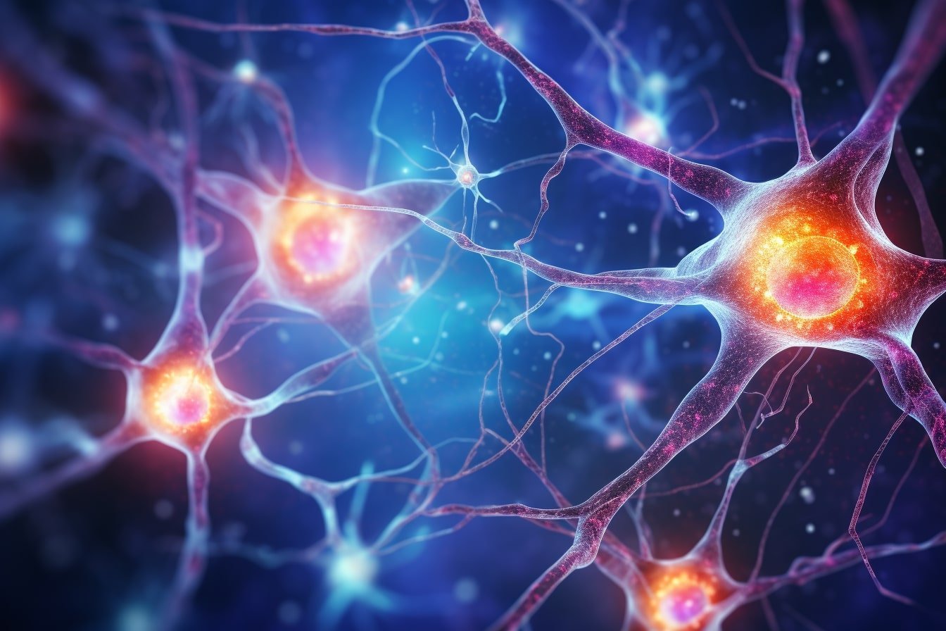
However, where the IGF that activates the receptor was coming from was unknown. Credit: Neuroscience News
These
hormones can enter the brain through the bloodstream from the liver or
can be synthesized directly in neurons and glial cells within the brain.
They bind to receptors, including the IGF1-Receptor, activating signals
that modulate neuron growth and activity. Disruption of this signaling
pathway is involved in cognitive decline and diseases such as
Alzheimer’s.
To understand how IGF1 and IGF2 promote brain health, scientists
investigated the activation of this signaling pathway in the
hippocampus, an area of the brain critical for learning and memory.
Specifically,
they wanted to explore whether IGF signaling was active during synaptic
plasticity, the cellular process that strengthens connections between
neurons during memory formation and protects against cognitive decline.
To
do this, Max Planck scientists developed a biosensor that detected when
the IGF1-Receptor was active, allowing them to visualize the activity
of the signaling pathway involved in plasticity.
When
a synapse was undergoing plasticity, the scientists observed that the
IGF1-Receptor was robustly activated in the strengthening synapse and
nearby synapses. This receptor activation was critical for synaptic
growth and strengthening during plasticity. However, where the IGF that
activates the receptor was coming from was unknown.
Lead researcher and first author of the scientific publication, Dr.
Xun Tu, however, described how being able to visualize the receptor
activation during plasticity gave them a clue.
“The fact that the
activation of the IGF-Receptor was localized near the synapse undergoing
plasticity suggested that IGF1 or IGF2 might be produced in hippocampal
neurons and locally released during plasticity,” she explained.
To
explore this hypothesis, the scientists tested whether IGF1 and IGF2
were produced and could be released from hippocampal neurons.
Interestingly, they found a region-specific difference in the production
of IGF1 and IGF2. One group of neurons in the hippocampus, CA1 neurons,
produced IGF1; another group, CA3 neurons, produced IGF2 (see picture).
When
either CA1 or CA3 neurons were activated in a way that mimicked
synaptic plasticity, IGF wasreleased. Importantly, when the scientists
disrupted the ability of the neurons to produce IGF, the activation of
the IGF1-Receptor during plasticity and synaptic growth and
strengthening was blocked.
Senior author on the publication and Max Planck Scientific Director, Dr. Ryohei Yasuda, summarized the findings.
“This
work reveals a local, autocrine mechanism in neurons that is critical
for brain plasticity. When a synapse undergoes plasticity, IGF is
released locally to activate the IGF1-Receptor on the same neuron.
Disrupting this mechanism impairs the plasticity, highlighting its
critical role in maintaining cognitive health.”
This discovery of
this new mechanism sheds light on how memories are encoded in the brain
and highlights the importance of further study on the insulin
superfamily of hormones in the brain.
The
scientists hope that understanding the mechanism through which IGF
hormones facilitate brain plasticity, will lead to research into whether
targeting this signaling pathway could prevent cognitive decline and
combat diseases like Alzheimer’s.
Funding: This research was supported by Louis D
Srybnik Foundation Inc. and Foundation for the Art, Science, and
Education Inc., the National Institutes of Health (Grant Numbers:
R35NS116804, DP1NS096787, and R01MH080047), and the Max Planck Florida
Institute for Neuroscience. This content is solely the responsibility of
the authors and does not necessarily represent the official views of
the funders.
About this neuroplasticity research news
Author: Katie Edwards Source: Max Planck Institute Contact: Katie Edwards – Max Planck Institute Image: The image is credited to Neuroscience News
Your doctors and stroke hospital will completely fail at creating EXACT exercise protocols and EXACT antioxidant protocols. You're screwed because they will do nothing to help you navigate the best course of action. You are surrounded by incompetent stroke medical 'professionals'. Guidelines like this are the lazy version of professionalism. This would never be allowed in the business world, firings would be the result.
MedicalXpress Breaking News-and-Events | May 20, 2019
An international team of researchers
representing several institutions in Japan and the United States has
published promising findings that may stand to benefit people living
with the specter of Alzheimer's and other neurodegenerative diseases, as
well as age-related cognitive decline.
Advertisement
In their paper published in PNAS, "Leptin in hippocampus
mediates benefits of mild exercise by an antioxidant on neurogenesis and
memory," Yook and colleagues present results from a series of
experiments—murine and in vitro—that elucidate the role of leptin in
cognitive function. Leptin is a hormone that is produced in adipose
tissue and in the hippocampus, the part of the brain where memory and
spatial learning are processed.
The relationship between exercise and improved cognitive function is well established. Likewise, certain dietary supplements,
for example, docasahexaenoic acid (DHA) have also shown promise in
improving cognition and in slowing or halting cognitive decline. The
effect of both exercise and supplementation with an antioxidant on
plasticity and cognitive function within the hippocampus has until now
been largely unexplored, however. Previous research has demonstrated
that leptin in particular is a promising therapeutic target for
neurodegenerative diseases such as Alzheimer's.
Yook and colleagues sought to answer the particular question of
whether mild exercise (ME) combined with the antioxidant supplement
astaxanthin (AX) might confer benefit in terms of cognitive function and
neuronal plasticity; and whether or not the two interventions—AX and ME
together—could have a synergistic effect beyond the additive effects of
either treatment administered separately. The investigators
"hypothesized that ME-enhanced hippocampal neurogenesis and memory might be further improved with dietary AX via mediation by a neurotrophic factor
such as h-LEP [hippocampal leptin]." To test their hypothesis, they
conducted a series of experiments on mice and in vitro using human brain
cell lines.
The first experiment examined the performance of four groups of
wild-type mice on standard tests used to evaluate murine memory and
spatial learning. The mice comprised the following groups: sedentary
with placebo (SE+PL); mild exercise with placebo (ME+PL); sedentary with
astaxanthin (SE+AX); and mild exercise with astaxanthin (ME+AX). The
SE+PL group performed the worst while the ME+PL and SE+AX groups both
performed better. The best performers were the mice in the ME+AX group, a
finding that supports the enhanced effects of both interventions on
memory and spatial learning. The test data were further reinforced by
cell counts of Ki67-positive cells and BrdU/NeuN cells, both measures of
adult hippocampal neurogenesis (AHN), that showed "a strong additional
effect" at work when AX and ME were combined.
To better understand the role of LEP in the changes observed in the
mouse brains, the researchers undertook a DNA microarray and gene expression analysis,
looking at up- and down-regulated genes in relation to the various
groups of mice, and particularly where the genes overlapped between
treatment groups. Their results, especially with regard to the
antioxidant ABHD3 gene and the LEP gene, confirm the synergistic effect of AX and ME on spatial memory and AHN.
Importantly, the study authors also found after further analysis that
circulating plasma leptin levels remained unchanged among treatment
groups, a finding which demonstrates that h-LEP—that is, leptin found in
the hippocampus—is the specific target molecule responsible for the
improvements demonstrated by combined ME+AX therapy. At the protein
level, h-LEP and LEPRa (a leptin receptor), were also correlated with
improvements in spatial memory, while the AKT/STAT3 signaling pathways
were implicated in these improvements as well.
For the in vitro experiment, the investigators used human
neuroblastoma cell lines—cells known to endogenously synthesize
leptin—to observe the effect of exposing them directly to varying
amounts of AX. They noticed a direct dose-dependent response with regard
to expressed leptin, as well as up-regulation in the ABHD3 and LEP genes.
Finally, to establish whether or not leptin is required to achieve
the synergistic effect seen with the AX+ME mice, Yook and colleagues
repeated their earlier mouse experiment on ob/ob knockout mice,
using leptin-deficient animals bred for obesity and diabetes research.
They found that leptin deficiency did indeed play a role, as these mice
performed poorly relative to the wild-type mice, thus confirming leptin
as the crucial component of the AX+ME synergy observed in the prior
experiment.
To further confirm the mediative effect of leptin in the brain, the scientists injected the ob/ob
mice with leptin over the course of 4 weeks, finding that the
synergistic effects of AX and ME were restored in these mice. The
researchers also observed increased levels of proteins pIGF1R and pP13K
in ME+PL and ME+AX groups, independently of whether or not the mice were wild-type or knockout.
The researchers offer further discussion of several details of their
study, for example, commenting that "our results of increased leptin and
IGF1R support the possibility that the enhancement of AHN and memory
function by ME+AX may be due to the interplay of both leptin and IGF1R
expression," and also noting a correlation between memory increase and
an increase in levels of the hippocampal receptor LEPRa.
While the results shown here in murine models are certainly
promising, what might this mean in a clinical context for humans? For
one thing, mild exercise for humans has been characterized in this paper
as that which is typical of a yoga or tai chi session, which puts it
within reach of many people. Another factor is the ready availability of
the relatively inexpensive nutritional supplement astaxanthin.
Ultimately, the authors conclude that "our findings advance the
notion that ME combined with a dietary antioxidant such as AX, which
induces endogenous h-LEP, may be an effective nonpharmacological
strategy for preventing or improving cognitive function and brain
health, and for slowing cognitive decline. This strategy may be
particularly useful in vulnerable individuals, including the elderly."
Sounds like something useful for strokes also. But nothing will be done for at least 50 years because we have no one executing a stroke strategy, failure on a grand scale of all our stroke associations.
Brain Neurotrauma: Molecular, Neuropsychological, and Rehabilitation Aspects. Boca Raton (FL): CRC Press; 2015. Chapter 7. Frontiers in Neuroengineering.
Excerpt
Growing
interest in post-traumatic brain plasticity events has fueled
investigations of therapeutic approaches that promote endogenous
neurorepair. Insulin-like growth factor-1 (IGF-1) is a polypeptide
hormone with critical roles in regulating brain plasticity mechanisms.
This chapter summarizes literature related to how expression of IGF-1
and its signaling components are altered after traumatic brain injury
(TBI). To understand the potential effects of changes in endogenous
IGF-1, the major roles of IGF-1 in CNS function are reviewed, with
attention to how these IGF-mediated events may impact the response to
TBI. In light of the multiplicity of CNS functions mediated by IGF-1,
supplementation of endogenous IGF-1 may provide neuroprotection and
promote neuronal repair in the injured brain. Coupled with a handful of
preclinical studies in TBI, a larger literature in other CNS injuries
such as stroke, hypoxic ischemia and spinal cord injury demonstrates
potential beneficial effects of IGF-1 following injury. TBI
pathophysiology is multifaceted, including primary and secondary events.
Primary injury results from the mechanical forces including
acceleration, deceleration, and impact forces at the moment of injury,
producing diffuse or focal pathology. This initial phase is
characterized by tissue deformation, membrane depolarization, disruption
of blood vessels and axons, ischemia, and cell membrane damage
(Beauchamp et al., 2008; Dietrich et al., 1994; Gaetz, 2004). Secondary
injury evolves from this early damage over a period of hours to days and
even weeks to months, characterized by a complex network of biochemical
events (Dikmen et al., 2009; Farkas and Povlishock, 2007; McIntosh et
al., 1999). Excitatory amino acids and inflammatory cytokines released
early in the secondary injury cascade lead to altered calcium
homeostasis. Excessive intracellular calcium can signal various
biochemical pathways initiating inflammation, free radical generation,
and cytoskeletal damage. Increased calcium can activate proteases
including calpains and caspases. Once activated, these proteases can
cause widespread cell damage via cytoskeletal protein degradation and
necrotic or apoptotic cell death pathways initiated within hours and
continuing for days after brain injury. Secondary injury responses
ultimately culminate in white matter damage and neurodegeneration
contributing to behavioral morbidity. In response to destructive events,
the brain also has the capacity to promote cell repair through various
compensatory mechanisms commonly referred to as neuroplasticity. Altered
growth factor signaling, synaptogenesis, angiogenesis, neurogenesis,
and gliogenesis are among these posttrauma brain remodeling events
(Kernie and Parent, 2009; Schoch et al., 2012; Stein and Hoffman, 2003;
Yu et al., 2008). Expression and release of endogenous neurotrophic
factors is altered by various forms of central nervous system (CNS)
injuries including TBI. An increase in their expression is considered as
one of the mechanisms to promote neuroprotection and neurorepair after
damage (Guan et al., 2003). After TBI, expression of growth factors such
as neurotrophin 4/5, nerve growth factor, basic fibroblast growth
factor, brain-derived neurotrophic factor (BDNF), and IGF-1 are
increased (Conte et al., 2003; Madathil et al., 2010; Royo et al.,
2006). Many of these growth factors play important roles in brain
development and thus their increased expression after brain injury can
recapitulate many of the processes involved in brain growth,
accelerating neuronal repair. Despite the improved understanding of TBI
pathology, no therapeutic approach for treatment has yet been proved
efficacious. Pharmacological approaches under research for TBI can be
grouped as either neuroprotective or neuroreparative depending on their
mode of action. Neuroprotective strategies that promote neuronal
survival are focused mainly on attenuating acute damage from glutamate
excitotoxicity, free radicals, or calcium influx. Neurorepair approaches
promote neuroregeneration or neuroplasticity events. IGF-1, because of
the multiplicity of its actions, provides a combined approach by
attenuating cell death and promoting brain repair events (Aberg et al.,
2000, 2006; Anderson et al., 2002; Lopez-Lopez et al., 2004).
M.A. Silva-Couto, Department of Physical Therapy, Federal University of São Carlos, Rodovia Washington Luis, Km 235, Monjolinho,
São Carlos, São Paulo, Brazil 13565-905.
C.L. Prado-Medeiros, Department of Physical Therapy, Federal University of São Carlos.
A.B. Oliveira, Department of Physical Therapy, Federal University of São Carlos.
C.C. Alcântara, Department of Physical Therapy, Federal University of São Carlos.
A.T. Guimarães, Department of Physical Therapy, Federal University of São Carlos.
T.F. Salvini, Department of Physical Therapy, Federal University of São Carlos.
R. Mattioli, Department of Physical Therapy, Federal University of São Carlos.
T.L. Russo, Department of Physical Therapy, Federal University of São Carlos.
Abstract
Background and Purpose
The muscle weakness that is exhibited post-stroke is due to a
multifactorial etiology involving central nervous system and
skeletal muscle changes. Insulin-like growth
factor I (IGF-1) and IGF binding protein 3 (IGFBP-3) have been described
as biomarkers
of neuromuscular performance in many conditions.
However, no information about these biomarkers is available for chronic
hemiparetic
subjects. Thus, the purpose of the present study
was to investigate possible factors involved to muscle weakness in
chronic
post-stroke subjects, such as serum IGF-1 and
IGFBP-3 concentrations, muscle volume and neuromuscular performance of
knee
flexors and extensors in chronic hemiparetic
post-stroke subjects.
Methods A
cross-sectional study was performed on 14 post-stroke subjects who were
paired with healthy controls. Mobility, functionality,
balance and quality of life were recorded as
outcome measures. The knee flexor and extensor muscle volumes and
neuromuscular
performance were measured by nuclear magnetic
resonance, dynamometry and electromyography. The serum concentrations of
IGF-1
and IGFBP-3 were quantified by ELISA.
Results The
hemiparetic group had low concentrations of serum IGF-1 (25%) and
IGFBP-3 (40%); reduced muscle volume in the vastus
medialis (32%), vastus intermedius (29%), biceps
femoris (16%), semitendinosus and semimembranosus (12%); reduced peak
torque,
power and work of the knee flexors and
extensors; and altered agonist and antagonist muscle activation compared
to controls.
Conclusions Low serum IGF-1 and IGFBP-3 concentrations, deficits in neuromuscular performance, selective muscle atrophy, and decreased
agonist muscle activation are presented in chronic post-stroke subjects.