Changing stroke rehab and research worldwide now.Time is Brain! trillions and trillions of neurons that DIE each day because there are NO effective hyperacute therapies besides tPA(only 12% effective). I have 523 posts on hyperacute therapy, enough for researchers to spend decades proving them out. These are my personal ideas and blog on stroke rehabilitation and stroke research. Do not attempt any of these without checking with your medical provider. Unless you join me in agitating, when you need these therapies they won't be there.

What this blog is for:

My blog is not to help survivors recover, it is to have the 10 million yearly stroke survivors light fires underneath their doctors, stroke hospitals and stroke researchers to get stroke solved. 100% recovery. The stroke medical world is completely failing at that goal, they don't even have it as a goal. Shortly after getting out of the hospital and getting NO information on the process or protocols of stroke rehabilitation and recovery I started searching on the internet and found that no other survivor received useful information. This is an attempt to cover all stroke rehabilitation information that should be readily available to survivors so they can talk with informed knowledge to their medical staff. It lays out what needs to be done to get stroke survivors closer to 100% recovery. It's quite disgusting that this information is not available from every stroke association and doctors group.

Showing posts with label objective diagnosis. Show all posts

Sunday, June 28, 2026

Blood pressure control may be the key to optimizing stroke treatment after thrombectomy

How the hell are you objectively measuring reperfusion success? I've seen a lot of reports that declared reperfusion a failure because the patient didn't recover. You blithering idiots are ignoring the neuronal cascade of death in the first week and thus letting die hundreds of millions to billions of neurons! No wonder stroke recovery never gets better with this level of stupidity!

 Blood pressure control may be the key to optimizing stroke treatment after thrombectomy

Blood pressure management after thrombectomy for acute ischemic stroke may require a change in approach. The HOPE clinical trial-short for Hemodynamic Optimization of Cerebral Perfusion after Endovascular Therapy-led by the Sant Pau Research Institute (IR Sant Pau), has shown that adapting blood pressure targets to the degree of cerebral reperfusion significantly improves patients' functional recovery without increasing the risk of complications.

Until now, we have applied fairly uniform strategies after thrombectomy, but probably not all patients need the same approach. Our results suggest that adjusting blood pressure according to the degree of reperfusion can have a direct impact on recovery."

Dr. Pol Camps-Renom, head of the Cerebrovascular Diseases Research Group at IR Sant Pau and one of the study coordinators

The findings, presented during a plenary session at the annual European Stroke Organisation conference-the leading European scientific society dedicated to stroke-and now published in JAMA Neurology position this work among the most important recent contributions in the stroke field. They have the potential to guide new hemodynamic management strategies after thrombectomy.

Reopening the artery does not always translate into recovery

Mechanical thrombectomy has been a major advance in the treatment of large-vessel occlusion stroke because it can restore blood flow in previously blocked arteries. However, a well-known paradox remains in clinical practice: despite successful angiographic reperfusion, a substantial proportion of patients-around half-do not achieve satisfactory functional recovery in the medium term.(Really? you don't know about the 

the neuronal cascade of death in the first week killing off hundreds of millions to billions of neurons! You're that stupid?)

This phenomenon, known as "clinically ineffective reperfusion," reflects the fact that reopening the vessel does not always result in effective restoration of cerebral perfusion at the tissue level. Mechanisms involved include reperfusion injury, microcirculatory dysfunction, loss of cerebral autoregulation, and hemorrhagic transformation, all of which can compromise brain tissue viability even after a technically successful intervention.

"Many times we can reopen the artery, but the brain tissue does not respond as expected," explains Dr. Pol Camps-Renom. "The reason is that microvascular perfusion and autoregulatory mechanisms may be impaired, and this is where factors such as blood pressure become critical."

As a result, blood pressure control during the hours following thrombectomy has become a key component of clinical management because it directly influences the balance between maintaining adequate perfusion and avoiding hemorrhagic complications. However, the evidence available so far has been limited and, at times, contradictory. Previous trials based on uniform intensive blood pressure reduction strategies have not demonstrated consistent benefits and have even suggested possible adverse effects.

An individualized approach based on reperfusion physiology

The HOPE trial introduces a different approach based on the concept that hemodynamic management should be adapted to each patient's physiological condition after thrombectomy. The study included 440 patients treated at 11 Spanish hospitals, who were randomly assigned either to a conventional strategy or to blood pressure management tailored to the degree of reperfusion achieved.

Unlike previous trials, HOPE implemented a differentiated strategy according to the final angiographic result. Patients with near-complete or complete reperfusion were treated with lower blood pressure targets to reduce the risk of reperfusion injury, whereas patients with incomplete reperfusion maintained higher blood pressure levels to preserve cerebral perfusion.

This approach recognizes that the brain may be in extremely diverse hemodynamic states, in which both excessively high blood pressure and overly aggressive reductions can be harmful. For this reason, the protocol included close monitoring during the first 72 hours, with dynamic treatment adjustments.

Better functional recovery without increased complications

This strategy resulted in a significant and consistent improvement in clinical outcomes. At 90 days, 60.0% of patients in the intervention group achieved functional independence, compared with 47.1% in the control group, representing an absolute difference of 13.3 percentage points, a clinically meaningful improvement. In addition, the overall analysis showed a favorable trend toward better levels of recovery, reinforcing the consistency of the benefit.

In terms of safety, the strategy was associated with a lower incidence of hemorrhagic transformation, without increasing mortality or serious complications, confirming a favorable balance between efficacy and safety. "We have shown that it is possible to improve patient recovery without adding risk," adds Dr. Joan Martí-Fàbregas, another investigator involved in the study. "This balance between efficacy and safety is probably one of the most relevant aspects of the findings."

Toward a paradigm shift in post-stroke management

The results of the HOPE trial point toward a more individualized model for blood pressure control after thrombectomy. In a setting where previous trials had produced neutral or unfavorable results, this study introduces a physiology-based approach that can optimize the balance between perfusion and hemorrhagic risk.

Beyond its findings, HOPE provides key elements for the design of future studies, including the stratification of therapeutic targets and prolonged hemodynamic monitoring. The study also reinforces the idea that stroke treatment does not end with recanalization but continues during the hours that follow. "Rather than applying rigid targets, the key is to better understand each patient's physiology," concludes Dr. Camps-Renom.

Although the trial was stopped before reaching the planned sample size, its results demonstrate a clinically meaningful effect size. Nevertheless, additional studies will be required to confirm these findings before they can be broadly incorporated into routine clinical practice.

Overall, the HOPE trial positions blood pressure control as a key component in optimizing stroke treatment after thrombectomy and opens the door to more precise strategies tailored to individual patients.

Source:
Journal reference:

Camps-Renom, P., et al. (2026) Personalized Blood Pressure Targeting After Endovascular Therapy for Acute Ischemic Stroke: A Randomized Clinical Trial. JAMA Neurology. DOI: 10.1001/jamaneurol.2026.1706. https://jamanetwork.com/journals/jamaneurology/fullarticle/2850074

Wednesday, May 8, 2024

INTEnsive ambulance-delivered blood pressure Reduction in hyper-ACute stroke Trial (INTERACT4): study protocol for a randomized controlled trial

 I don't see where they objectively determine if it is a bleed or a clot. Because if it's a clot then why would you want to lower blood pressure since you want more oxygenated blood to circulate in the brain?

INTEnsive ambulance-delivered blood pressure Reduction in hyper-ACute stroke Trial (INTERACT4): study protocol for a randomized controlled trial

  • Study protocol
  • Open access
  • Published:

You have full access to this open access article

Trials Aims and scope Submit manuscript
INTEnsive ambulance-delivered blood pressure Reduction in hyper-ACute stroke Trial (INTERACT4): study protocol for a randomized controlled trial

Abstract

Background

Early pre-hospital initiation of blood pressure (BP) lowering could improve outcomes for patients with acute stroke, by reducing hematoma expansion in intracerebral hemorrhage (ICH), and time to reperfusion treatment and risk of intracranial hemorrhage in ischemic stroke (IS). We present the design of the fourth INTEnsive ambulance-delivered blood pressure Reduction in hyper-ACute stroke Trial (INTERACT4).

Methods

A multi-center, ambulance-delivered, prospective, randomized, open-label, blinded endpoint (PROBE) assessed trial of pre-hospital BP lowering in 3116 hypertensive patients with suspected acute stroke at 50+ sites in China. Patients are randomized through a mobile phone digital system to intensive BP lowering to a target systolic BP of < 140 mmHg within 30 min, or guideline-recommended BP management according to local protocols. After the collection of in-hospital clinical and management data and 7-day outcomes, trained blinded assessors conduct telephone or face-to-face assessments of physical function and health-related quality of life in participants at 90 days. The primary outcome is the physical function on the modified Rankin scale at 90 days, analyzed as an ordinal outcome with 7 categories. The sample size was estimated to provide 90% power (α = 0.05) to detect a 22% reduction in the odds of a worse functional outcome using ordinal logistic regression.

Discussion

INTERACT4 is a pragmatic clinical trial to provide reliable evidence on the effectiveness and safety of ambulance-delivered hyperacute BP lowering in patients with suspected acute stroke.

Trial registration

ClinicalTrials.gov NCT03790800. Registered on 2 January 2019; Chinese Trial Registry ChiCTR1900020534. Registered on 7 January 2019. All items can be found in this protocol paper.

Thursday, November 23, 2023

New Map Shows How Our Neurons Communicate

This should be required to use in stroke patients, it would give us an objective diagnosis of the damaged /dead neurons.

New Map Shows How Our Neurons Communicate

Summary: Researchers from Cleveland Clinic and OHSU have unveiled a pioneering technique for charting the intricate conversations occurring within our brains. Such insights are key to decoding behavioral alterations in neurological disease patients.

The innovative tool, CaMPARI, allows scientists to witness brain activity in real-time, marking active neurons red and inactive ones green. This breakthrough could offer pathways to better treatments and understanding of diseases like Alzheimer’s.

Key Facts:

  1. The study, using the CaMPARI system, can map real-time brain activity by highlighting active neurons in red and inactive ones in green.
  2. This research is significant for understanding behavior and personality changes in Alzheimer’s disease and related disorders.
  3. The team’s findings, recently published in Nature Communications, have the potential to shape the future of cognitive neuroscience, with the promise of improved treatment options.

Source: Cleveland Clinic

A research team led by Cleveland Clinic and Oregon Health and Science University (OHSU) has developed a new method for mapping how the parts of the brain “speak” to each other, critical to understanding behavior changes in patients with neurological disease.

Diseases like Alzheimer’s disease change how patients communicate and act, affecting their relationships and well-being. Cleveland Clinic’s Hod Dana, PhD, is collaborating with Jacob Raber, PhD, an OHSU behavioral neuroscientist, on mapping out the electrical paths that connect and coordinate the parts of the brain needed to complete different tasks.

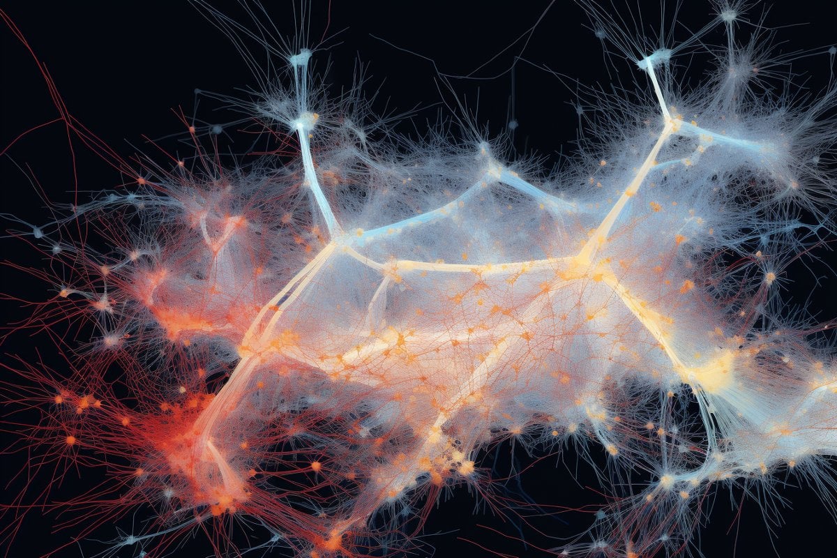
This shows neurons.
Decision-making, forming a memory or completing a task all involve brainwaves, signaling pathways that use cells called neurons. Credit: Neuroscience News

Friday, November 18, 2022

Wearable Sensors for Stroke Rehabilitation

We need this so we can get an objective diagnosis of our movement problems. Then we could map protocols that fix those problems to them and have repeatable recovery options. But no, no one in stroke seems be thinking like that at all. We get crapola guidelines instead, we seem to have no intelligence in the stroke medical world at all.


Wearable Sensors for Stroke Rehabilitation

  • 3 Accesses

Abstract

In this chapter, we provide a review of the current applications of wearable sensors in the field of stroke rehabilitation. Four key points are discussed in this review. First, wearable sensors are a viable solution for monitoring movement during rehabilitation exercises and clinical assessments, but more work needs to be done to derive clinically relevant information from sensor data collected during unstructured activities. Second, wearable technologies provide critical information related to the performance of activities in daily life, information that is not necessarily captured during in-clinic assessments. Third, wearable technologies can provide feedback and motivation to increase movement in the home and community settings. Finally, technologies are rapidly emerging that can complement “traditional” wearable sensors and sometimes replace them as they provide less obtrusive means of monitoring motor function in stroke survivors. These developing technologies, as well as readily available wearable sensors, are transforming stroke rehabilitation, their development is progressing at a fast pace, and their use so far has allowed us to gather important information, that we would have not been able to collect otherwise, which has tremendous potential to further advance stroke rehabilitation.

Sunday, October 30, 2022

A framework for clinical utilization of robotic exoskeletons in rehabilitation

You mention limited guidance on the use of these but don't actually solve the problem.

1. What is the objective diagnosis that would indicate use of each one of these. 

2. What is the EXACT PROTOCOL for their use?

3. What is the expected result from following that protocol?

A framework for clinical utilization of robotic exoskeletons in rehabilitation

Abstract

Exoskeletons are externally worn motorized devices that assist with sit-to-stand and walking in individuals with motor and functional impairments. The Food & Drug Administration (FDA) has approved several of these technologies for clinical use however, there is limited evidence to guide optimal utilization in every day clinical practice. With the diversity of technologies & equipment available, it presents a challenge for clinicians to decide which device to use, when to initiate, how to implement these technologies with different patient presentations, and when to wean off the devices. Thus, we present a clinical utilization framework specific to exoskeletons with four aims.

These aims are to assist with clinical decision making of when exoskeleton use is clinically indicated, identification of which device is most appropriate based on patient deficits and device characteristics, providing guidance on dosage parameters within a plan of care and guidance for reflection following utilization. This framework streamlines how clinicians can approach implementation through the synthesis of published evidence with appropriate clinical assessment & device selection to reflection for success and understanding of these innovative & complex technologies.

Background

The evolution towards evidenced based practice in physical therapy has progressed over the past 25 years, however many barriers to effective translation to clinical practice persist [1]. One critical barrier is when a novel intervention or technology is introduced, there is a paucity of evidence and processes to guide clinicians on how it can be integrated into their everyday clinical practice.

In the current manuscript, we will discuss the clinical use of robotic exoskeletons, which have come into commercial availability since 2011. In the context of this manuscript, exoskeletons are defined as externally worn devices that assist with sit-to-stand and gait training in individuals with motor and functional impairments. They have tremendous potential to assist in the delivery of rehabilitative care through improved efficiency, decreased cost with ability to achieve a high stepping dosage and intensity, and decreased therapist-burden and risk of injury compared to other gait training strategies [2, 3]. The field of robotic technologies is rapidly evolving, with a projected growth of 26% over the next 5 years [4]. Exoskeletons currently approved for clinical use by the US FDA include RewalkTM, Ekso™, Indego™, Hybrid Assistive Limb (HAL) TM for medical use (lower limb type), Rewalk Restore™, B-Temia Keeogo + ™ and Honda Walking Assist Device (WAD)TM.(5, 6) Table 1 describes the FDA-approved device features including level of assistance, resistance modulation, joint control, type of feedback, and stepping actuation. Exoskeletons currently are not considered standard of care in rehabilitation, however patients often seek facilities offering these advanced technologies. Given the emerging evidence of clinical utility, patient interest, and anticipated growth of the field, it is critically important clinicians can effectively evaluate and implement the use of these devices.

Table 1 Comparison of FDA Approved Exoskeleton Devices. Summary of current devices in marketplace with difference in joints controlled, location and type of support provided, resistance or assistance capabilities, method of stepping actuation, and minimum walking function required

B LE = bilateral lower extremity device, U LE = unilateral lower extremity device, FP = fully powered; device provides majority of power at joints and user needs little to no volitional strength to utilize; PA = partially assistive; device provides customized partial assistance to augment deficits to improve gait.

Depending on the rehabilitation facility, clinicians may have access to only one of these devices while others may have multiple options. Regardless of the device availability, practitioners must systematically assess the technology’s features related to their patient’s impairments and functional level to determine if utilization is indicated. Table2 describes the outcomes from randomized control trials to date that have focused on use of FDA approved devices compared to conventional care.

Table 2 Clinical outcomes for trials including diagnoses approved by FDA. Summary of objective outcome measures pre to post intervention from clinical trials investigating FDA approved diagnoses

Specifically, this manuscript focuses on diagnoses approved for use by the FDA. Studies which have investigated the sub-acute and chronic stroke populations included persons with single or unilateral stroke, with a majority including individuals greater than 55 years of age [7,8,9,10,11, 14, 15]. In the incomplete spinal cord injury (SCI) population, most investigations are single group interventional studies or pilot randomized trials. These studies mostly focus on inclusion of participants with incomplete (AIS C or D) injuries with upper motor neuron signs and sufficient upper extremity strength to use an assistive device. Studies focusing on participants with cervical level injuries, AIS A and B injury classification, and lower motor neuron injuries are limited and with varying sample sizes of 9–52 subjects [12, 13]. It should be noted, the aim in many of these studies was to obtain FDA approval with a primary focus on establishing safety with one primary efficacy outcome. Thus in many cases, the true functionality and clinical effectiveness of these devices has not been investigated. Furthermore, these studies also do not focus on dosing, progression strategies, or rehabilitation principles critical to therapeutic intervention [16,17,18].

Adding more uncertainty to application of the available literature, the clinical practice guideline (CPG) for improving walking function in chronic neurological diagnoses, recommended against utilizing robotic interventions [17]. Ten of the eleven studies referenced were not the FDA approved devices focused on in this current manuscript, and eight of the studies focused on treadmill-based robots, specifically the Lokomat [17] These conclusions should be taken with caution, given the substantial differences in functionality and physical demand between the treadmill-based robots and the overground exoskeletons of current focus. Thus, understanding the current literature along with synthesis of knowledge from clinical experience regularly utilizing exoskeletons in practice was critical in developing this framework.

In this four-step framework, we focus specifically on clinical application, rather than exoskeleton use for personal mobility. As authors, we are in a unique position to propose a comprehensive framework to assist in this systematic evaluation due to having extensive experience utilizing a wide array of these exoskeletons during the research and development phase, FDA clinical trials, as well as extensive use in everyday clinical practice [2, 11, 14, 19, 20].

Framework

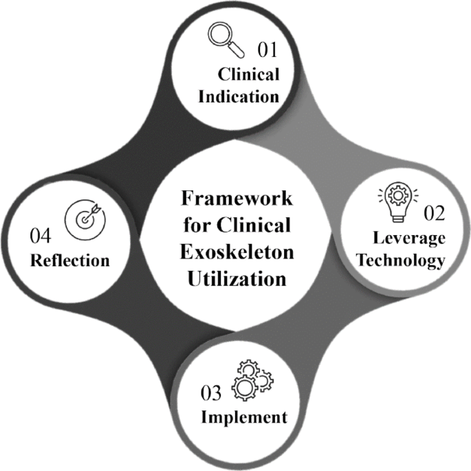
Fig. 1
figure 1

4-step clinical exoskeleton framework. Framework structures clinical decision making surrounding appropriate patient identification, leveraging suitable technology to match patient needs, implementing into a plan of care and clinical reflection to guide further use

Step 1: Clinical indications for exoskeleton use

Clinicians performing evaluations may identify a patient is suitable for exoskeleton utilization at the beginning of an episode of care, or when challenges arise during gait training within a conventional plan of care. Often a patient’s clinical presentation does not match the exact inclusion/exclusion criteria described in the published literature. This should not preclude a clinician from considering incorporation of exoskeleton technology in the plan of care. Inclusion criteria can serve as a baseline for understanding which patient populations and presentations have been investigated to date. Because technology and software development often outpace scientific research, frequently the device investigated is an older version with fewer features or modes than what is available currently.

Thursday, October 6, 2022

NYU researchers partner with FDA to identify biomarkers for stroke rehabilitation

What a pile of shit! Do you not understand that biomarkers DO ABSOLUTELY NOTHING to get survivors recovered. A valid question: Are you a blithering idiot or not?

NYU researchers partner with FDA to identify biomarkers for stroke rehabilitation


A unique collaboration between the engineering sector (NYU Tandon), the clinical sector (NYU Langone), and the regulatory sector (the U.S. Food and Drug Administration) will bring biomarker data to bear on neurorehabilitation technology.

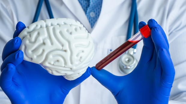
Person in doctors coat holds a model brain next to a red vial.

BROOKLYN, New York, Wednesday, September 28, 2022 — Stroke is the leading cause of age-related motor disabilities and is becoming more prevalent in younger populations as well. But while there is a burgeoning marketplace for rehabilitation devices that claim to accelerate recovery, including robotic rehabilitation systems, recommendations for how and when to use them are based mostly on subjective evaluation of the sensorimotor capacities of patients who use them. (What you really need is an objective diagnosis which leads directly to EXACT PROTOCOLS THAT DELIVER RECOVERY! Call it by the correct term; objective diagnosis.)

S. Farokh Atashzar, assistant professor of Electrical & Computer Engineering and Mechanical & Aerospace Engineering at the NYU Tandon School of Engineering and the director of the Medical Robotics and Interactive Intelligent Technologies (MERIIT) Lab; in collaboration with John-Ross Rizzo, associate professor of Biomedical Engineering at NYU Tandon and Ilse Melamid Associate Professor of rehabilitation medicine at the NYU School of Medicine, are working with Dr. Ramin Bighamian from the U.S. Food and Drug Administration (FDA) to design a regulatory science tool (RST) based on data from biomarkers in order to improve the review processes for such devices and how best to use them. 

The team will design and validate a robust recovery biomarker enabling a first-ever stroke rehabilitation RST based on exchanges between regions of the central and peripheral nervous systems. 

“There is currently no approved RST for assessing the efficacy of rehabilitative devices for post-stroke motor recovery,” explained Atashzar. “This unique collaboration will shed light on how neurological markers can demystify the complex patterns of neural communication, realizing an objective ‘neurophysiological window to degradation in the interaction between anatomy, activation, and pathway in post-stroke patients.”

The research team will also promote FDA regulatory science on next-generation brain-machine interfaces. Additionally, the project will include seminars and undergraduate research to promote STEM education with a focus on engaging students from underrepresented groups.

The project is supported by the U.S. National Science Foundation.  

“This project exemplifies how, as we build our expertise in areas like neural engineering, we are cultivating a highly collaborative research atmosphere — across disciplines such as data science and medicine, between Tandon and other schools at NYU, and spanning the lab and the classroom — all with the greater good in the mind of helping patients,” said Jelena Kovačević, Dean of NYU Tandon. “The importance of this work with the NYU School of Medicine speaks for itself, but it is also heartening to see validation of it from both the FDA and NSF.”

Saturday, September 18, 2021

Concurrent validity of human pose tracking in video for measuring gait parameters in older adults: a preliminary analysis with multiple trackers, viewing angles, and walking directions

 Your doctor and therapists need to use something like this to get an objective diagnosis of your gait problems. With that objective diagnosis they can then go to that database of stroke rehab protocols and select the ones that fix those disabilities.  One of my PTs said to; 'Walk this way'. Totally fucking useless no diagnosis of the problems I had and he expected me to figure out how to correct them by myself. I left him shortly after that. 

Concurrent validity of human pose tracking in video for measuring gait parameters in older adults: a preliminary analysis with multiple trackers, viewing angles, and walking directions


Abstract

Background

Many of the available gait monitoring technologies are expensive, require specialized expertise, are time consuming to use, and are not widely available for clinical use. The advent of video-based pose tracking provides an opportunity for inexpensive automated analysis of human walking in older adults using video cameras. However, there is a need to validate gait parameters calculated by these algorithms against gold standard methods for measuring human gait data in this population.

Methods

We compared quantitative gait variables of 11 older adults (mean age = 85.2) calculated from video recordings using three pose trackers (AlphaPose, OpenPose, Detectron) to those calculated from a 3D motion capture system. We performed comparisons for videos captured by two cameras at two different viewing angles, and viewed from the front or back. We also analyzed the data when including gait variables of individual steps of each participant or each participant’s averaged gait variables.

Results

Our findings revealed that, i) temporal (cadence and step time), but not spatial and variability gait measures (step width, estimated margin of stability, coefficient of variation of step time and width), calculated from the video pose tracking algorithms correlate significantly to that of motion capture system, and ii) there are minimal differences between the two camera heights, and walks viewed from the front or back in terms of correlation of gait variables, and iii) gait variables extracted from AlphaPose and Detectron had the highest agreement while OpenPose had the lowest agreement.

Conclusions

There are important opportunities to evaluate models capable of 3D pose estimation in video data, improve the training of pose-tracking algorithms for older adult and clinical populations, and develop video-based 3D pose trackers specifically optimized for quantitative gait measurement.

Background

Clinically established techniques for examining gait quality in older adults typically require technologies such as motion capture systems which are expensive and time consuming, require specialized expertise and staff to operate, and are not widely available for clinical use. As a result, gait monitoring practices have mainly involved cross-sectional gait assessments in laboratory settings or under experimental conditions which do not reflect the cognitive and physical demands of natural walking or usual locomotion [1].

With the advent of commercially available depth cameras, specifically the Kinect sensor (Microsoft, Redmond, WA), researchers were able to monitor natural walking of participants [2,3,4,5,6,7]. However, the Kinect camera has a limited depth of field (0.5 to 4.5 m) which can only capture few steps. This limitation, along with concerns about cost and potential hardware obsolescence (the sensor was commercially unavailable for an extended period until a newer version was released) motivate adopting other technologies for the purpose of natural gait monitoring. Although other depth sensing cameras are available, it would be ideal if technologies can make use of regular videos from cameras that are ubiquitous, such as surveillance cameras.

Advances in computer vision technology and human pose estimation in image/video data can address these limitations. A number of algorithms have been developed for human pose tracking that are capable of automated analysis of human walking using only standard RGB camera videos [8,9,10,11,12,13,14,15]. These algorithms use deep learning models that are trained on a large corpus of annotated videos, resulting in models capable of detecting body segments (head, hands, knees, feet, etc.) in new videos outside of the training dataset. These packages are freely available and can be used to process videos of human walking in any setting with minimal cost and technical expertise [15]. Gait parameters can subsequently be computed from the sequence of tracked body parts [16]. However, for use in clinical applications, there is a need to validate gait variables calculated from pose tracking data against gold standard methods for measuring human gait data, e.g., three-dimensional (3D) motion capture systems [15].

Previous studies on the validation of video pose tracking algorithms mainly used a single pose tracking algorithm, mainly OpenPose [8], in sagittal view, and healthy young adults [9, 11, 12, 15, 17]. Less is known about the performance of other publicly available pose tracking algorithms such as AlphaPose [13] or Detectron [14] particularly for pose tracking of gait in a frontal view and in older adult populations. There are several reasons that this analysis is valuable and necessary: i) comparison of different pose trackers allows researchers to choose the most appropriate one for the purpose, ii) recording walks in a frontal view allows the capture of more steps and an analysis of stability in the frontal plane, and iii) pose tracking algorithms require validation in older adults as their posture and gait are different to that in young adults and is characterized by lower speed, and greater variability [18].

The aim of this study was, therefore, to investigate the concurrent validity of spatiotemporal gait measurement in the frontal plane based on three common pose trackers (AlphaPose [13], OpenPose [8], and Detectron [14]) against a 3D motion capture system by doing a correlation analysis between the gait variables calculated from the two systems in older adults.

More at link.