Changing stroke rehab and research worldwide now.Time is Brain! trillions and trillions of neurons that DIE each day because there are NO effective hyperacute therapies besides tPA(only 12% effective). I have 523 posts on hyperacute therapy, enough for researchers to spend decades proving them out. These are my personal ideas and blog on stroke rehabilitation and stroke research. Do not attempt any of these without checking with your medical provider. Unless you join me in agitating, when you need these therapies they won't be there.

What this blog is for:

My blog is not to help survivors recover, it is to have the 10 million yearly stroke survivors light fires underneath their doctors, stroke hospitals and stroke researchers to get stroke solved. 100% recovery. The stroke medical world is completely failing at that goal, they don't even have it as a goal. Shortly after getting out of the hospital and getting NO information on the process or protocols of stroke rehabilitation and recovery I started searching on the internet and found that no other survivor received useful information. This is an attempt to cover all stroke rehabilitation information that should be readily available to survivors so they can talk with informed knowledge to their medical staff. It lays out what needs to be done to get stroke survivors closer to 100% recovery. It's quite disgusting that this information is not available from every stroke association and doctors group.

Showing posts with label progesterone. Show all posts

Monday, September 1, 2025

Application of hair HPG axis hormone levels before onset in predicting the risk of stroke

With no explanation of what should be done to prevent a stroke; THIS RESEARCH WAS TOTALLY FUCKING USELESS! You're fired!

Does anyone in the stroke medical world actually think?

 Application of hair HPG axis hormone levels before onset in predicting the risk of stroke


  • 1Rehabilitation Center, The First Affiliated Hospital with Nanjing Medical University, Nanjing, China
  • 2School of Biological Science and Medical Engineering, Southeast University, Nanjing, China
  • 3Department of Rehabilitation Medicine, The Eighth Affiliated Hospital of Southern Medical University (The First People’s Hospital of Shunde Foshan), Foshan, China

Background: There have been many studies on the relationship between sex hormones and stress, mood, blood pressure, etc., but its impact on the incidence of stroke remains unknown.

Objective: To investigate the expression levels of hypothalamic–pituitary-gonadal (HPG) axis related hormones such as testosterone (T) and progesterone (P) in hair before stroke and their effects on the risk of stroke.

Methods: 48 patients with stroke were recruited from November 2022 to May 2023 as the observation group and 35 healthy subjects were recruited as the control group. There was no obvious difference in age, gender and BMI between the two groups (p > 0.05). T and P levels in hair were tested by LC-MS/MS, and the correlation with the risk of stroke was analyzed.

Results: The T and P levels of hair before the onset of stroke in the observation group were significantly lower than those in the control group (p < 0.01). The T level of men’s hair before the onset of stroke in the observation group was significantly lower than those in the control group (p < 0.05). The T and P levels of women’s hair before the onset of stroke in the observation group were significantly lower than those in the control group (p < 0.01). The results of ROC curve showed that the cut-off value of T level in men’s hair before the onset of stroke was 4.35 pg/mg, the AUC was 0.690 (0.545, 0.835), the sensitivity was 62.50%, and the specificity was 82.61%. The cut-off value of T level in women’s hair before the onset of stroke was 5.00 pg/mg, the AUC was 0.818 (0.658, 0.978), the sensitivity was 75.00%, and the specificity was 83.33%. The cut-off value of P level in women’s hair before the onset of stroke was 8.00 pg/mg, the AUC was 0.891 (0.754, 1.000), the sensitivity was 81.25%, and the specificity was 100.00%.

Conclusion: This preliminary report is the first to suggest that HPG axis hormones such as T and P in hair could have predictive value in screening for stroke risk.

1 Introduction

Cerebrovascular diseases have now ascended to the foremost cause of mortality in China, with stroke constituting the single most prevalent etiology of disability, exerting a substantial burden on both individuals and society (1). Stroke is a preventable and manageable disease, and early screening combined with active intervention can markedly enhance patient prognosis (2).

Emerging evidence suggests associations between hypothalamic–pituitary-gonadal (HPG) axis hormones including testosterone (T), estradiol (E2), and progesterone (P) and stroke incidence (34). Haya et al. (5) conducted a 29-year follow-up study involving 4,615 adult males and 4,724 adult females to investigate endogenous hormone profiles, revealing that extremely low serum T concentrations were significantly associated with elevated ischemic stroke risk in males. However, this study faced methodological limitations, including the absence of precise temporal data on stroke onset in community-based populations, which has led to the need for studies in larger samples. Furthermore, the requirement for continuous serological monitoring and extended follow-up until stroke occurrence resulted in prolonged study duration, substantial costs, and potential confounding from unadjusted variables such as aging.

Hormone levels in human hair serve as endogenous biomarkers for retrospective assessment of hypothalamic–pituitary-adrenocortical (HPA) and HPG axis activity (6). The measurement of hormone levels in hair is retrospective, with 1 cm of hair corresponding to approximately 1 month of hormonal accumulation, and a reliable retrospective window extending up to 6 months (7). Compared to traditional biological matrices such as blood, serum, and urine, hair hormone analysis offers distinct advantages, including cumulative representation of long-term exposure, non-invasive sampling, and the ability to reflect baseline levels over extended periods (similar to how hemoglobin A1c is used to assess serum glucose levels) (79). While clinical studies on hair hormones have proliferated (1011), current research predominantly focuses on associations between hair hormone levels and psychiatric, emotional, and sleep-related disorders. For instance, Wright et al. (12) demonstrated in a systematic review that cortisol quantification in hair can evaluate chronic stress exposure in elderly populations. Deng et al. (11) investigated functional characteristics of the HPA and HPG axes, as well as their interplay, in patients with schizophrenia by analyzing expression levels of eight biomarkers in hair samples. Similarly, Wang et al. (13) identified a significant correlation between pre-onset HPA axis hyperactivity and post-stroke emotional disorders.

Current research lacks substantial evidence on the correlation between pre-stroke HPG axis hormone levels in hair and stroke risk. Considering the influence of age and sex on HPG axis activity (14), this study investigates the relationship between the expression levels of HPG axis hormones in the hair and the risk of stroke onset among individuals aged over 50, across different genders. The findings aim to provide novel insights and methodologies for early stroke screening and intervention strategies.

2 Materials and methods

This study was reviewed and approved by the Ethics Committee of Jiangsu Provincial People’s Hospital (2022-SR-553), and was prospectively registered with the Chinese Clinical Trial Registry (ChiCTR2200065803). All participants provided written informed consent to participate in the study.

2.1 Participants

Study participants included 48 stroke patients (32 males, 16 females) hospitalized at Jiangsu Provincial People’s Hospital and its Qixia Rehabilitation Branch between November 2022 and May 2023. Subjects in the stroke group had a mean age of 64.67 ± 7.53 years, with a mean disease duration of 31.50 days (IQR 33.00). Stroke group included 35 ischemic and 13 hemorrhagic cases. A control group of 35 healthy individuals (23 males, 12 females; mean age 65.69 ± 8.90 years) was recruited. No statistically significant differences were observed between groups in age, sex, or body mass index (BMI) (p > 0.05) (Table 1).
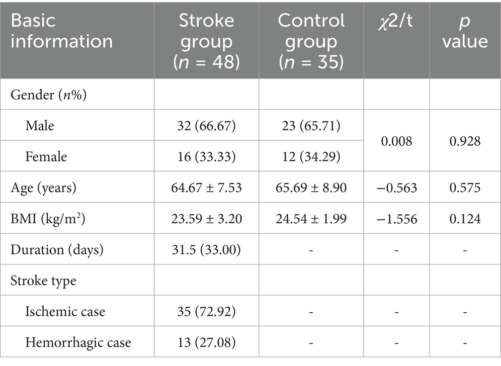
Table 1
www.frontiersin.org

Table 1. Comparison of general data between the two groups [M (IQR)] or M ± SD.

Inclusion Criteria for Stroke Group were as follows: ① Age 50–80 years; ➁ Stroke diagnosis confirmed by neuroimaging and compliant with diagnostic guidelines established by the Chinese Stroke Society; ➂ First-ever unilateral stroke; ➃ Disease duration ≤ 3 months; ➄ Clinically stable with vital signs within normal ranges; ➅ Postmenopausal status (confirmed by amenorrhea ≥ 12 months) for female participants.

Inclusion Criteria for Control Group were as follows: ① Age 50–80 years; ➁ No history of stroke or major neurological disorders; ➂ Postmenopausal status for female participants.

Exclusion Criteria (Both Groups) were as follows: ① Auditory comprehension deficits or inability to follow verbal commands; ➁ Concurrent with other central nervous system lesions (e.g., traumatic brain injury, brain neoplasms); ➂ History of reproductive system disorders or surgeries; ➃ Substance abuse, hormone therapy within the past year, or chronic alcoholism; ➄ Occipital hair length < 3 cm; ➅ Recent hair treatments affecting analysis.

2.2 Sample collection

Hair samples were collected from all subjects on their enrollment day. Hair samples were collected by cutting 1 cm of hair from the occipital region, located at the back of the subject’s head near the scalp. Specimens were wrapped in aluminum foil, labeled, and stored at room temperature protected from light.

2.3 Hormonal analysis

Hormonal concentrations in hair were analyzed via high-performance liquid chromatography–tandem mass spectrometry (LC-MS/MS), where an Agilent 1,200 liquid chromatograph (Agilent Technologies, Inc., United States) was combined with an API 3200 Q-TRAP mass spectrometer (Applied Biosystems, Inc., United States), following a previously developed method (15). Hair samples corresponding to the period before the onset of lesions were selected based on the disease course (If the duration of the patient’s disease is 30 days at the time of admission, the 1 cm of hair closest to the scalp is the total amount of hormones accumulated in the hair in the month when the stroke occurred, and the 2 cm close to the scalp is the total amount of hormones accumulated in the hair in the month before the stroke occurred). Hair samples were rinsed with 5 mL of methanol for 2 min, and then heated to 50°C for drying. In a 2 mL centrifuge tube, hair samples were weighed after measuring 1–2 mm in length and cut into powder. After incubation in methanol for 24 h at 25°C, the mixture was centrifuged at 1.2 × 104 r/min for 5 min. Transfer 800 μL of supernatant into another clean centrifuge tube and evaporate nitrogen at 40°C. The residue was then redissolved in 50 mL of mobile phase for LC-MS/MS analysis.

There was good performance, with limits of detection and quantification of 0.3 and 1.0 pg/mg for T, and 0.2 and 0.5 pg/mg for P, respectively. The recovery, intra-day, and inter-day coefficients of variation met the requirements. Analyses were conducted at the Jiangsu Provincial Key Laboratory of Biomaterials and Devices, Southeast University.

2.4 Statistical analysis

SPSS 25.0 software was used for data analysis. Normality of continuous variables was assessed via the Shapiro–Wilk test. Normally distributed data are presented as mean ± standard deviation, while non-normally distributed data are expressed as median (interquartile range, IQR). Group comparisons for normally distributed continuous variables utilized independent samples t-tests, whereas non-parametric Mann–Whitney U tests were applied to non-normally distributed data. Receiver operating characteristic (ROC) curve analysis was conducted to evaluate the predictive efficacy and the cutoff values of T and P for stroke risk. Categorical variables are reported as frequencies and percentages, with between-group comparisons performed using chi-square tests. A two-tailed p < 0.05 was considered statistically significant.

3 Results

3.1 Comparison of T and P expression levels between the two groups

The T and P levels of hair before the onset of stroke were significantly lower in the stroke group compared to the healthy control group (T: 3.60 vs. 6.90 pg/mg, Z = −3.52, p = 0.001; P: 5.85 vs. 9.80 pg/mg, Z = −3.08, p = 0.002). The median reductions in T and P levels were 48% and 40%, respectively (Table 2).

Sunday, November 17, 2019

Combination therapy with vitamin D3, progesterone, omega-3 fatty acids and glutamine reverses coma and improves clinical outcomes in patients with severe traumatic brain injuries: A case series

You can ask your doctors if anything here would help in stroke recovery. Doesn't know, then the correct and only answer is for them to contact researchers to get an answer.  Not doing anything, call the president and ask what the fuck is the responsibility of the stroke doctors. Delivering the status quo of stroke rehab means that the stroke president also needs to be fired.  Or you could look up these:

Combination therapy with vitamin D3, progesterone, omega-3 fatty acids and glutamine reverses coma and improves clinical outcomes in patients with severe traumatic brain injuries: A case series

How to cite this article:
Matthews LR, Danner OK, Ahmed YA, Dennis-Griggs DM, Frederick A, Clark C, Moore R, DuMornay W, Childs EW, Wilson KL. Combination therapy with vitamin d3, progesterone, omega 3-fatty acids and glutamine reverses coma and improves clinical outcomes in patients with severe traumatic brain injuries: A case series. International Journal of Case Reports and Images 2012;4(3):143–149.


Abstract


Introduction: Traumatic brain injury (TBI) is a major public health problem and a leading cause of death and disability in the United States. Management of patients with TBI has changed very little over the last 20 years.
Case Series: A case series of three patients with severe TBIs who were aggressively treated with vitamin D3, progesterone, omega 3-fatty acids, and enteral glutamine for six weeks, termed neuroceutical augmentation for traumatic brain injury (NATBI), with very favorable outcomes.
Conclusion: A large clinical study trial using these four supplements (NATBI) together is warranted.

Keywords: Traumatic brain injury, Vitamin D3, Omega-3 fatty acids, Loveza, Progesterone, Cerebral edema, Glutamine

Introduction

Traumatic brain injury (TBI) is a major public health problem. According to CDC it affects over 1.7 million people annually in U.S. with 275,000 hospitalizations and 52,000 deaths. [1] The medical cost for treating TBI patients in the United States in 2010 was $76.5 billion and rising annually. [1] Primary causes for TBI include the following: motor vehicle crashes, falls, assaults and sports or recreation-related injuries (concussions). Finding the right treatment to reduce mortality rates and improve the clinical outcomes in TBI patients has been elusive.
Management of patients with TBI has changed very little over the last 20 years. Advancements in the treatment of TBI requires great understanding of the biochemical mechanisms of the brain during a normal resting state as well as the metabolism after a severe traumatic event. Brain metabolism is markedly altered during TBI. After the initial insult to the brain, the brain's metabolism is altered and can increase up to 140% of its normal metabolism.
Vitamin D (a steroid hormone) and omega-3 fatty acids (an essential fatty acid) are both very powerful anti-inflammatory agents that reduce cerebral edema and swelling. Glutamine becomes an essential amino acid during stress and produces the extra glucose (via the Cori cycle) that is used by the injured brain and the extra glucose used by the immune response system to fight off infection during stress. Progesterone (also a steroid hormone) is a neuroprotector of injured brain cells and potentiates the effect of vitamin D.
These agents are all immune modulators which work synergistically to prevent secondary brain injury by limiting or decreasing inflammation; an increasing well-recognized cause of ongoing brain swelling after a primary injury. They are also neuroprotectors that makes the neurons more resistant to stress, ischemia, hypothermia, hyperthermia, hypoglycemia, hyperglycemia, hypotension, and hypertension. Immune modulation with nutritional supplements is a rapidly advancing field with a very promising future in treating TBI as well as other critically injured/ill patients.
We present a case series of three patients with severe TBIs who were aggressively treated with vitamin D3, progesterone, omega-3 fatty acids and enteral glutamine for up to six weeks, termed neuroceutical augmentation for traumatic brain injury (NATBI), with very favorable outcomes. [2] [3] [4] [5] Patients in a coma with severe TBI (Glascow Coma Score <8) who were admitted to a Level I trauma center were evaluated in a prospective observational study. Patients were treated with a neuroceutical combination of vitamin D3, omega-3 fatty acids, progesterone, and glutamine initially via a nasogastric tube and later orally for six weeks. Primary outcomes were mortality rate and return to recovery which was defined as a Glascow Coma Score (GCS) of 10 or greater.NATBI protocol works on multiple levels and neuroprotective pathways in TBI patients by down regulating cytokine production, preventing oxidative stress (free radical oxygen formation), decreasing cerebral edema, and inflammation, thus limiting secondary brain injury in contradistinction to progesterone therapy alone (Protect III study). [3] [4] [5] In addition, our NATBI regimen is relatively inexpensive, safe, and very effective at reducing brain and systemic inflammation post-injury.
Leslie R Matthews1, Omar K Danner1, Y A Ahmed2, Diane M Dennis-Griggs1, Alexis Frederick1, Clarence Clark1, Ronald Moore1, Wilson DuMornay1, Ed W Childs1, Kenneth L Wilson1
1Morehouse School of Medicine, Department of Surgery, 720 Westview Drive, SW, Atlanta, GA, USA.
2Department of Epidemiology, King Saud University, College of Pharmacy, Riyadh, Kingdom of Saudia of Arabia.

doi:10.5348/ijcri-2013-03-281-CS-2

Address correspondence to:
Leslie Ray Matthews
MD, FACS, 720 Westview Drive
SW, Atlanta
Georgia 30228
USA
Phone: (404) 6162391
Fax: (404) 6161417
Email: lematthews@msm.edu

Access full text article on other devices

Access PDF of article on other devices

[HTML Abstract]   [PDF Full Text] [Print This Article]
[Similar article in Pumed] [Similar article in Google Scholar]


Monday, January 29, 2018

14 Powerful Ways to Form New Synapses in the Brain

Is your doctor suggesting any of these to help your stroke recovery? Unless they are written as protocols, they are completely fucking useless in guaranteeing new synapses. Screaming at your incompetent? doctor may be required!

14 Powerful Ways to Form New Synapses in the Brain 


1. Omega-3 Fatty Acids, Uridine and Choline

2. Low Level Laser Therapy

3. Bacopa

4. Exercise

5. Magnesium Threonate

6. Intermittent Fasting

7. Ginkgo Biloba

8. Motor Learning

9. Resveratrol

10. Piracetam

11. Quercetin

12. Intranasal Insulin

13. Progesterone

14. Antioxidant Nutrients

15. BONUS: 4 Things to Avoid

Details at link. You might want to ask your doctor about these. 

Friday, November 24, 2017

Effects of Female Sex Steroids Administration on Pathophysiologic Mechanisms in Traumatic Brain Injury

8 posts on estrogen all the way back to Jan. 2012 just to show you how fucking incompetent your stroke medical world is. 9 posts on progesterone  back to Jan. 2012 show the same incompetence. You doctor can explain why nothing has been done. 

Effects of Female Sex Steroids Administration on Pathophysiologic Mechanisms in Traumatic Brain Injury


  • Mohammad Khaksari
  • Zahra Soltani
  • Nader Shahrokhi
  1. 1.Physiology Research Center, Institute of NeuropharmacologyKerman University of Medical SciencesKermanIran
  2. 2.Endocrinology and Metabolism Research Center, Institute of Basic and Clinical Physiology SciencesKerman University of Medical SciencesKermanIran
  3. 3.Neuroscience Research Center, Institute of NeuropharmacologyKerman University of Medical SciencesKermanIran
Review


Abstract

Secondary brain damage following initial brain damage in traumatic brain injury (TBI) is a major cause of adverse outcomes. There are many gaps in TBI research and a lack of therapy to limit debilitating outcomes in TBI or enhance the neurogenesis, despite pre-clinical and clinical research performed in TBI. Females show harmful outcomes against brain damage including TBI less than males, independent of different TBI occurrence. A significant reduction in secondary brain damage and improvement in neurologic outcome post-TBI has been reported following the use of progesterone and estrogen in many experimental studies. Although useful features of sex steroids including progesterone have been identified in TBI clinical trials I and II, clinical trials III have been unsuccessful. This review article focuses on evidence of secondary injury mechanisms and neuroprotective effects of estrogen and progesterone in TBI. Understanding these mechanisms may enable researchers to achieve greater success in TBI clinical studies. It seems that the design of clinical studies should be revised due to translation loss of animal studies to clinical studies. The heterogeneous and complex nature of TBI, the endogenous levels of sex hormones at the time of taking these hormones, the therapeutic window of the drug, the dosage of the drug, the selection of appropriate targets in evaluation, the determination of responsive population, gender and age based on animal studies should be considered in the design of TBI human studies in future.

Saturday, December 17, 2016

Progesterone sharpens temporal response profiles of sensory cortical neurons in animals exposed to traumatic brain injury

Followup should be done since there are conflicting results for stroke and TBI, but that will never occur until our fucking failures of stroke associations are destroyed.
http://www.ingentaconnect.com/content/cog/ct/pre-prints/content-ct-1705_allitt_et_al
Download / Buy Article:
Open access content The full text is Open Access.
 Download
(PDF 1,170 kb)
 

Abstract:

Traumatic brain injury (TBI) initiates a cascade of pathophysiological changes that are both complex and difficult to treat. Progesterone (P4) is a neuroprotective treatment option that has shown excellent preclinical benefits in the treatment of TBI but these benefits have not translated well in the clinic. We have previously shown that P4 exacerbates the already hypoactive upper cortical responses in the short-term post-TBI and does not reduce upper cortical hyper-activity in the long-term, and we concluded that there is no tangible benefit to sensory cortex firing strength. Here we examined the effects of P4 treatment on temporal coding resolution in the rodent sensory cortex in both the short-term (4 days) and long-term (8 weeks) following impact acceleration-induced TBI. We show that; in the short-term post-injury TBI has no effect on sensory cortex temporal resolution and that P4 also sharpens the response profile in all cortical layers in the uninjured brain and all layers other than layer 2 in the injured brain. In the long-term TBI broadens the response profile in all cortical layers despite firing rate hyperactivity being localised to upper cortical layers and P4 sharpens the response profile in TBI animals in all layers other than L2 and has no long-term effect in the Sham brain. These results indicate that P4 has long-term effects on sensory coding that may translate to beneficial perceptual outcomes. The effects seen here, combined with previous beneficial pre-clinical data, emphasise that P4 is still a potential treatment option in ameliorating TBI induced disorders.
Keywords: Cortical laminae; Electrophysiology; Progesterone; Sensory cortex; Traumatic brain injury
DOI: https://doi.org/10.3727/096368916X694229
Affiliations: Department of Physiology, Monash University, Clayton VIC, Australia
Appeared or available online: December 7, 2016

Tuesday, July 26, 2016

A clinical trial of progesterone for severe traumatic brain injury

Does this invalidate this research?

Progesterone Changes VEGF and BDNF Expression and Promotes Neurogenesis After Ischemic Stroke

 Who the fuck is going to answer that question and write up a stroke protocol?
http://www.mdlinx.com/internal-medicine/medical-news-article/2014/12/15/traumatic-brain-injury/5803762/?

Progesterone has been associated with robust positive effects in animal models of traumatic brain injury (TBI) and with clinical benefits in two phase 2 randomized, controlled trials. Authors investigated the efficacy and safety of progesterone in a large, prospective, phase 3 randomized clinical trial. Primary and secondary efficacy analyses showed no clinical benefit of progesterone in patients with severe TBI. These data stand in contrast to the robust preclinical data and results of early single–center trials that provided the impetus to initiate phase 3 trials.

Methods

  • Authors conducted a multinational placebo–controlled trial, in which 1195 patients, 16 to 70 years of age, with severe TBI (Glasgow Coma Scale score, <=8 [on a scale of 3 to 15, with lower scores indicating a reduced level of consciousness] and at least one reactive pupil) were randomly assigned to receive progesterone or placebo.
  • Dosing began within 8 hours after injury and continued for 120 hours.
  • The primary efficacy end point was the Glasgow Outcome Scale score at 6 months after the injury. The Extended Glasgow Outcome Scale may be widely used and accepted but it still has no objective measurement except for the death answer.

Results

  • Proportional–odds analysis with covariate adjustment showed no treatment effect of progesterone as compared with placebo (odds ratio, 0.96; confidence interval, 0.77 to 1.18).
  • The proportion of patients with a favorable outcome on the Glasgow Outcome Scale (good recovery or moderate disability) was 50.4% with progesterone, as compared with 50.5% with placebo.
  • Mortality was similar in the two groups.
  • No relevant safety differences were noted between progesterone and placebo.
Go to PubMed Go to Abstract Print Article Summary Cat 2 CME Report

Monday, April 25, 2016

Very early administration of progesterone for acute traumatic brain injury

A negative trial that was published. The Extended Glasgow Outcome Scale may be widely used and accepted but it still has no objective measurement except for the death answer.
Does this invalidate this research?

Progesterone Changes VEGF and BDNF Expression and Promotes Neurogenesis After Ischemic Stroke

With NO stroke leadership or strategy we will never know that answer.


Very early administration of progesterone for acute traumatic brain injury


Abstract

BACKGROUND:

Traumatic brain injury (TBI) is a major cause of death and disability worldwide. Progesterone has been shown to improve neurologic outcome in multiple experimental models and two early-phase trials involving patients with TBI.

METHODS:

We conducted a double-blind, multicenter clinical trial in which patients with severe, moderate-to-severe, or moderate acute TBI (Glasgow Coma Scale score of 4 to 12, on a scale from 3 to 15, with lower scores indicating a lower level of consciousness) were randomly assigned to intravenous progesterone or placebo, with the study treatment initiated within 4 hours after injury and administered for a total of 96 hours. Efficacy was defined as an increase of 10 percentage points in the proportion of patients with a favorable outcome, as determined with the use of the stratified dichotomy of the Extended Glasgow Outcome Scale score at 6 months after injury. Secondary outcomes included mortality and the Disability Rating Scale score.

RESULTS:

A total of 882 of the planned sample of 1140 patients underwent randomization before the trial was stopped for futility with respect to the primary outcome. The study groups were similar with regard to baseline characteristics; the median age of the patients was 35 years, 73.7% were men, 15.2% were black, and the mean Injury Severity Score was 24.4 (on a scale from 0 to 75, with higher scores indicating greater severity). The most frequent mechanism of injury was a motor vehicle accident. There was no significant difference between the progesterone group and the placebo group in the proportion of patients with a favorable outcome (relative benefit of progesterone, 0.95; 95% confidence interval [CI], 0.85 to 1.06; P=0.35). Phlebitis or thrombophlebitis was more frequent in the progesterone group than in the placebo group (relative risk, 3.03; CI, 1.96 to 4.66). There were no significant differences in the other prespecified safety outcomes.

CONCLUSIONS:

This clinical trial did not show a benefit of progesterone over placebo in the improvement of outcomes in patients with acute TBI. (Funded by the National Institute of Neurological Disorders and Stroke and others; PROTECT III ClinicalTrials.gov number, NCT00822900.).

Tuesday, January 12, 2016

Progesterone Changes VEGF and BDNF Expression and Promotes Neurogenesis After Ischemic Stroke

And we will never understand  how this works until we actually get a stroke strategy and sponsor research to answer these simple questions. But this won't occur because we have NO stroke strategy or stroke leadership in any part of stroke and this will fall thru the cracks.
http://link.springer.com/article/10.1007/s12035-015-9651-y
  • Chao Jiang 
  • , Fangfang Zuo
  • , Yuejuan Wang
  • , Hong Lu
  • , Qingwu Yang
  • , Jian Wang 
$39.95 / €34.95 / £29.95 *
* Final gross prices may vary according to local VAT.
Get Access

Abstract

Studies have shown that progesterone enhances functional recovery after ischemic stroke, but the underlying mechanisms are not completely understood. Therefore, we investigated the effect of progesterone on vascular endothelial growth factor (VEGF), brain-derived neurotrophic factor (BDNF), and neurogenesis in a rodent stroke model. Rats underwent permanent middle cerebral artery occlusion (pMCAO) and then received intraperitoneal injections of progesterone (15 mg/kg) or vehicle at 1 h followed by subcutaneous injections at 6, 24, and 48 h. We examined VEGF and BDNF expression by Western blotting and/or immunostaining and microvessel density by lectin immunostaining. Neurogenesis in the subventricular zone was determined by immunostaining of Ki67 and doublecortin, and double BrdU/Nestin immunostaining. We calculated brain water content with the wet-dry weight method on day 3 and assessed neurologic deficits with the modified neurological severity score on days 1, 3, 7, and 14. Progesterone-treated rats showed a significant decrease in VEGF expression, but an increase in BDNF expression, compared with that of vehicle-treated pMCAO rats on day 3 post-occlusion. Progesterone did not alter the microvessel density, but it reduced brain water content compared with that in vehicle-treated rats on day 3 post-occlusion. Progesterone treatment increased the numbers of newly generated neurons in the subventricular zone and doublecortin-positive cells in the peri-infarct region on day 7 post-occlusion. In addition, progesterone improved neurologic function on days 7 and 14 post-occlusion. Our data suggest that the enhancement of endogenous BDNF and subsequent neurogenesis could partially underlie the neuroprotective effects of progesterone.

Keywords

Brain-derived neurotrophic factor Cerebral ischemia Neurogenesis Progesterone Vascular endothelial growth factor