Use the labels in the right column to find what you want. Or you can go thru them one by one, there are only 33,991 posts. Searching is done in the search box in upper left corner. I blog on anything to do with stroke. DO NOT DO ANYTHING SUGGESTED HERE AS I AM NOT MEDICALLY TRAINED, YOUR DOCTOR IS, LISTEN TO THEM. BUT I BET THEY DON'T KNOW HOW TO GET YOU 100% RECOVERED. I DON'T EITHER BUT HAVE PLENTY OF QUESTIONS FOR YOUR DOCTOR TO ANSWER.
Changing stroke rehab and research worldwide now.Time is Brain!trillions and trillions of neuronsthatDIEeach day because there areNOeffective hyperacute therapies besides tPA(only 12% effective). I have 523 posts on hyperacute therapy, enough for researchers to spend decades proving them out. These are my personal ideas and blog on stroke rehabilitation and stroke research. Do not attempt any of these without checking with your medical provider. Unless you join me in agitating, when you need these therapies they won't be there.
What this blog is for:
My blog is not to help survivors recover, it is to have the 10 million yearly stroke survivors light fires underneath their doctors, stroke hospitals and stroke researchers to get stroke solved. 100% recovery. The stroke medical world is completely failing at that goal, they don't even have it as a goal. Shortly after getting out of the hospital and getting NO information on the process or protocols of stroke rehabilitation and recovery I started searching on the internet and found that no other survivor received useful information. This is an attempt to cover all stroke rehabilitation information that should be readily available to survivors so they can talk with informed knowledge to their medical staff. It lays out what needs to be done to get stroke survivors closer to 100% recovery. It's quite disgusting that this information is not available from every stroke association and doctors group.
Your competent? doctor can tell you all about chemokines and their role in your recovery, right? Or are you going to say nothing and let incompetence fester? And not initiating human testing is an even worse offense!
Chemokines are traditionally known for their roles in immune cell recruitment during inflammation, but emerging evidence suggests that they may also directly regulate cellular states within the central nervous system. Specifically, it remains unclear whether CXCL16 affects microglial functional states in ischemic stroke. Here, we demonstrated that recombinant CXCL16 (rCXCL16) modulated the expression of inflammation- and repair-associated markers in primary microglia and in the ischemic brain. Functionally, microglia pretreated with rCXCL16 increased HT-22 cell viability and reduced apoptosis in an indirect co-culture system. Consistently, in vivo administration of rCXCL16 reduced infarct size, restored neurobehavior performance, and suppressed apoptosis in experimental stroke in mice. These findings identify rCXCL16 as a modulator of microglial responses and suggest that its neuroprotective effects are associated with reduced inflammatory marker expression and attenuation of apoptotic injury after ischemic stroke.
Repeating the reporting on this since repetition may be the only way to get thru the thick heads in your stroke hospitals! Print this out and slap your doctor with it, asking when s/he will initiate research in humans to prove this out!
Do you
prefer your doctor, hospital and board of director's incompetence NOT
KNOWING? OR NOT DOING? Your choice; let them be incompetent or demand
action!
The brain evidently can regenerate itself better than previously assumed after injuries or certain autoimmune diseases. Using a mouse model, researchers at the University of Zurich have demonstrated that special supporting and nourishing cells repopulate damaged areas of the brain by initially sending only newly formed cell nuclei there.
Glial cells are supporting and nourishing cells in the brain. Star-shaped glial cells called astrocytes are vital to the functioning of neurons. They supply the nerve cells with nutrients, help to regulate blood flow and keep brain tissue healthy. It had long been assumed that when astrocytes are lost – as happens, for instance, in brain injuries or autoimmune diseases such as rare neuromyelitis optica spectrum disorder, in which the body's own antibodies destroy these cells – the adult brain cannot fully replace them.
Regenerative astrocytes repair damaged tissue
A new study by co-lead authors Marina Herwerth and Matthias Wyss from the Institute of Pharmacology and Toxicology at the University of Zurich (UZH) has now overturned that assumption: their research team headed by Bruno Weber discovered a specialized group of "regenerative" astrocytes in the brains of living mice that step in on the perimeter of the damaged area of the brain to rebuild the cells.
The findings of our study reveal a previously unknown ability of the adult brain to repair itself. They point toward new ways of supporting recovery from ailments involving the loss of astrocytes."
Bruno Weber, University of Zurich
Only cell nuclei migrate
The researchers used two-photon microscopy to observe the brains of living mice in real time over a period of several weeks and mapped which genes switch on in which areas of the brain. This way they were able to identify the special astrocytes that take care of rebuilding injured tissue. But those cells don't just divide, they also perform a remarkable feat: "they send the newly formed nuclei of their daughter cells gliding across long distances to repopulate the damaged area of the brain and knit the astrocyte network back together," Weber explains.
Starting points for targeted regeneration
The discovery of how adult brain cell nuclei migrate through the long star-shaped extensions of astrocytes to injured tissue expands comprehension of how the brain organizes and regenerates itself after certain injuries. If those mechanisms can be selectively activated, that could help to more effectively repair damaged brain tissue, restore astrocyte networks and thus improve recovery after certain brain disorders. "We were able to identify numerous genes and signaling pathways that are temporarily activated during repair. They could serve as starting points in the future for influencing post-disease and -injury regeneration processes," Weber stresses.
Herwerth, M., et al. (2026). Focal astrocyte loss reveals nuclear translocation during lesion repopulation. Nature Neuroscience. DOI: 10.1038/s41593-026-02354-5. https://www.nature.com/articles/s41593-026-02354-5
How long will it take for your competent? doctor/hospital to get human testing going for stroke? NEVER?
Do you
prefer your doctor, hospital and board of director's incompetence NOT
KNOWING? OR NOT DOING? Your choice; let them be incompetent or demand
action!
Your stroke medical 'professionals' have been creating protocols on astrocytes for over a decade, right!
Summary: Researchers discovered a previously unknown mechanism through which the adult mammalian brain repairs itself following focal injuries or autoimmune damage. Using two-photon microscopy and longitudinal gene mapping in living mouse models, researchers identified a specialized population of “regenerative” astrocytes capable of repopulating damaged brain regions.
Rather than relying solely on classical cell body division at the site of injury, these specialized astrocytes situated along the lesion perimeter send newly formed daughter cell nuclei gliding long distances through their star-shaped cellular extensions. These migrating cell nuclei repopulate the depleted lesion zone, re-establishing functional astrocyte networks.
This discovery overturns long-held assumptions regarding the limited regenerative capacity of adult glial networks, revealing molecular signaling pathways that could serve as therapeutic targets for traumatic brain injury and autoimmune conditions such as neuromyelitis optica spectrum disorder (NMOSD).
Key Facts
Overturning Dogma on Glial Regeneration: Demonstrates that the adult central nervous system possesses a previously unrecognized capability to replace lost astrocytes and restore damaged tissue architecture.
Mechanism of Long-Distance Nuclear Migration: Specialized regenerative astrocytes remain at the lesion boundary and send newly generated cell nuclei gliding across long astrocytic extensions into the depleted injury core.
Rebuilding Functional Glial Networks: Astrocytes perform vital homeostatic functions, including nutrient supply to neurons, blood flow regulation via end-feet, and extracellular ion balance, making their network reconstruction essential for neuronal survival.
Targeted Clinical Applications: Holds therapeutic relevance for neurotraumatic brain injuries and rare autoimmune conditions like neuromyelitis optica spectrum disorder (NMOSD), where autoantibodies selectively destroy astrocytes.
Molecular Targets for Therapeutics: The team identified specific genes and signaling pathways temporarily activated during nuclear migration, providing potential targets for pharmacological interventions to accelerate brain repair.
Source: University of Zurich
The brain evidently can regenerate itself better than previously assumed after injuries or certain autoimmune diseases. Using a mouse model, researchers at the University of Zurich have demonstrated that special supporting and nourishing cells repopulate damaged areas of the brain by initially sending only newly formed cell nuclei there.
Glial cells are supporting and nourishing cells in the brain. Star-shaped glial cells called astrocytes are vital to the functioning of neurons. They supply the nerve cells with nutrients, help to regulate blood flow and keep brain tissue healthy.
It had long been assumed that when astrocytes are lost – as happens, for instance, in brain injuries or autoimmune diseases such as rare neuromyelitis optica spectrum disorder, in which the body’s own antibodies destroy these cells – the adult brain cannot fully replace them.
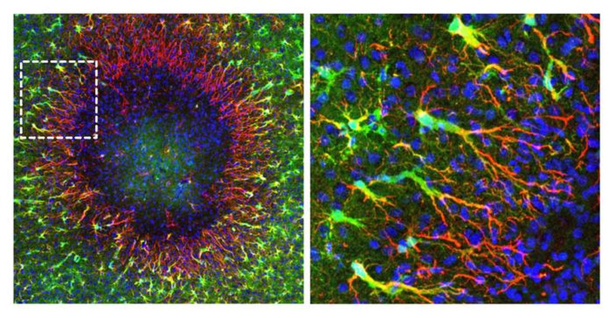
The image on the left shows a brain lesion (diameter: just under 0.5 mm). Around the perimeter of the lesion, the newly discovered “regenerative” astrocytes begin to seal the defect by forming long cellular extensions (shown in red). Newly formed cell nuclei (shown in blue) migrate along the cellular extensions toward the damaged area. Unaltered astrocytes (shown in green) surround the lesion area. The image on the right shows an enlargement of the marked area in the left image. Credit: Institute of Pharmacology and Toxicology, University of Zurich
Regenerative astrocytes repair damaged tissue
A new study by co-lead authors Marina Herwerth and Matthias Wyss from the Institute of Pharmacology and Toxicology at the University of Zurich (UZH) has now overturned that assumption: their research team headed by Bruno Weber discovered a specialized group of “regenerative” astrocytes in the brains of living mice that step in on the perimeter of the damaged area of the brain to rebuild the cells.
“The findings of our study reveal a previously unknown ability of the adult brain to repair itself. They point toward new ways of supporting recovery from ailments involving the loss of astrocytes,” Weber says.
Only cell nuclei migrate
The researchers used two-photon microscopy to observe the brains of living mice in real time over a period of several weeks and mapped which genes switch on in which areas of the brain. This way they were able to identify the special astrocytes that take care of rebuilding injured tissue. But those cells don’t just divide, they also perform a remarkable feat: “they send the newly formed nuclei of their daughter cells gliding across long distances to repopulate the damaged area of the brain and knit the astrocyte network back together,” Weber explains.
Starting points for targeted regeneration
The discovery of how adult brain cell nuclei migrate through the long star-shaped extensions of astrocytes to injured tissue expands comprehension of how the brain organizes and regenerates itself after certain injuries. If those mechanisms can be selectively activated, that could help to more effectively repair damaged brain tissue, restore astrocyte networks and thus improve recovery after certain brain disorders.
“We were able to identify numerous genes and signaling pathways that are temporarily activated during repair. They could serve as starting points in the future for influencing post-disease and -injury regeneration processes,” Weber stresses.
Key Questions Answered:
Q: How do “regenerative” astrocytes differ from standard cell division during tissue repair?
A: Instead of whole cells migrating or simply dividing locally, these specialized astrocytes remain at the perimeter of the damaged area. They divide and send the newly formed nuclei of their daughter cells gliding long distances through their extended cellular processes directly into the injured zone to rebuild the network.
Q: What conditions cause the loss of astrocytes in the adult brain?
A: Astrocytes are lost during traumatic brain injuries, strokes, and specific neuroinflammatory or autoimmune conditions, most notably Neuromyelitis Optica Spectrum Disorder (NMOSD), where the body’s immune system produces autoantibodies that target and destroy astrocytes.
Q: How was this nuclear migration observed in real time?
A: Researchers at the University of Zurich used in vivo two-photon microscopy in living mouse models over several weeks. This allowed them to track living cells, observe nuclear movement through astrocytic branches, and map corresponding gene expression changes as the tissue repaired itself.
Editorial Notes:
This article was edited by a Neuroscience News editor.
Journal paper reviewed in full.
Additional context added by our staff.
About this neuroscience research news
Author: Kurt Bodenmueller Source: University of Zurich Contact: Kurt Bodenmueller – University of Zurich Image: The image is credited to Institute of Pharmacology and Toxicology, University of ZurichOriginal Research: Open access.
“Focal astrocyte loss reveals nuclear translocation during lesion repopulation” by Marina Herwerth, Matthias T. Wyss, Nicola B. Schmid, Anna Lasne, Jacqueline Condrau, Luca Ravotto, José María Mateos Melero, Andres Kaech, Gustav Bredell, Carolina Thomas, Rachel Kim, Petra Kukanja, Vladyslav L. Korobeynyk, Christine Stadelmann, Thomas Misgeld, Jeffrey L. Bennett, Sebastian Jessberger, Aiman S. Saab, Shane A. Liddelow & Bruno Weber. Nature Neuroscience DOI:10.1038/s41593-026-02354-5
A competent? doctor and hospital WOULD INSURE HUMAN TESTING OCCURS! Do you have competence among in your stroke medical 'professionals'?
Do you prefer your doctor, hospital and board of director's incompetence NOT KNOWING? OR NOT DOING? Your choice; let them be incompetent or demand action!
Summary: Researchers
discovered a previously unknown mechanism through which the adult
mammalian brain repairs itself following focal injuries or autoimmune
damage. Using two-photon microscopy and longitudinal gene mapping in
living mouse models, researchers identified a specialized population of
“regenerative” astrocytes capable of repopulating damaged brain regions.
Rather
than relying solely on classical cell body division at the site of
injury, these specialized astrocytes situated along the lesion perimeter
send newly formed daughter cell nuclei gliding long distances through
their star-shaped cellular extensions. These migrating cell nuclei
repopulate the depleted lesion zone, re-establishing functional
astrocyte networks.
This discovery
overturns long-held assumptions regarding the limited regenerative
capacity of adult glial networks, revealing molecular signaling pathways
that could serve as therapeutic targets for traumatic brain injury and
autoimmune conditions such as neuromyelitis optica spectrum disorder
(NMOSD).
Key Facts
Overturning Dogma on Glial Regeneration:
Demonstrates that the adult central nervous system possesses a
previously unrecognized capability to replace lost astrocytes and
restore damaged tissue architecture.
Mechanism of Long-Distance Nuclear Migration:
Specialized regenerative astrocytes remain at the lesion boundary and
send newly generated cell nuclei gliding across long astrocytic
extensions into the depleted injury core.
Rebuilding Functional Glial Networks:
Astrocytes perform vital homeostatic functions, including nutrient
supply to neurons, blood flow regulation via end-feet, and extracellular
ion balance, making their network reconstruction essential for neuronal
survival.
Targeted Clinical Applications:
Holds therapeutic relevance for neurotraumatic brain injuries and rare
autoimmune conditions like neuromyelitis optica spectrum disorder
(NMOSD), where autoantibodies selectively destroy astrocytes.
Molecular Targets for Therapeutics:
The team identified specific genes and signaling pathways temporarily
activated during nuclear migration, providing potential targets for
pharmacological interventions to accelerate brain repair.
Source: University of Zurich
The
brain evidently can regenerate itself better than previously assumed
after injuries or certain autoimmune diseases. Using a mouse model,
researchers at the University of Zurich have demonstrated that special
supporting and nourishing cells repopulate damaged areas of the brain by
initially sending only newly formed cell nuclei there.
Glial
cells are supporting and nourishing cells in the brain. Star-shaped
glial cells called astrocytes are vital to the functioning of neurons.
They supply the nerve cells with nutrients, help to regulate blood flow
and keep brain tissue healthy.
It had
long been assumed that when astrocytes are lost – as happens, for
instance, in brain injuries or autoimmune diseases such as rare
neuromyelitis optica spectrum disorder, in which the body’s own
antibodies destroy these cells – the adult brain cannot fully replace
them.
Regenerative astrocytes repair damaged tissue
A
new study by co-lead authors Marina Herwerth and Matthias Wyss from the
Institute of Pharmacology and Toxicology at the University of Zurich
(UZH) has now overturned that assumption: their research team headed by
Bruno Weber discovered a specialized group of “regenerative” astrocytes
in the brains of living mice that step in on the perimeter of the
damaged area of the brain to rebuild the cells.
“The findings of our study reveal a previously unknown ability of the
adult brain to repair itself. They point toward new ways of supporting
recovery from ailments involving the loss of astrocytes,” Weber says.
Only cell nuclei migrate
The
researchers used two-photon microscopy to observe the brains of living
mice in real time over a period of several weeks and mapped which genes
switch on in which areas of the brain. This way they were able to
identify the special astrocytes that take care of rebuilding injured
tissue. But those cells don’t just divide, they also perform a
remarkable feat: “they send the newly formed nuclei of their daughter
cells gliding across long distances to repopulate the damaged area of
the brain and knit the astrocyte network back together,” Weber explains.
Starting points for targeted regeneration
The
discovery of how adult brain cell nuclei migrate through the long
star-shaped extensions of astrocytes to injured tissue expands
comprehension of how the brain organizes and regenerates itself after
certain injuries. If those mechanisms can be selectively activated, that
could help to more effectively repair damaged brain tissue, restore
astrocyte networks and thus improve recovery after certain brain
disorders.
“We were able to identify
numerous genes and signaling pathways that are temporarily activated
during repair. They could serve as starting points in the future for
influencing post-disease and -injury regeneration processes,” Weber
stresses.
Key Questions Answered:
Q: How do “regenerative” astrocytes differ from standard cell division during tissue repair?
A:
Instead of whole cells migrating or simply dividing locally, these
specialized astrocytes remain at the perimeter of the damaged area. They
divide and send the newly formed nuclei of their daughter cells gliding
long distances through their extended cellular processes directly into
the injured zone to rebuild the network.
Q: What conditions cause the loss of astrocytes in the adult brain?
A:
Astrocytes are lost during traumatic brain injuries, strokes, and
specific neuroinflammatory or autoimmune conditions, most notably
Neuromyelitis Optica Spectrum Disorder (NMOSD), where the body’s immune
system produces autoantibodies that target and destroy astrocytes.
Q: How was this nuclear migration observed in real time?
A:
Researchers at the University of Zurich used in vivo two-photon
microscopy in living mouse models over several weeks. This allowed them
to track living cells, observe nuclear movement through astrocytic
branches, and map corresponding gene expression changes as the tissue
repaired itself.
Editorial Notes:
This article was edited by a Neuroscience News editor.
Journal paper reviewed in full.
Additional context added by our staff.
About this neuroscience research news
Author: Kurt Bodenmueller Source: University of Zurich Contact: Kurt Bodenmueller – University of Zurich Image: The image is credited to Institute of Pharmacology and Toxicology, University of Zurich
Original Research: Open access. “Focal astrocyte loss reveals nuclear translocation during lesion repopulation”
by Marina Herwerth, Matthias T. Wyss, Nicola B. Schmid, Anna Lasne,
Jacqueline Condrau, Luca Ravotto, José María Mateos Melero, Andres
Kaech, Gustav Bredell, Carolina Thomas, Rachel Kim, Petra Kukanja,
Vladyslav L. Korobeynyk, Christine Stadelmann, Thomas Misgeld, Jeffrey
L. Bennett, Sebastian Jessberger, Aiman S. Saab, Shane A. Liddelow &
Bruno Weber. Nature Neuroscience DOI:10.1038/s41593-026-02354-5
Have your competent? doctor decipher this since choline is helpful in brain health and reducing your dementia risk. Your doctor better know of all these 17 research articles! OR COMPLETE FUCKING INCOMPETENCE!
A 5,090-person cohort and complementary mouse experiments trace how a diet-linked microbial pathway could reshape atrial structure, electrical activity, and autonomic control.
A recent study published in The Journal of Clinical Investigation suggests that trimethylamine N-oxide (TMAO), produced when gut microbes convert nutrients such as choline into trimethylamine (TMA), which is then oxidized in the liver, is independently associated with prevalent atrial fibrillation (AF) in humans and may promote AF susceptibility, onset, and progression in mice. By inhibiting muscarinic receptor 2 signaling, TMAO may disrupt autonomic regulation, with increased sympathetic tone proposed as one mechanism contributing to AF. These findings suggest that diet, through its effects on the gut microbiome and TMAO production, may influence AF susceptibility, although dietary effects were tested only in mice, and the human findings came from a cardiovascular referral cohort.
AF remains a major contributor to cardiovascular disease (CVD)-related illness and death worldwide. Elevated TMAO levels have been associated with CVD-related changes, including cardiac fibrosis and inflammation. However, the biological mechanisms through which TMAO may promote AF remain unclear. An improved understanding of the gut microbiome-related changes that influence AF development could help researchers develop more targeted treatments to reduce the global burden of AF and CVD. Future studies will need to determine whether therapeutic strategies targeting TMAO can safely reduce AF and how differences in gut microbiome composition, liver metabolism, and kidney filtration affect circulating TMAO levels.
About the study
In the present study, researchers investigated whether TMAO generated through the gut microbiome could promote AF development. To do so, they quantified TMAO, choline, and betaine levels in plasma samples obtained from 5,090 individuals from the Cleveland Clinic GeneBank who were undergoing elective cardiac catheterization, using liquid chromatography-electrospray ionization-tandem mass spectrometry (LC-ESI-MS/MS). Individuals with a myocardial infarction during the preceding four weeks or elevated troponin I at enrollment were excluded.
The team also used genetically engineered mice expressing the CREM-IbΔC-X variant of the human cyclic adenosine monophosphate (cAMP) response element modulator gene to investigate AF development. These animals received TMAO- or choline-supplemented diets in separate experiments. The researchers conducted transesophageal electrical pacing studies to determine AF inducibility among wild-type C57BL/6J mice fed TMAO-supplemented or standard diets. They placed electrodes in the esophagus of the animals to deliver electrical impulses and promote arrhythmias in the heart. They separately used serial needle-electrode electrocardiograms (ECGs) to monitor the first onset of paroxysmal AF and progression to persistent AF, defined as AF detected across 10 consecutive ECG recordings, in CREM-IbΔC-X mice receiving different diets, with wild-type mice included in separate control experiments.
The team also explored the effects of iodomethylcholine (IMC), a selective inhibitor of choline trimethylamine-lyase (CutC/D), on TMAO levels. They analyzed microbial DNA from cecal samples, used echocardiography to assess the effects of choline and IMC on cardiac structure, and performed cardiac electrical mapping to examine the electrophysiological effects of choline supplementation. They exposed human and murine cardiac cells, including fibroblasts and cardiomyocytes, to physiologically relevant TMAO levels and examined interleukin-1β (IL-1β) and NLRP3 expression. They also performed murine ECG experiments using MCC950, a chemical compound that inhibits the NOD-, LRR- and pyrin domain-containing protein 3 (NLRP3) inflammasome.
The researchers used logistic regression to estimate odds ratios (ORs) for prevalent AF. The models accounted for variables such as age, sex, smoking habits, comorbidities, and laboratory findings, including high-sensitivity C-reactive protein (hs-CRP) and estimated glomerular filtration rate (eGFR).
Results
The team found that plasma TMAO, betaine, and choline levels were independently and significantly associated with AF prevalence. For TMAO, the adjusted odds ratio comparing the highest with the lowest concentration tertile was 1.7 (95% confidence interval, 1.3-2.1). The TMAO- or choline-supplemented CREM-IbΔC-X mice developed AF earlier than chow-fed controls, without significant differences in body mass or appreciable liver-related pathologies.
Viva Saliva Book: The ultimate guide to saliva Book preview Download eBookCompilation of the top interviews, articles, and news in the last year.Download the latest edition
The genetically modified animals also showed higher plasma TMAO levels after choline supplementation. These findings suggest that TMAO may contribute to AF onset and progression in mice, and that reducing gut microbial production of TMAO may help lower AF susceptibility in this model. Mice fed TMAO diets showed an increased likelihood of developing AF in the transesophageal pacing study. In fact, mice receiving TMAO supplementation showed an 11-fold increase in AF inducibility in both sexes compared with controls.
Choline supplementation altered the structure and function of the heart. Left atrial size was significantly increased at five and eight weeks, while IMC treatment attenuated the enlargement at eight weeks. In choline-supplemented mice, optical mapping revealed a nonsignificant reduction in conduction velocity but significantly shortened action-potential duration at 80% repolarization and reduced cardiac wavelength. In HEK293 cells engineered to express muscarinic receptor 2 (M2R), TMAO inhibited receptor signaling in the presence of the M2R agonist carbachol. Together with higher heart rates in choline-fed mice, this finding supported a possible role for autonomic dysfunction in promoting AF, although sympathetic activity was not directly measured.
While choline accelerated AF onset, IMC treatment delayed the onset of paroxysmal and persistent AF and reduced TMAO levels by suppressing the microbial conversion of choline to TMA under both aerobic and anaerobic conditions, thereby reducing subsequent TMAO formation in the liver. Choline altered gut microbial communities in association with AF. IMC, on the other hand, attenuated these changes by reversing the loss of gut microbiome diversity and reducing the choline-associated increase in overall Firmicutes abundance, although individual species showed differing patterns. An exploratory analysis also linked Parvibacter caecicola to the timing of paroxysmal AF onset in mice, although the authors noted that this association requires further investigation. TMAO also did not increase NLRP3 or IL-1β expression at physiologically relevant concentrations, and MCC950 did not delay AF development, suggesting that NLRP3 inflammasome activation was not a major mechanism in this mouse model.
Conclusions
The findings demonstrate that higher plasma TMAO levels were independently associated with prevalent AF in humans, whereas direct TMAO exposure or gut microbial production of TMA from dietary choline promoted AF onset and progression in mouse models. However, the human analysis was observational and assessed existing rather than incident AF, while the mechanistic and therapeutic findings came from mice and cell experiments. No human dietary or IMC intervention was tested, and AF recurrence following IMC withdrawal was not examined.
Together, the preclinical findings support clinical investigation of TMAO-lowering approaches as potential strategies for AF prevention. In future studies, researchers should explore different molecules that can reduce TMAO levels and determine how gut microbiome composition, hepatic FMO3 activity, and renal clearance influence circulating TMAO levels and treatment responses in humans.
Journal reference:
Arjunan, S. et al. (2026). Gut microbial trimethylamine N-oxide generation promotes risk of atrial fibrillation via muscarinic receptor-mediated autonomic dysfunction. The Journal of Clinical Investigation. DOI: 10.1172/JCI201684, https://www.jci.org/articles/view/201684
After your competent? doctor gets human testing going then s/he can create the protocols that fill those cavities with axon pathfinding and dendritic branching! Not understanding any of this IS PURE INCOMPETENCE from your doctor!
An injectable biomaterial turned stroke-damaged areas into
hubs of repair, helping mouse brains grow new blood vessels and nerve
fibers while restoring near-normal movement.
A stroke can
leave behind more than damaged brain cells. In severe cases, it creates
an empty cavity where living tissue once carried signals, supplied
blood, and controlled movement. Duke University researchers are now testing an injectable material designed to turn that biological void into a place where repair can begin.
In
mice, the treatment drew immune cells into the stroke cavity and helped
organize them into a coordinated healing response. New blood vessels
spread through the injured area, nerve fibers became more abundant, and
the animals regained motor abilities that approached those of healthy
mice.
The findings were published in Cell Biomaterials.
The material was injected directly into the damaged region five days
after the stroke, meaning it was tested as a repair strategy rather than
an emergency treatment.
Why Stroke Damage Is So Hard to Repair
Most
strokes occur when a clot cuts off blood flow to part of the brain.
Clot-dissolving drugs and procedures that physically remove the blockage
can save threatened tissue when delivered quickly. Once brain cells
have died, however, restoring circulation cannot bring them back.
A
major ischemic stroke may destroy enough tissue to leave a fluid-filled
cavity. Rehabilitation can train surviving brain networks to take on
new roles, but medicine currently has no established way to reconstruct
the missing region itself.
“Once brain tissue has been lost,
restoring blood flow is no longer enough,” said Tatiana Segura, the
Robert Plonsey Distinguished Professor of Biomedical Engineering at
Duke. “Our goal is to engineer the injured space so that immune,
vascular, and neural repair processes can begin to work together.”
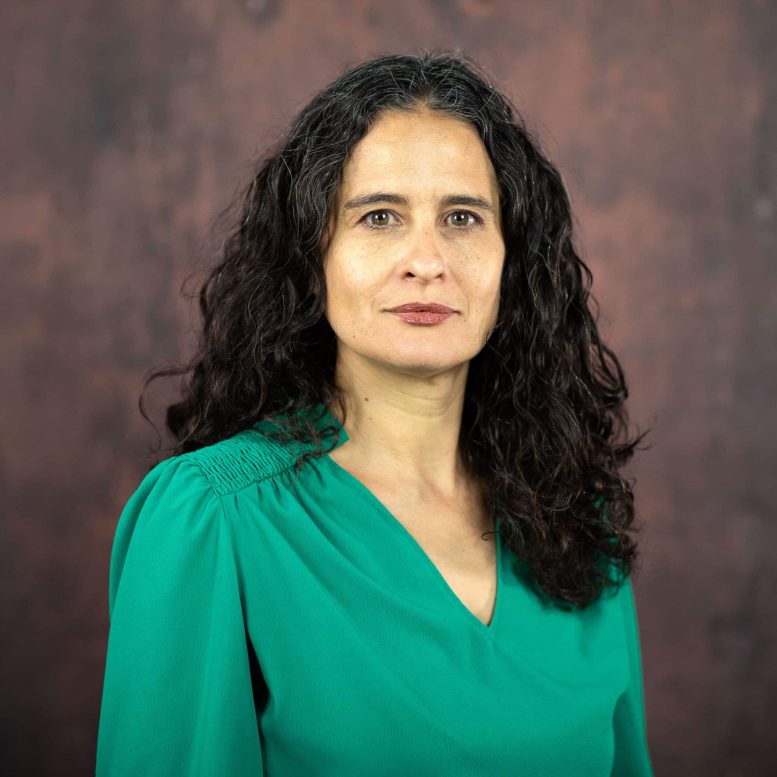
Tatiana Segura. Credit: Duke University
An Injectable Scaffold for Brain Repair
Rather
than trying to manufacture replacement brain tissue, Segura’s team
developed a temporary framework that encourages the body to do more of
the rebuilding itself.
The treatment is based on MAPS, or
microporous annealed particle scaffolds. These injectable materials are
assembled from tiny hydrogel particles that connect after delivery while
leaving open spaces between them. Unlike a solid gel, the porous
structure gives cells room to enter, move, and form new tissue.
Microporous scaffolds can support cellular infiltration and blood vessel
growth without waiting for the entire material to break down first.
Those
instructions came from astrocytes, star-shaped cells that support
neurons, help regulate the brain’s environment, and react rapidly to
injury. Astrocytes communicate partly by releasing extracellular
vesicles, or EVs. These nanoscale packages transport proteins, lipids, and genetic material between cells.
Researchers
grew astrocytes in the laboratory and exposed them to different
signaling molecules. They then collected the EVs produced under those
conditions and tested whether the packages could attract immune cells
and encourage tissue repair.
Simply releasing EVs into the damaged
brain would allow many of them to disperse. To keep the signals where
they were needed, the researchers chemically attached the vesicles to
the hydrogel particles.
Turning the Scaffold Into a Signaling Hub
This
design transformed the scaffold into more than a physical support. It
became a localized signaling hub where incoming cells could repeatedly
encounter molecular instructions.
“We are not simply placing a
material into the brain,” Segura said. “We are engineering a local
environment that can coordinate several parts of the repair response.”
EVs
produced after astrocytes were exposed to IL-4 and C1q generated the
strongest results. The combination attracted macrophages and a
surprisingly persistent population of neutrophils into the stroke
cavity.
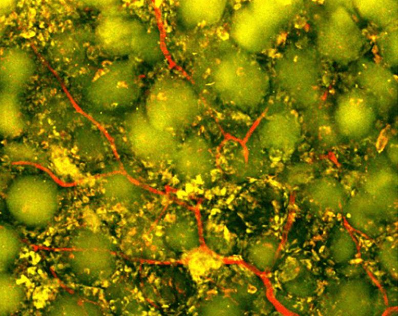
Two-photon
imaging at day 27 using Ly6G-green fluorescent protein (GFP) reporter
mice to visualize neutrophils. IL-4/C1q-EV + MAPS implants exhibited
dense vascularization and focal accumulation of GFP+ cells within
scaffold pores. Credit: Duke University
Immune Cells Take on a Surprising Role
Neutrophils
are among the immune system’s fastest responders. After a stroke, they
are often associated with inflammation and additional tissue damage,
especially during the early phase of injury. Yet immune cells do not
always have a single fixed role. Their behavior can change depending on
timing, location, and the molecular signals surrounding them.
Inside
the engineered scaffold, neutrophils appeared to become part of the
repair process rather than merely contributing to destruction.
The
researchers tested that possibility by depleting the immune-cell
population rich in neutrophils. Blood vessel formation dropped sharply,
and the scaffold underwent far less remodeling. The experiment showed
that these cells were not simply present at the injury site. They were
helping drive the response.
“This result changes how we think
about neutrophils after stroke,” said Shangjing Xin, lead scientist of
the study and a postdoctoral fellow in the Segura Laboratory. “Their
role appears to depend on when they arrive, where they are located, and
the signals they receive from their surroundings. Our study demonstrates
a potential engineering strategy to recruit and retain these cells at
the right time.”
New Blood Vessels and Nerve Fibers Emerge
The
treatment produced visible changes throughout the damaged region. Blood
vessels grew across the cavity, potentially creating the circulation
needed to support living tissue. Researchers also detected more axonal
fibers within and around the injury. Axons are the long projections
neurons use to carry electrical signals to other cells.
Those biological changes were accompanied by improved movement.
During
a grid-walking test, scientists measured how often the mice misplaced a
front paw while crossing an uneven surface. Animals treated with the
optimized scaffold made fewer errors over time. By eight weeks, their
performance could not be statistically distinguished from that of
healthy control mice, and the improvement continued through the end of
the study.
The scaffold itself proved essential. When researchers
delivered the EVs without MAPS, they did not observe comparable blood
vessel growth. The result suggests that the treatment depended on both
components: the biological messages carried by the vesicles and the
porous structure that concentrated those messages while giving cells
space to organize.
Toward a Scalable Human Stroke Therapy
The
study relied on EVs collected from primary rat astrocytes, which would
not be a practical source for a widely available human therapy.
Segura’s
laboratory is now exploring astrocytes made from human-induced
pluripotent stem cells. These cells can be produced from reprogrammed
adult cells and expanded in the laboratory, potentially offering a more
scalable and clinically relevant source of EVs. Researchers may also be
able to adjust the conditions under which the astrocytes grow to better
control the messages their vesicles carry.
“You do not restore an
ecosystem simply by containing the initial damage,” Segura said. “You
have to create the conditions that allow life to return. That is how we
think about the stroke cavity. The material is not intended to reproduce
the brain itself but to create an environment where the body’s own
cells can enter, communicate, and participate in rebuilding vascularized
tissue.”
Reference: “IL-4/C1q activated astrocyte-derived
extracellular vesicles promote stroke infarct recovery by recruiting
peripheral leukocytes” by Shangjing Xin, Lucy Zhang, Nhi V. Phan,
Mengying An, Ligen Shi, S. Thomas Carmichael and Tatiana Segura, 21 July
2026, Cell Biomaterials. DOI: 10.1016/j.celbio.2026.100543
Do you really think anyone in stroke is competent enough to get human testing going? I don't, everything in stroke IS A COMPLETE FUCKING FAILURE! Prove me wrong; failure is defined as not getting to 100% recovery! Don't try your tyranny of low expectations on me. Here; oc1dean@gmail.com, I'll print it verbatim with my reply. Have at it, or are you afraid to engage with a stroke-addled survivor?
Of course your competent? doctor can inform these researchers of earlier work.
When someone has a stroke caused by a blood clot, doctors can quickly restore blood flow. However, they can’t easily replace the brain tissue that gets lost. Recovering this tissue usually means relying on rehab to help the remaining brain circuits adapt.
A team of biomedical engineers at Duke University has built an injectable biomaterial that could change stroke recovery.
Rebuilding the Brain’s Neighborhood
The research team isn’t attempting to rebuild the brain directly. Instead, they are setting up a scaffolding system to let the body do the work for them. They achieve this system using tiny hydrogel microparticles called MAPS. When the material is injected into the cavity caused by a stroke, it creates a porous structure for cells to grow on.
“Once brain tissue has been lost, restoring blood flow is no longer enough,” said Tatiana Segura, the Robert Plonsey Distinguished Professor of Biomedical Engineering at Duke. “Our goal is to engineer the injured space so that immune, vascular, and neural repair processes can begin to work together.”
The team attached specific signals to these particles so that the body’s immune cells could help. These signals come from astrocytes, which are star-shaped cells in the brain.
“We are not simply placing a material into the brain,” Segura added. “We are engineering a local environment that can coordinate several parts of the repair response.”
Surprising Helpers
The team found that certain signaling molecules attracted helpful immune cells, including the most common type of white blood cell: neutrophils. Usually, neutrophils cause inflammation right after a stroke. However, that wasn’t the case in this scenario.
“This result changes how we think about neutrophils after stroke,” said Shangjing Xin, lead scientist of the study and a postdoctoral fellow in the Segura Laboratory. “Their role appears to depend on when they arrive, where they are located, and the signals they receive from their surroundings. Our study demonstrates a potential engineering strategy to recruit and retain these cells at the right time.”
In mouse tests, this treated scaffold helped grow new blood vessels and improved movement. By eight weeks, the mice performed like healthy controls on a coordination test.
Right now, the work is still preclinical. The team is looking at using human cells next to make it scalable.
“You do not restore an ecosystem simply by containing the initial damage. You have to create the conditions that allow life to return. That is how we think about the stroke cavity,” Segura said. “The material is not intended to reproduce the brain itself, but to create an environment where the body’s own cells can enter, communicate and participate in rebuilding vascularized tissue.”