Neurological conditions like hemiplegia following stroke
or tetraplegia following spinal cord injury, result in a massive
compromise in motor function. Each of the two conditions can leave
individuals dependent on caregivers for the rest of their lives. Once
medically stable, rehabilitation is the main stay of treatment. This
article will address rehabilitation of upper extremity function. It is
long known that moving the affected limb is crucial to recovery
following any kind of injury. Overtime, it has also been established
that just moving the affected extremities does not suffice, and that the
movements have to involve patient’s participation, be as close to
physiologic movements as possible, and should ideally stimulate the
entire neuromuscular circuitry involved in producing the desired
movement. For over four decades now, functional electrical stimulation
(FES) is being used to either replace or retrain function. The FES
therapy discussed in this article has been used to retrain upper
extremity function for over 15 years. Published data of pilot studies
and randomized control trials show that FES therapy produces significant
changes in arm and hand function. There are specific principles of the
FES therapy as applied in our studies: (i) stimulation is applied using
surface stimulation electrodes, (ii) there is minimum to virtually no
pain during application, (iii) each session lasts no more than 45–60
min, (iv) the technology is quite robust and can make up for specificity
to a certain extent, and (v) fine motor function like two finger
precision grip can be trained (i.e., thumb and index finger tip to tip
pinch). The FES therapy protocols can be successfully applied to
individuals with paralysis resulting from stroke or spinal cord injury.
Introduction
Application of functional electrical stimulation (FES)
for therapeutic purposes in rehabilitation settings dates back to the
1960’s when
Liberson et al. (1961)
used an FES system to stimulate the peroneal nerve to correct foot drop
by triggering a foot switch, a single-channel electrical stimulation
device stimulated the common peroneal nerve via a surface electrode,
producing ankle dorsiflexion during the swing phase of gait (
Liberson et al., 1961).
This led to the first commercially available FES system with surface
stimulation electrodes. Since then FES technology has been researched
extensively to evaluate its benefits in diverse neurological conditions,
and using an array of application techniques (
Baldi et al., 1998;
Field-Fote, 2001;
Popovic et al., 2005,
2011,
2012,
2016;
Yan et al., 2005;
Frotzler et al., 2008;
Griffin et al., 2009;
Daly et al., 2011;
Kapadia et al., 2011,
2013,
2014a;
Giangregorio et al., 2012;
Malešević et al., 2012;
Martin et al., 2012;
Kawashima et al., 2013;
Lee et al., 2013;
Sadowsky et al., 2013;
Ho et al., 2014;
Kapadia N. et al., 2014;
Popović, 2014;
Sharif et al., 2014;
Bauer et al., 2015;
Howlett et al., 2015;
Vafadar et al., 2015;
Buick et al., 2016;
Cuesta-Gómez et al., 2017;
Fu et al., 2019;
Straudi et al., 2020).
The two common uses of FES are to replace function (i.e., as an
orthotic device) and to retrain function (i.e., as a therapeutic
device). In this article we will limit ourselves to the therapeutic
application of FES.
In the therapeutic application (FES therapy), FES is
used as a short-term treatment modality. The expectation is that, after
training with the FES system, the patients will be able to voluntarily
perform the trained activities without FES (i.e., patients are expected
to regain voluntary function). To date, a few high-quality randomized
controlled trials have been performed, proving the efficacy of FES
therapy over other rehabilitation techniques (
Sharififar et al., 2018;
Yen et al., 2019).
This paucity in multicenter randomized controlled trials and the
limited access to systems that can properly deliver FES therapy might
have affected its uptake in clinical settings (
Ho et al., 2014;
Auchstaetter et al., 2016).
Fortunately, both these issues are being addressed as new FES systems
that are specifically developed for FES therapy are being introduced, as
well as large scale multicenter randomized controlled trials are being
planned to further confirm the efficacy of this rehabilitation modality.
This article will provide readers with the details on how
transcutaneous multichannel FES therapy for the upper extremity can be
applied in clinical trials and as such the same methodology can be used
in clinical practice by physiotherapists and occupational therapists.
The FES methodology discussed here has been developed
with the intent to be user friendly, robust and to be able to produce
better functional gains than the presently available best-practice
rehabilitation techniques. The FES system used in our laboratory is a
surface stimulation system with up to 4 stimulation channels that can
produce gross motor function as well as precision grips such as two
finger pinch grip. However, the methodology of FES application discussed
here is pertinent to any multichannel transcutaneous FES device. We
have used transcutaneous FES to retrain reaching and grasping in
individuals with both spinal cord injury and stroke (
Thrasher et al., 2008;
Kapadia and Popovic, 2011;
Kapadia et al., 2011,
2013;
Popovic et al., 2012;
Hebert et al., 2017).
The results obtained in both patient populations indicate functional
improvements after 8–14 weeks of therapy (20–48 h of stimulation).
Patients showed reduced dependency on caregivers, and some even became
independent in their activities of daily living.
This article will extensively detail how FES was applied
in these previously successful clinical trials to retrain reaching and
grasping functions in individuals who sustained a spinal cord injury or a
stroke.
Materials and Equipment
The FES system we used was a four channel surface
stimulation device consisting of a software, a portable stimulator with a
programmed chip card, self-adhesive stimulation electrodes, and various
man-machine interfaces, such as push buttons, sliding potentiometers (
Mangold et al., 2005), accelerometers (
Widjaja et al., 2011), EMG/biofeedback sensor, joysticks (
Sayenko et al., 2013), foot switches (
Popovic et al., 2001b), gait phase detection system (
Pappas et al., 2004) and brain–machine interface (
Márquez-Chin et al., 2009).
This FES system has been extensively used in clinical trials by
researchers both in North America and in Europe. Its unique capability
is the ability to program stimulation protocols customized to a
patient’s needs in less than 10–15 min.
Software
The software of our FES system allows one to
specify/alter all stimulation parameters: frequency, minimum and maximum
intensity, pulse duration, ramp time, synchronization and order of
stimulations, type of user interactions and number of repetitions. The
sensory, motor, functional and maximum thresholds are set using the
continuous stimulation mode where the stimulation frequency and pulse
duration are pre-set to values of 40 Hz and 200 μs, respectively.
Methods
Clinical Applications
To date, approximately 150 spinal cord injury and 50
stroke patients have been treated using transcutaneous FES in our
facilities, ranging from pilot clinical trials to randomized controlled
trials. The FES system has been primarily used as a therapeutic device
for retraining reaching and grasping. More recently FES was successfully
applied to an individual with cervical spondylotic myelopathy to
retrain upper extremity function with very promising results (
Popovic et al., 2016).
Neuroprosthesis for Grasping in Spinal Cord Injury Patients (University Health Network REB # 02-032, REB # 09-007, REB # 09-008)
In case of patients with spinal cord injury the upper
extremity retraining program is designed based on the level and extent
of injury. For example, in C1–C5 cervical incomplete injuries initially
FES might be used to retrain proximal function and then once the patient
is able to position their arm in the working space then distal function
can be trained. The FES protocols for retraining proximal function in
SCI remain the same as stroke (please refer to the next section on
stroke for details). In patients with low cervical injury (C6 and
below), proximal upper extremity function might be preserved, and FES
might then be used to retrain distal function right from the beginning.
Also, it is important to note that again based on level of injury
patient with SCI may have difficulty with both hand closing and opening
and will typically need to be trained for both.
Over the years, various grasping protocols have been
identified and designed allowing for a wide variety of grasping patterns
to be trained with a great deal of fidelity. Currently, the grasping
patterns that can be successfully retrained using a transcutaneous multi
-channel FES system are:
(1) Palmar Grasp (holding a ball)
(2) Lateral Grasp (holding a tray)
(3) Tripod grip (thumb, index, and middle finger: holding a pen)
(4) Two finger opposition (thumb and index finger: holding a peg)
(5) Lateral Pinch (thumb and index finger: holding a credit card)
(6) Two finger lateral pinch (index and middle finger: smoker’s grip)
(7) Lumbrical grip (all four fingers with the thumb: holding a closed book).
It is important to mention that FES therapy has the
capability to help stroke and spinal cord injury patients relearn how to
voluntarily perform all of the above grasps bilaterally and
simultaneously, using surface FES system.
We have conducted a number of clinical studies using
this FES technology the most recent one being a randomized controlled
trial in incomplete cervical SCI patients (
Popovic et al., 2011).
Individuals allocated to the intervention group in this trial received
FES stimulation protocols specifically designed for their upper
extremity functional deficits. Individualized stimulation sequences were
developed for each patient. The commonly trained grasping patterns were
power and precision grasp where power grasp was used mainly to grasp
larger everyday objects and the precision grip was used mainly to
manipulate smaller objects. Power grasp was generated by partly flexing
the fingers and the thumb in flexion and slight opposition. Lateral
pinch was generated by fully flexing the fingers followed by the thumb
flexion. Muscles that were stimulated during therapy were the following:
• Wrist flexors: flexor carpi radialis and flexor carpi ulnaris;
• Wrist extensors: extensor carpi radialis (longus and brevis) and ulnaris;
• Finger flexors: flexor digitorum superficialis and flexor digitorum profundus;
• Finger extensors: extensor digitorum;
• Thumb abductors: median nerve, or abductor pollicis brevis and longus;
• Thumb flexors: flexor pollicis brevis and flexor pollicis longus;
• Thumb oppositors: opponens pollicis;
• Metacarpophalangeal flexors and interphalangeal joint extensors: lumbricals.
The FES protocol allowed for individuals with little to
no voluntary movement at the wrist and fingers to be able to perform
simple tasks while being stimulated with the FES. This is what
differentiates FES from other therapies. In the early stages of FES
therapy, all the movements were performed with the help of FES. The
treatment plan and instruction to participants were as follows:
(1) “Imagine hand opening” (or any movement that the therapist would like to train).
(2) “Try to perform the movement using your own muscle strength.”
(3) After trying for about 10 s: “Now, try to perform the movement with the help of FES.”
Hence, emphasis was put on participants voluntarily
attempting the movement while being stimulated with the FES. During
therapy when the participants started showing an ability to voluntarily
contract certain muscle groups FES for those muscle groups was reduced
to a minimum and gradually withdrawn completely. The available channel
was then used on other muscle groups that were still weak and needed to
be trained. The order in which muscle groups were sequentially
“reactivated” was subject-dependent. FES was always delivered while the
participants were performing functional tasks, such as grasping a mug,
pouring water, holding a pen, etc.
The distinctiveness of this intervention is that FES is
not primarily intended for muscle strengthening. Instead, it is used to
retrain the neuromuscular system to execute tasks that it is unable to
carry out voluntarily. Movements were performed against gravity and
sometimes against light manual resistance. The number of repetitions was
determined based on individual participant’s strength and endurance. In
general, all participants spent 30–45 min out of 1-h session performing
activities of daily living with FES. The stimulation parameters used
were the following: (a) balanced, biphasic, current regulated electrical
pulses; (b) pulse amplitude from 8 to 50 mA (typical values 15–30 mA);
(c) pulse width 250 μs; and (d) pulse frequency 40 Hz (
Popovic et al., 2011).
During the intervention, the therapist, at their discretion, adjusted
the placement of electrodes and guided the hand movements. The therapist
ensured that the movements were functional. Occasionally FES would be
combined with conventional rehabilitation strategies including
strengthening exercises, stretching exercises, etc.
Neuroprosthesis for Grasping in Stroke Patients (University Health Network REB # 02-032).
The most important difference between FES training in
spinal cord injury and stroke patients; is that stroke patients have
difficulty opening their hand as they often exhibit flexor synergy and
high levels of tone in the finger flexors. In stroke patients therefore,
the focus of the therapy is on hand opening and relaxing the fingers.
In spinal cord injury patient’s the focus of the FES therapy is on
finger flexion and grasping tasks as weakness of the finger flexors is a
bigger problem. Below are the methods of FES application in clinical
trials conducted in individuals who suffered a stroke (
Popovic et al., 2005;
Thrasher et al., 2008;
Kapadia et al., 2013).
For individuals allocated to the FES therapy group,
treatment began by proximal shoulder muscle training. The muscles that
were stimulated were deltoid, biceps, and triceps. Typically,
participants would recover proximal function first. As soon as they
gained functional strength in the proximal muscles, FES for those
muscles would be discontinued and applied to distal muscles of the
forearm and hand. The most difficult and time-consuming task was to
train voluntary extension of the fingers. This is crucial to be able to
get one’s hand around the objects that need to be manipulated. Once the
participants were able to successfully open their hand with FES
assistance, low amplitude stimulation of the finger flexors was used to
signal hand closing. Stimulation parameters used to stimulate the
muscles and nerves were the same as used for individuals with spinal
cord injury (See section on “Neuroprosthesis for Grasping in Spinal Cord
Injury Patients).
In the early stages of the treatment, the arm/hand tasks
were performed predominantly with the help of FES. As participants
showed improvement stimulation was gradually reduced to a minimum and
eventually phased out. Typical treatment session lasted for about 45
min, including the donning and doffing of electrodes. During all FES
sessions the physiotherapist guided the movements and provided
assistance as appropriate to carry out the intended movement in as close
to physiological manner as possible.
Over the years the FES-reaching protocols have expanded to cover various functional reach patterns:
(1) Sideways reaching
(2) Sideways reaching with hand opening
(3) Forward reaching and retrieving
(4) Forward reaching and retrieving with hand opening
(5) Reaching over opposite shoulder
(6) Reaching over opposite shoulder to forward reaching to sideways reaching
(7) Reaching over opposite knee
(8) Hand to mouth
All of these protocols can easily be paired with the
FES-grasping protocols for the spinal cord injury population to train
reaching and grasping together.
Practical Considerations for Therapist
In most of the clinical trials, FES sessions of 45–60
min were delivered 3–5 days a week, for 8–16 weeks, for a total of about
40 sessions. In our clinical experience, we found that patients are
able to tolerate a maximum of one 60 min session per day and within the
session typically we are able to stimulate one movement pattern for
approximately 10–15 repetitions before fatigue sets in, however, it is
important to note that this frequency is individual based and may vary
based on extent of injury, chronicity and status of neuro-muscular
system. Self-adhesive surface stimulation electrodes were used during
therapy. All the patients were treated by registered physiotherapists or
occupational therapists. In all instances, each phase of the FES was
triggered by the treating Physiotherapist or Occupational therapist
using a push button. All FES sessions incorporated functional tasks
during FES sessions. All FES training was in combination with
conventional physiotherapy or occupational therapy techniques selected
based on individual patient needs. Also, irrespective of the population,
patients were required to concentrate and actively make an attempt to
carry out the movement while being assisted by FES, as described above.
The stepwise directions to conduct an upper extremity
FES training session with a transcutaneous multi-channel FES device are
as follows:
(1) Identify the functions to be trained (reaching and/or grasping).
(2) Select the order of the tasks to be re-trained:
typically, start with gross motor tasks (proximal muscles) in early
stages of therapy followed by fine movement control (distal muscles).
(3) For each task identify the muscles to be stimulated: at any given time either only simple
reaching or grasping tasks such as touching mouth or palmar grasp can
be trained or more complex tasks such as reaching + grasping can be
trained based on number of channels available for stimulation.
(4) First identify the optimal electrodes positioning:
For a given function, find the motor point; the electrode position
where a maximal contraction is obtained with minimum stimulation current
delivered. We recommend finding the motor point using a smaller
electrode, by trying several positions on the bulk of the muscles to be
stimulated. This allows for finding an electrode position with minimal
secondary and unintentional stimulation of other muscles and/or nerves.
Once you find the optimal electrode position(s) for a muscle, mark it
with a pen/marker, and identify position(s) for other muscles.
(5) Apply self-adhesive electrodes over the motor points of the muscles identified.
Note:
In case one has a stimulator that has galvanically isolated stimulation
channels, one can apply the following: all electrodes on one aspect of
the forearm can be “grounded” using a single return/anode electrode,
i.e., all muscles on the palmar aspect of the forearm can be grounded
using one electrode just proximal to the ventral aspect of the wrist
joint and similarly all electrodes on the dorsal aspect of the forearm
can be grounded using one electrode over the dorsal aspect of the wrist.
If the stimulator does not have galvanically isolated stimulation
channels one should not use this “common ground” strategy.
If
you use non-alternate and asymmetrical pulses waveform (with the
negative depolarizing pulse always on the same electrode, and the
positive balancing pulse at a lower amplitude), then you will have an
“active” electrode to be positioned on the motor point, and a “passive”
or return/anode electrode under which there is no effective stimulation
(setting typically used for smaller muscles). If you use alternate
and/or symmetrical pulse waveform, then both electrodes are “active” and
will trigger contractions similarly (setting typically used for larger
muscles). The choice between one or two active electrode(s) is based on
the muscle size (one active electrode is preferable where there is no
space on the bulk of the muscle to position two electrodes). Also,
having a single “active” electrode ensures greater specificity of the
muscle and muscle volume that is stimulated.
(6) Identify and record the different stimulation thresholds:
Identify sensory threshold (when the patient feels the current for the
first time), motor threshold (when a palpable or a visible contraction
is produced), functional threshold (when the desired functional movement
is produced) and maximum threshold (beyond which the patient does not
tolerate an increase in current amplitude).
Note:
It is important to define the thresholds with the same current
characteristics (pulse width and frequency) as the one used during FES
therapy, because it has an impact on the comfort and efficiency of the
stimulation.
(7) Explain to the patient what to expect when the FES in turned on
Example: “First your hand will close and then it will open.”
(8) Turn on the stimulator and adjust the current intensities for all muscles to the levels determined previously
(intensity should not exceed the determined maximum threshold). Trigger
the FES protocol a few times so the patient has a clear understanding
of what to expect with each phase of FES. Once the patient has a clear
understanding of the protocol, select the functional object to be used
during training. If needed, assist the patient to bring their hand close
to/around the object to be manipulated.
(9) Instruct the patient that she/he has to make an active attempt to perform the intended movement.
Example:
For a grasp/release task, ask the patient to close the hand to grasp
the object and, after the patient has attempted for about 5–10 s, assist
with FES. Once the patient is able to grasp the object with assistance
from FES, complete the functional task, for example transfer object from
point A to point B. Following successful object transfer, instruct the
patient to release the object and after about 5–10 s of the patient
unsuccessfully attempting to release the object trigger the FES sequence
for hand opening.
(10) Repeat this protocol 10–15 times.
Then, select another protocol and perform the next task for 5–7 min or
as appropriate for that task. Execute 3–6 different protocols in a 1-h
session, with active stimulation for 30–40 min (depending on patient’s
fatigue and therapist’s expertise with the system). The 1-h therapy
duration includes positioning of the electrodes and all relevant
preparations for therapy initiation and therapy completion.
(11) Rest time should be given when the patient asks for it and/or when muscle fatigue sets in.
(12) When the therapy is completed, turn off the stimulator, remove the electrodes and inspect the skin underneath for any redness.
Note:
Occasionally redness may be present from the electrode sticking on the
skin, but it should dissipate in less than 24 h. Ask patient to monitor
area and re-inspect at the next session.
The selection of stimulation sequences is done based on
clinical assessments which typically include use of standardized
assessment tools like Graded Redefined Assessment of Strength,
Sensibility and Prehension, Toronto Rehabilitation Institute- Hand
Function Test and Spinal Cord Independence Measure Self-care Sub-scores
in spinal cord injury (
Popovic et al., 2011) and Action Research Arm Test and Fugl Meyer assessment – upper extremity scores in stroke (
Hebert et al., 2017).
Limitations
There are certain limitations to this technology. The
limb muscles that are intended for FES treatment have to be accessible
for placement of the stimulation electrodes (
Popovic et al., 2001a).
There should not be a major degree of lower motor neuron injury or
nerve-root damage of the stimulated muscle. In a number of patients with
spinal cord injury, there may be a variable amount of peripheral nerve
damage (
Doherty et al., 2002)
(motoneurons and nerve-roots) that restricts the application of FES.
Also, the patient has to be cognitively able to follow the instructions
and actively participate in the therapy process. The patient should not
have any contraindications for FES application like metal implants at
the site of stimulation, pace-maker, open wound or rash at the site of
electrode placement, uncontrolled autonomic dysreflexia, etc.
Besides, with programmable surface stimulation devices,
one would need an inter-professional team including bio medical
engineers who are proficient in programming the stimulation protocols.
This programming limitation may not apply to the more sophisticated
newer FES systems. Presently there are commercially available FES
systems that can deliver FES therapies discussed in this article. The
reader is encouraged to find a device that delivers FES therapies and is
approved by the local regulatory body. Systems that do not have
neuroplasticity and neuromodulation in their indication for use defined
by the regulatory body should be avoided, as these stimulators are for
muscle strengthening and improving range of motion, and not for FES
therapy discussed in this article.
Results
To date, in our laboratory transcutaneous FES therapy
has been successfully applied to over 200 patients with either stroke or
spinal cord injury. Based on the outcomes of these studies, it can be
said that short duration FES therapy combined with conventional
occupational therapy and physiotherapy has the ability to produce
positive changes in these patients (
Popovic et al., 2005,
2011,
2016;
Thrasher et al., 2008;
Kapadia and Popovic, 2011;
Kapadia et al., 2013).
The underlying mechanism responsible for these changes include central
modulation effects. Stimulation induces cortical plasticity by
modulating the ascending pathways through the Ia muscle fiber afferents (
Chipchase et al., 2011).
Additionally, somatosensory inputs to the motor cortex are essential
for motor learning and control, and play critical roles in the motor
recovery process (
Vidoni et al., 2010;
Pan et al., 2018).
Stimulation above the motor threshold increases excitability of
corticomotor pathway by activating sensory axons and recruiting synaptic
motoneurons and motor reflex (
Chipchase et al., 2011).
FES therapy in combination with conventional PT and OT techniques
harnesses the benefits of neuroplasticity thereby improving function and
enhancing participant independence with activities of daily living.
In the randomized controlled trial carried out in
individuals with subacute (<6 months post injury) incomplete
traumatic C3–C7 spinal cord injury, it was found that the individuals
who received 40 h of FES therapy had far greater improvements on the
Self Care Sub-scores of the Functional Independence Measure and Spinal
Cord Independence Measure as compared to individuals who received 40 h
of conventional occupational therapy (
Popovic et al., 2011).
These gains were retained, or further improvement was observed, in the
FES therapy group at the time of 6 months follow up assessment (
Popovic et al., 2011).
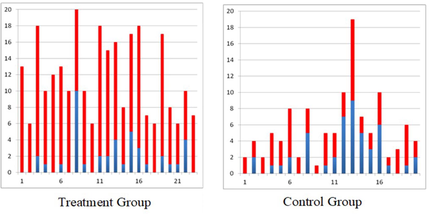
To date we have obtained similar results in all individuals with
sub-acute incomplete spinal cord injury who received 40 h of FES therapy
(
Figure 1).
Similar results were obtained in
the randomized controlled trial carried out in individuals with acute
(2–7 weeks post) severe stroke with a total arm and hand score no more
than 2 on the Chedoke McMaster Stages of Motor Recovery (less than 15
points on Fugl Meyer Assessment Upper Limb Sub-score) (
Thrasher et al., 2008;
Hebert et al., 2017;
Marquez-Chin et al., 2017).
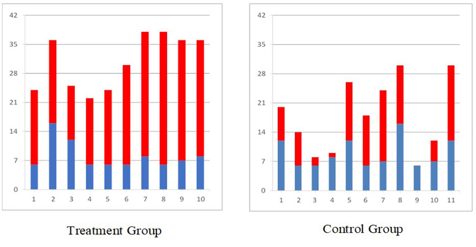
The individuals who received 12–16 weeks of FES therapy for the arm and
hand had statistically better improvement on the Self-care sub-score of
the Functional Independence Measure (
Figure 2),
Fugl Meyer Assessment, Barthel Index, and Chedoke McMaster Stages of
Motor Recovery as compared to individuals who received conventional
occupational therapy and physiotherapy for the same duration. Detailed
results of this study are published elsewhere.
In another clinical trial in chronic severe pediatric stroke population (
Kapadia N. et al., 2014),
where all four participants received a total of 48 h of FES therapy,
statistically significant improvements were observed on the Quality of
Upper Extremity Skills Test as well as on various sub components of the
Rehabilitation Engineering Laboratory Hand Function Test (this is the
Toronto Rehabilitation Institute- Hand Function Test with a scoring
system adapted for stroke).
Discussion
Short duration multichannel surface FES is a viable and
safe treatment modality that can be successfully applied in patients
with neurological conditions. It is important to note that we did not
formally investigate safety and feasibility in our clinical trials
mainly because transcutaneous FES has been applied in clinical trials
for over 5 decades now without any reports of major adverse events.
However, given that we have applied FES to over 200 patients over the
past 15+ years we are able to confidently say that transcutaneous FES is
both safe and feasible. Across all of our clinical trials we did not
encounter any serious adverse events and we have been able to
successfully retain our study participants for the duration of the
research therapy. Discussed here is an in-depth application of
transcutaneous multi-channel FES therapy of the upper extremity in
spinal cord injury and stroke patients. In order to obtain maximum
benefits of this therapy there are some general points to keep in mind.
The goal of this manuscript is not to explore the
mechanism of improvement in individuals with stroke and spinal cord
injury following FES as this is a methods paper and as such these
mechanisms have been widely discussed in literature (
Quandt and Hummel, 2014;
Hara, 2015;
Luo et al., 2020;
Marquez-Chin and Popovic, 2020).
We do, however, recommend some basic principles of FES application on
the widely accepted belief that mechanism of improvement with this
therapy is based on the principles of neuroplasticity (
Nagai et al., 2016).
First and foremost it is strongly recommended that therapy should be
started as soon as the medical condition of the patient is stabilized,
i.e., preferably in the acute or sub-acute phase post-injury. Secondly,
active participation of the patient during treatment is critical. Along
with the FES, patients have to make an active attempt to execute the
target movement. Third, the movements carried out should be functional
and should follow a physiological pattern as closely as possible
(movements similar to those of able-bodied individuals). Fourth, therapy
should be combined with conventional rehabilitation modalities
(example: stretching and strengthening) to reap maximum benefits.
Lastly, while no specific dosing study has been conducted, our group
recommends delivering at least three 1 h sessions per week. However, our
group does not recommend more than one session per day, as this often
exhausts the patient and prevents them from actively participating in
the second session. In total, at least 20 sessions are needed to obtain
clinically relevant changes, however, it is often recommended that
patients have 40 or more hours of therapy to maximize outcomes and
experience greater gains in function.
It should be noted that, in certain very acute or
chronic spinal cord injury cases, a strengthening phase is necessary
prior to the functional training phase because the muscles are minimally
responsive to stimulation at first (
Popovic et al., 2002) due to initial spinal shock (
Galeiras Vázquez et al., 2017) or due to long-term disuse (
Popovic et al., 2002).
It is important to bear in mind that although FES
therapy has not been extensively tested in individuals with cervical
complete spinal cord injury, those that have been trained with the
system have shown remarkable improvements that were much more profound
than those produced with conventional therapy (
Popovic et al., 2006).
This evidence merits conducting more comprehensive clinical trials with
FES therapy in cervical complete spinal cord injury patients.
In conclusion, the most attractive feature of
multichannel surface stimulators is that they are non-invasive, often
programmable and allow for various muscles/muscle groups to be
stimulated simultaneously in physiological patterns. They have a high
level of fidelity and are able to produce global upper-limb motions as
well as fine finger movements like two pinch grip (thumb and index
finger) and tripod grip (thumb, index, and middle finger) using surface
stimulation electrodes.
The specific surface stimulator used in our clinical
studies, is not widely available any longer, however, the methodological
considerations discussed above remain the same irrespective of the type
of stimulation device. Any stimulator that can produce protocols
discussed in this article can be used for FES therapy. Although the new
stimulators used for the FES therapy come with guidelines for locating
motor points, therapists should be mindful that motor points can
anatomically vary between individuals. If required, the first session
should be dedicated to finding correct stimulation points, after which
these can be marked down for future sessions.
As important as it is to assist weak muscles with FES
during execution of functional tasks, it is equally important that once
functional voluntary strength is recovered (at least 3/5 on Manual
Muscle Testing), stimulation is withdrawn from those muscles and the
patient is encouraged to voluntarily control the muscles themselves. The
available FES channels can then be applied to other weaker muscle
groups that still need retraining. In some cases, with severe
spasticity, manual stretching of the tight muscles prior to stimulation
may yield better results.
Data Availability Statement
The datasets generated for this study are available on request to the corresponding author.


 Naaz Kapadia
 Bastien Moineau1,5,6 and
  Milos R. Popovic