Changing stroke rehab and research worldwide now.Time is Brain! trillions and trillions of neurons that DIE each day because there are NO effective hyperacute therapies besides tPA(only 12% effective). I have 523 posts on hyperacute therapy, enough for researchers to spend decades proving them out. These are my personal ideas and blog on stroke rehabilitation and stroke research. Do not attempt any of these without checking with your medical provider. Unless you join me in agitating, when you need these therapies they won't be there.

What this blog is for:

My blog is not to help survivors recover, it is to have the 10 million yearly stroke survivors light fires underneath their doctors, stroke hospitals and stroke researchers to get stroke solved. 100% recovery. The stroke medical world is completely failing at that goal, they don't even have it as a goal. Shortly after getting out of the hospital and getting NO information on the process or protocols of stroke rehabilitation and recovery I started searching on the internet and found that no other survivor received useful information. This is an attempt to cover all stroke rehabilitation information that should be readily available to survivors so they can talk with informed knowledge to their medical staff. It lays out what needs to be done to get stroke survivors closer to 100% recovery. It's quite disgusting that this information is not available from every stroke association and doctors group.

Showing posts with label cognitive deficits. Show all posts

Friday, March 6, 2026

Early Tackle Football Exposure Linked to Later-Life Clinical Impairment

 I played a tiny bit prior to high school, as a freshman practiced a couple weeks and decided it was not for me.

Early Tackle Football Exposure Linked to Later-Life Clinical Impairment


 Younger age at first exposure to tackle football was not associated with CTE presence or severity but was linked to worse informant-reported cognitive, neurobehavioral, and neuropsychiatric outcomes among deceased players aged 60 years and older. Earlier tackle football exposure was linked to worse later-life clinical outcomes but not chronic traumatic encephalopathy (CTE) pathology, according to results published in the Journal of Neurotrauma Risk for CTE is increased with repetitive head impacts sustained during contact sports. However, prior studies have indicated that several neurodegenerative and cerebrovascular pathologies can result from repetitive head impacts. Some of this heterogeneity may be due to the age at first exposure to contact sports. Researchers from Boston University Chobanian & Avedisian School of Medicine and Harvard Medical School analyzed data from 677 deceased male former American football players from the Understanding Neurologic Injury and Traumatic Encephalopathy (UNITE) Brain Bank. The donors were evaluated for CTE pathology, cognitive function, and behavior on the basis of age at first exposure to tackle football. Prospective, population-based studies of former American football players are necessary to better understand the relationship between younger [age at first exposure] to tackle football and clinical impairments. Participants were stratified by age at death: 277 died before age 60, and 400 died at age 60 and older. Those who died before age 60 years had a mean (SD) age at death of 39.9 (12.4) years compared with 74.6 (8.2) years among those who died at or after age 60 (P<.001). The younger group had a younger mean (SD) age at first exposure (10.1 [3.00] vs 11.9 [2.7] years;P<.001), fewer mean (SD) years of play (11.1 [5.7] vs 13.0 [5.7] years; P<.001), and were less likely to have played football professionally (26.1% vs 45.8%; P<.001). The proportion of White participants was 74.6% in the younger group and 87.3% in the older group (P=.002). Overall, 471 donors (69.6%) had CTE. Evidence of CTE was observed among 57.0% of the younger and 78.3% of the older participants (P<.001). The severity of CTE increased with age (P < .001). Among those younger than 60 years, CTE stages were I (36.7%), II (31.0%), III (29.7%), and IV (2.5%) compared with 4.5%, 8.3%, 43.6%, and 43.6%, respectively, among those 60 years and older. The younger cohort also had lower rates of Alzheimer disease (1.8% vs 30.3%; P<.001) and Lewy body disease (P<.001) with brainstem (1.1% vs 10.8%) or limbic or neocortical (1.8% vs 12.3%) predominance compared with the older cohort, respectively. In the younger cohort, age at first exposure did not predict any cognitive, neurobehavioral, or neuropsychiatric outcomes (all P ³.292). In the older cohort, however, age at first exposure was associated with the composite cognitive (standardized b, -0.133;P=.009), neurobehavioral (standardized b, -0.119; P=.035), and neuropsychiatric (standardized b, -0.113; P=.048) scores. Each year older at first exposure was associated with lower odds of impairment on the Behavior Rating Inventory of Executive Function for Adults (BRIEF-A) Metacognition Index (OR, 0.774; P =.028), Cognitive Difficulties Scale (OR, 0.775; P =.003), and Behavioral Regulation Index (OR, 0.831; P =.044).

No association was observed between age at first exposure and CTE presence or stage.

Study limitations include the retrospective reporting of clinical outcomes.

The study authors concluded, “Prospective, population-based studies of former American football players are necessary to better understand the relationship between younger [age at first exposure] to tackle football and clinical impairments.”

Disclosures: Some study authors declared affiliations with biotech, pharmaceutical, and/or device companies. Please see the original reference for a full list of authors’ disclosures.

References:

Nosek SB, Gil SG, Abdolmohammadi B, et al. Younger age of first exposure to American football is associated with worse informant-reported clinical outcomes in older age brain donors. J Neurotrauma. Published online February 13, 2026. doi:10.1177/08977151261424707

Monday, February 3, 2025

Cerebral small vessel disease linked to cognitive deficits in older adults

 Your competent? doctor needs to address and fix this problem before it causes another stroke.
Cerebral small vessel disease includes various conditions in which small blood vessels in the brain are damaged, often causing stroke.

Cerebral small vessel disease linked to cognitive deficits in older adults

Key takeaways:

  • Differences in neuropsychological testing were not significant at a 4-year follow up.
  • Logical memory function in all participants increased over the full 8-year follow-up interval.

In a small cohort of adults in South Korea, the presence of cerebral small vessel disease was associated with color reading and visual deficits, but not memory, across an 8-year follow-up interval, according to new research.

“Although [the] majority of [cerebral small vessel disease (cSVD)] manifests sub-clinically, it is a major source of cognitive impairment during aging process leading to mild cognitive impairment and dementia,” Ali Tanweer Siddiquee, MPH, of the Institute of Human Genomic Study and College of Medicine at Korea University Ansan Hospital, and colleagues wrote in The Lancet Regional Health. “Cognitive dysfunction caused by cSVD may account for two-thirds of all vascular dementia.”Brain illustration

According to new research from South Korea, cerebral small vessel disease was associated with a decline in certain cognitive functions across an 8-year follow up for individuals aged 49 to 79 years. Image: Adobe Stock

Siddiquee and colleagues examined the longitudinal relationship between cSVD and cognitive decline in adults from their middle age to their elderly years.

Their population-based, prospective cohort study collected data from the Korean Genome and Epidemiology Study, commenced in South Korea in 2001.

The study included 2,454 individuals aged 40 to 79 years, free of dementia and other cerebrovascular conditions. Participants underwent both MRI and a comprehensive neuropsychological battery at baseline between 2011 and 2014. All participants were subjected to the same testing in two separate 4-year cycles (2015–2018 and 2019–2022), during which cognitive function testing analysis was also conducted. The neuropsychological battery included verbal and visual memory, verbal fluency, Digit Symbol–coding, Trail Making Test–A, and Stroop Tests.

Small vessel disease was defined by the presence of age-related white matter change, lacunes and cerebral microbleeds on MRI at baseline, with the main outcomes defined as adjusted mean differences of cognitive test performances by cSVD groups over time.

Covariates that were likely to exert influence over both cSVD and cognition, measured at baseline, included lifestyle, health, exercise, substance use habits, BMI and blood pressure.

Out of 2,454 participants, 908 registered signs of cSVD via MRI at baseline. These participants were aged approximately 3 years older than those who did not have cSVD.

Scores were not significantly different among the cognitive test battery, save for Trail Making Test–A, at baseline among cSVD groups.

By the 8-year follow-up, participants without cSVD performed significantly better than their cSVD counterparts in Stroop–color reading (mean difference = 1.19; 95% CI, 0.02–2.36) and visual reproduction-recognition (mean difference = 0.11; 95% CI, 0.01–0.21)].

Siddiquee and colleagues noted, however, that significant worsening between the groups was not observed at a 4-year follow up.

While no differential changes were recorded among cognitive testing by cSVD groups, the researchers observed a decrease over time in logical memory (Story Recall Tests) and decrease in Stroop-word reading in both cSVD groups, which were nearly identical.

Data additionally showed that the presence of cSVD within the brain was mostly in the frontal lobe followed by basal ganglia area.

“The presence of cSVD may be an important risk factor of cognitive decline, especially executive functioning, and thus, early prevention of cSVD would help preserving cognitive function in a normal aging population,” Siddiquee and colleagues wrote.

Sunday, December 8, 2024

Curcumin Reverses Cognitive Deficits through Promoting Neurogenesis and Synapse Plasticity via the Upregulation of PSD95 and BDNF in mice

 Does your competent? doctor have enough competence to get human testing going on this? NO? So, you don't have a functioning stroke doctor, do you? RUN AWAY!

Curcumin Reverses Cognitive Deficits through Promoting Neurogenesis and Synapse Plasticity via the Upregulation of PSD95 and BDNF in mice

Gaifen Li1

Email

Qiong Wu1

Chao Wang1

Pin Deng2

Jiaxin Li1

Zhiguang Zhai1

Yubo Li1

1 Institute of Basic Theory for Traditional Chinese Medicine, China Academy of Chinese Medical Sciences,

2 China Academy of Chinese Medical Sciences

Following prolonged exposure to hypoxic conditions, for example, due to ascent to high altitude, aging or stroke, cognitive deficits can develop. The exact nature and genesis of hypoxia-induced cognitive deficits remain unresolved. Curcumin has been reported to stimulate neurogenesis and reduce neuronal degeneration. This study aimed to investigate the effect of curcumin on cognitive deficits in hypoxic-brain injury mice and its potential mechanism. Eight-week-old male C57BL/6J mice were exposure to normobaric-hypoxia (13%O2) 14 days to establish hypoxic-brain injury models. Morris water maze and novel object recognition were used to detect the cognitive function of each mouse. Immunofluorescence assays, including Fluoro-Jade C (FJC) and bromodeoxyuridine (BrdU), were used to detect neuronal degeneration and neurogenesis. Thy1-YFP transgenic mice were used to detect synapse plasticity. Our results showed that curcumin administration rescued the impaired cognition of mice, shown as enhanced BrdU+ and dendritic spine in hippocampus. At the molecular level, curcumin was found to promote the expression of brain-derived neurotrophic factor (BDNF) and postsynaptic density protein 95 (PSD95). The results of primary hippocampal neuron detection showed that curcumin could promote dendritic growth. In conclusion, our study indicates that curcumin, increased BDNF and PSD95 expression and contacted with interneurons, salvaged of interneurons may normalize ambient neuroplasticity, resulting in the preservation of neurogenesis processes as well as contributing to improve cognitive performance.

Thursday, October 5, 2023

Loss of brain interneurons linked to cognitive deficits in aging

Is your doctor checking on this before automatically blaming the stroke for your cognitive deficits?

Loss of brain interneurons linked to cognitive deficits in aging

Normal aging is usually associated with a decline in memory, although it is unclear what factors play a role. In a new study, researchers studied specific interneurons, which serve as communication centers that connect other neurons, in the regions of the brain that are important for learning and memory.

Increasing age places people at risk, whether it is because of a normal decrease in cognitive ability or due to postoperative cognitive disorders. In the latter, the deficits can persist for many months after surgery especially when the patients are older than 60. Unfortunately, the underlying cellular mechanisms that cause these impairments are largely unknown.

Previous studies have shown that the region of the brain that is associated with learning and memory-;the hippocampus-; decreases in volume with age. Additionally, the levels of the molecule -aminobutyric acid, or GABA, and some of the interneurons that release it are also affected.

In the present study, the researchers focused on the hippocampal interneurons in a specific region, called the hilus of the dentate gyrus, that are characterized by their expression of somatostatin. This hormone has the ability to counteract the effects of growth hormones elsewhere in the body.

In the past, other researchers have found that one of the differences between cognitively impaired and unimpaired rats was that the former had a lower number of somatostatin-positive interneurons in the hilus of the dentate gyrus. We wanted to further investigate whether a loss of these neurons is really responsible for cognitive deficits, and whether it could thus serve as a model of aging in the hippocampus."

Uwe Rudolph (GNDP), professor of comparative biosciences

The researchers decreased the numbers of somatostatin-positive interneurons in the hippocampus of mice by using a toxin. They injected this toxin into the dentate hilus, so that the toxin would only be expressed in the somatostatin-positive interneurons, killing approximately 50% of these cells. Starting 3 weeks later, they conducted behavioral studies to test the learning abilities and memory of the mice.

The mice underwent three types of tests: whether they could remember and differentiate novel objects from familiar ones, navigate mazes using their short-term memories, and use their spatial learning to find a hidden platform in a pool of water. In all three cases, the mice that had decreased levels of somatostatin-positive interneurons struggled compared to those that did not receive the toxin.

The researchers also looked for changes in cellular signals that occur due to lower levels of somatostatin-positive interneurons. To do so, they focused on the microglia, which are immune cells that are among the first to respond when something goes wrong in the brain.

"We looked at microglial activation, which is a hallmark of the inflammation that is associated with aging and memory impairment," said Rajasekar Nagarajan, a postdoctoral researcher in the Rudolph lab. "We saw increased activation of microglia in the hippocampus of the mice that were injected with the toxin, even outside of the dentate gyrus."

In addition to determining the status of microglia, the team measured a protein called brain-derived neurotrophic factor, which is active in the hippocampus and plays a role in long-term memory. They found that there were lower BDNF levels in the hippocampal tissue of the toxin-injected mice. Furthermore, the researchers also found that these mice had fewer hippocampal dendritic spines, which are critical for learning and memory.

Unsurprisingly for the researchers, the results seen with the toxin-injected mice were essentially the same as with aged mice that were 18-19 months old and had not been injected with the toxin.

"18-month-old mice correspond to an age of approximately 60 years in humans. It roughly fits the time point at which we know that people more frequently develop neurocognitive deficits in response to surgery and anesthesia," Rudolph said. "Our results have shown that using this toxin to reduce the number of somatostatin-positive interneurons is sufficient to cause effects that resemble cognitive deficits in aging."

The researchers are excited that they can use these techniques to investigate aging-related defects without waiting for the mice to grow old, which is an expensive and time-consuming undertaking. "We will be using this method as a model to test which experimental compounds can be used to prevent or reverse both age-related and post-operative cognitive impairments," Nagarajan said.

Source:
Journal reference:

Nagarajan, R., et al. (2023) Genetic Ablation of Dentate Hilar Somatostatin-Positive GABAergic Interneurons is Sufficient to Induce Cognitive Impairment. Molecular Neurobiology. doi.org/10.1007/s12035-023-03586-3.

Monday, February 6, 2023

EEG-Neurofeedback as a Potential Therapeutic Approach for Cognitive Deficits in Patients with Dementia, Multiple Sclerosis, Stroke and Traumatic Brain Injury

 Maybe there is something in here, ask your doctor.

EEG-Neurofeedback as a Potential Therapeutic Approach for Cognitive Deficits in Patients with Dementia, Multiple Sclerosis, Stroke and Traumatic Brain Injury 

2nd Department of Neurology, AHEPA University Hospital, Aristotle University of Thessaloniki, 54621 Thessaloniki, Greece
*
Author to whom correspondence should be addressed.
†
These authors contributed equally to this work.
Life 2023, 13(2), 365; https://doi.org/10.3390/life13020365
Received: 15 December 2022 / Revised: 21 January 2023 / Accepted: 25 January 2023 / Published: 29 January 2023
(This article belongs to the Special Issue Applications of EEG in Neural Rehabilitation)

Abstract

Memory deficits are common in patients with dementia, such as Alzheimer’s disease, but also in patients with other neurological and psychiatric disorders, such as brain injury, multiple sclerosis, ischemic stroke and schizophrenia. Memory loss affects patients’ functionality and, by extension, their quality of life. Non-invasive brain training methods, such as EEG neurofeedback, are used to address cognitive deficits and behavioral changes in dementia and other neurological disorders by training patients to alter their brain activity via operant activity. In this review paper, we analyze various protocols of EEG neurofeedback in memory rehabilitation in patients with dementia, multiple sclerosis, strokes and traumatic brain injury. The results from the studies show the effectiveness of the ΕΕG-NFB method in improving at least one cognitive domain, regardless of the number of sessions or the type of protocol applied. In future research, it is important to address methodological weaknesses in the application of the method, its long-term effects as well as ethical issues.

1. Introduction

Cognitive deficits, such as memory loss and attention disorders, are very common nowadays because of the aging world population and the numerous neurodegenerative diseases that lead to cognitive impairment. Until recently, the treatment of cognitive deficits was based exclusively on the administration of appropriate medication. However, a deeper understanding of the nature of cognitive deficits combined with advances in technology has led to the development of methods and techniques aimed at improving both cognitive deficits and non-cognitive conditions, in order to improve the quality of life not only for the patients but also for their families.
A popular method is cognitive rehabilitation. Cognitive rehabilitation is a behavioral approach that aims to improve patients’ cognitive deficits, but also to provide assistance to both patients and their families in order to improve their daily lives. It can be carried out with paper and pencil exercises, but also with electronic programs, through which the patient practices various cognitive tasks [1].
Another method is neurofeedback (NFB). NFB is a biofeedback technique for training patients with neurological and psychiatric disorders to change brain activity through operant conditioning [2]. Through this method, the individual learns to enhance and inhibit specific electrophysiological parameters through the process of learning. Modification of the individual’s behavioral response is made possible through feedback and positive reinforcement [3]. EEG-NFB is investigated in patients with depression, post-traumatic stress, schizophrenia, addictions, attention deficit hyperactivity disorder, autism and learning disabilities, and it is correlated with short- or long-term symptom relief.
Neurophysiological factors for NFB training are not clearly defined. It is considered that two types of neuroplasticity are involved: Hebbian plasticity and homeostatic plasticity. Reformation of the neural membrane and synaptic potentiation because of EEG amplitude are characteristics of Hebbian neuroplasticity, while homeostatic plasticity plays the opposite role, in order to stabilize the neuronal activity and limit the expression of the Hebbian type. In any case, the neuronal mechanisms have not been clearly investigated until now [4,5].
There are three types of EEG-NFB: 1. slow cortical potentials; 2. coherence training; and 3. frequency training. The third type of training is the most commonly used and is aimed at modifying the power ratio of frequency bands. The frequency bands are divided into 1. delta, 2. theta, 3. alpha, 4. beta and 5. gamma [6].
These frequencies are used to classify brain oscillations [7]. They are created by the coordinated activity of cells and thus make it possible to communicate between different brain regions in a way that allows the brain to utilize the information it receives and then synthesize it [3]. Additionally, brain oscillations are connected with specific cognitive functions. Theta oscillations are connected with encoding retrieval, while alpha bands are connected with attention. Alpha and gamma oscillations are capable of suppressing factors that can reduce concentration [8].
The most commonly used EEG-NFB protocols for the therapeutic management of cognitive deficits are: 1.EEG-theta/beta ratio; 2. Sensorimotor Rhythm (SMR) (12–15 Hz) [9]. In the first protocol, theta band power (ranging between 4 and 7 Hz) divided by beta band ratio (ranging between 13 and 30 Hz) shows cortical and sub-cortical brain interactions [10]. The second protocol, SMR, includes rhythm with a mean frequency of 10 Hz, and it is recorded over sensorimotor cortices in C3 and C4 [11].
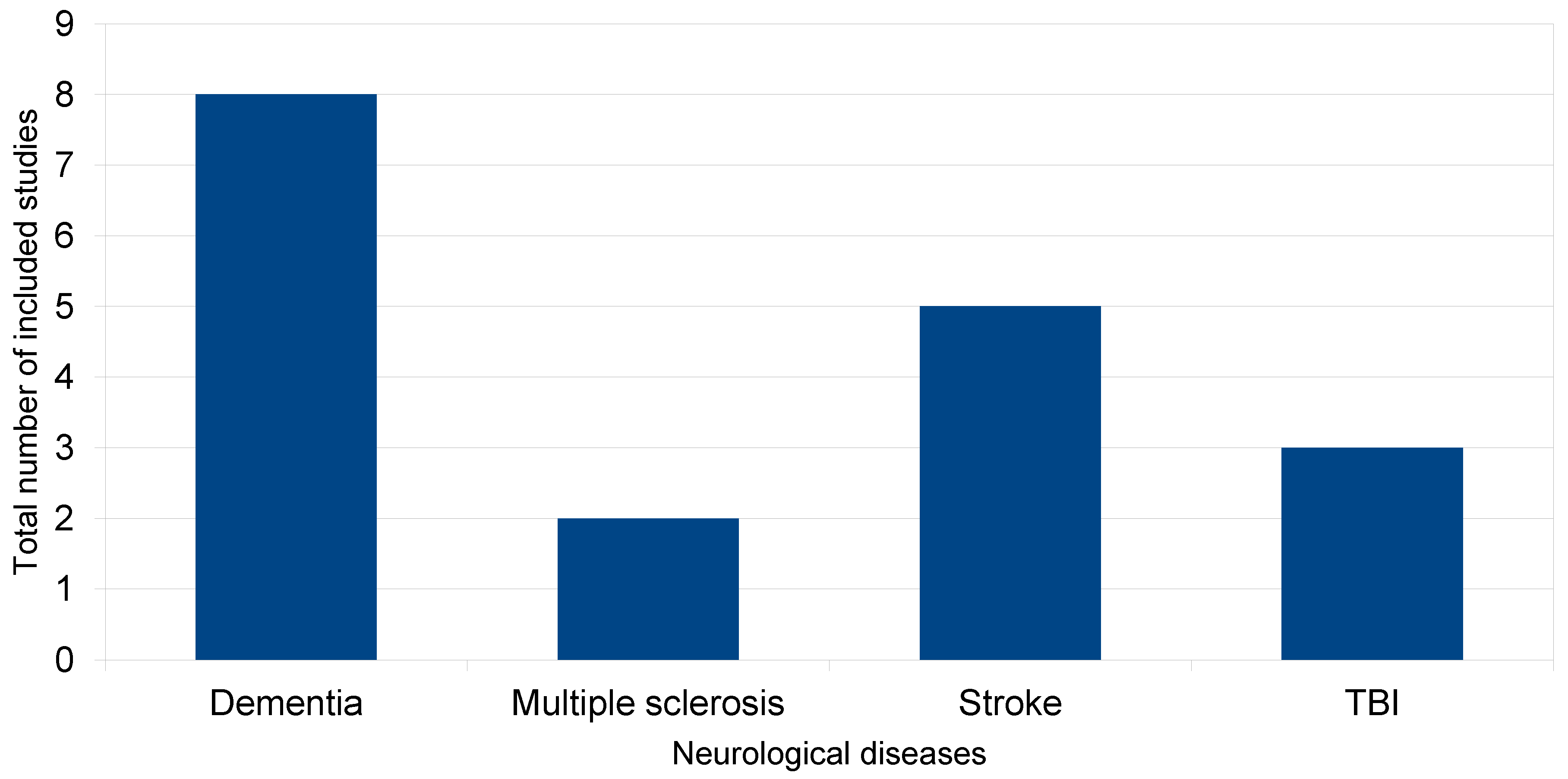
The effectiveness of EEG-NFB in cognitive rehabilitation is still under investigation and is mainly evaluated through comparisons of patients’ cognitive performance before and after their training in various EEG protocols. A review of studies using EEG-NFB as a therapeutic tool for treating cognitive deficits in patients with Alzheimer’s dementia (AD), mild cognitive disorder (MCI), stroke, multiple sclerosis (MS) and traumatic brain injury (TBI) is analyzed below. The novelty of our review lies in gathering findings on the effectiveness of EEG neurofeedback as a therapeutic method in the aforementioned four neurological disorders. Recent studies demonstrating the effectiveness of the method, regardless of whether EEG-NFB is used as a clinical tool or a wearable device, are reviewed.

Contribution of Our Review

  • Review of recent EEG-NFB studies in dementia, multiple sclerosis, strokes and TBI.
  • Therapeutic effectiveness of EEG-NFB regardless of how it is applied (clinical use of EEG or wearable device).
     
    More at link.

Saturday, October 8, 2022

Rehabilitation interventions for cognitive deficits in stroke survivors: A systematic review of randomized controlled trials

Well shit, if we had a database of all stroke research that was updated as research came in this type of systematic review would never need to be done. And you didn't even write up your findings as a protocol and distribute it to all 10 million yearly stroke survivors. I blame your mentors and senior researchers for such a failure in your objective.  The point of all stroke research is to get survivors recovered!

 Rehabilitation interventions for cognitive deficits in stroke survivors: A systematic review of randomized controlled trials


>

Abstract

Cognitive deficits are one of the most common impairments after stroke. It negatively affects physical and social functioning. Rehabilitation interventions for cognitive deficits post-stroke have taken less consideration. The present study aimed to provide an overview of the effects of various rehabilitation interventions on cognitive functions in patients with stroke. PUBMED, SCOPUS, PEDro, EMBASE, MEDLINE, and REHABDATA were searched for randomized controlled trials (RCTs) investigating the effects of rehabilitation interventions on cognitive domains poststroke until August 2021. The methodological quality of the selected studies was evaluated using the Cochrane Collaboration tool, and the effect sizes were calculated. Forty-four studies met the inclusion criteria. A total of 3561 individuals with stroke, 57.60% of whom were males. The mean age for all participants was 65.48 years. Eighteen RCTs were high, moderate  = 17), and low methodological quality (n = 9). The results showed evidence for the beneficial effects of many rehabilitation interventions on cognition in individuals with stroke.(And you did nothing with that evidence. Useless.) Rehabilitation plays a crucial role in improving cognitive functions in stroke patients with mild cognitive deficits. Virtual reality (VR), computer-based cognitive rehabilitation (CBCR), and non-aerobic exercises may promote cognitive functions in patients with stroke.

Wednesday, May 11, 2022

Multivariate profile and acute-phase correlates of cognitive deficits in a COVID-19 hospitalised cohort

 So 20 years of cognitive aging in severe COVID-19. Since you already lost 5 years that your doctor has no clue on how to recover you'll want to be vaccinated and boosted.

But WHOM is going to do the stroke research to recover 5 lost years of brain cognition due to your stroke?

Multivariate profile and acute-phase correlates of cognitive deficits in a COVID-19 hospitalised cohort

Doris A.ChatfieldbAnne ManktelowMPhilbAmyJollyaWilliamTrenderaPeter J.HellyeraMartina DelGiovaneaVirginia F.J.NewcombebJoanne G.OuttrimbBenWarnebJunaidBhattidLindaPointondAnneElmereNyarieSitholebfJohnBradleybghNathalieKingstonlStephen J.SawceriEdward T.Bullmorecdj…David K.Menonbck1
https://doi.org/10.1016/j.eclinm.2022.101417Get rights and content
Under a Creative Commons license
Open access

Summary

Background

Preliminary evidence has highlighted a possible association between severe COVID-19 and persistent cognitive deficits. Further research is required to confirm this association, determine whether cognitive deficits relate to clinical features from the acute phase or to mental health status at the point of assessment, and quantify rate of recovery.

Methods

46 individuals who received critical care for COVID-19 at Addenbrooke's hospital between 10th March 2020 and 31st July 2020 (16 mechanically ventilated) underwent detailed computerised cognitive assessment alongside scales measuring anxiety, depression and post-traumatic stress disorder under supervised conditions at a mean follow up of 6.0 (± 2.1) months following acute illness. Patient and matched control (N = 460) performances were transformed into standard deviation from expected scores, accounting for age and demographic factors using N = 66,008 normative datasets. Global accuracy and response time composites were calculated (G_SScore & G_RT). Linear modelling predicted composite score deficits from acute severity, mental-health status at assessment, and time from hospital admission. The pattern of deficits across tasks was qualitatively compared with normal age-related decline, and early-stage dementia.

Findings

COVID-19 survivors were less accurate (G_SScore=-0.53SDs) and slower (G_RT=+0.89SDs) in their responses than expected compared to their matched controls. Acute illness, but not chronic mental health, significantly predicted cognitive deviation from expected scores (G_SScore (p=​​0.0037) and G_RT (p = 0.0366)). The most prominent task associations with COVID-19 were for higher cognition and processing speed, which was qualitatively distinct from the profiles of normal ageing and dementia and similar in magnitude to the effects of ageing between 50 and 70 years of age. A trend towards reduced deficits with time from illness (r∼=0.15) did not reach statistical significance.

Interpretation

Cognitive deficits after severe COVID-19 relate most strongly to acute illness severity, persist long into the chronic phase, and recover slowly if at all, with a characteristic profile highlighting higher cognitive functions and processing speed.

Funding

This work was funded by the National Institute for Health Research (NIHR) Cambridge Biomedical Research Centre (BRC), NIHR Cambridge Clinical Research Facility (BRC-1215-20014), the Addenbrooke's Charities Trust and NIHR COVID-19 BioResource RG9402. AH is funded by the UK Dementia Research Institute Care Research and Technology Centre and Imperial College London Biomedical Research Centre. ETB and DKM are supported by NIHR Senior Investigator awards. JBR is supported by the Wellcome Trust (220258) and Medical Research Council (SUAG/051 G101400). VFJN is funded by an Academy of Medical Sciences/ The Health Foundation Clinician Scientist Fellowship. The views expressed are those of the authors and not necessarily those of the NHS, the NIHR or the Department of Health and Social Care.


Tuesday, October 19, 2021

An exercise ‘sweet spot’ reverses cognitive deficits of ageing by growth hormone induced neurogenesis

You can have your doctor read the 45 pages at the link.

An exercise ‘sweet spot’ reverses cognitive deficits of ageing by growth hormone induced neurogenesis

 Daniel G. Blackmore, Frederik J. Steyn, Alison Carlisle, Imogen O’Keeffe, King-Year Vien, Xiaoqing Zhou, Odette Leiter, Dhanisha Jhaveri, Jana Vukovic, Michael J. Waters, Perry F. Bartlett PII: S2589-0042(21)01244-X DOI: https://doi.org/10.1016/j.isci.2021.103275 Reference: ISCI 103275 To appear in: ISCIENCE Received Date: 21 July 2021 Revised Date: 9 September 2021 Accepted Date: 12 October 2021 Please cite this article as: Blackmore, D.G., Steyn, F.J., Carlisle, A., O’Keeffe, I., Vien, K.-Y., Zhou, X., Leiter, O., Jhaveri, D., Vukovic, J., Waters, M.J., Bartlett, P.F., An exercise ‘sweet spot’ reverses cognitive deficits of ageing by growth hormone-induced neurogenesis, ISCIENCE (2021), doi: https:// doi.org/10.1016/j.isci.2021.103275. This is a PDF file of an article that has undergone enhancements after acceptance, such as the addition of a cover page and metadata, and formatting for readability, but it is not yet the definitive version of record. This version will undergo additional copyediting, typesetting and review before it is published in its final form, but we are providing this version to give early visibility of the article. Please note that, during the production process, errors may be discovered which could affect the content, and all legal disclaimers that apply to the journal pertain.

Wednesday, January 20, 2021

Can cognitive rehabilitation improve attention deficits following stroke? -A Cochrane review summary with commentary

With no guidance from anyone in stroke you are completely on your own to figure out how to recover your cognitive deficits.

 Can cognitive rehabilitation improve attention deficits following stroke? -A Cochrane review summary with commentary

  NeuroRehabilitation , Volume 47(3) , Pgs. 355-357.

NARIC Accession Number: J85137.  What's this?
ISSN: 1053-8135.
Author(s): Hazelton, Christine.
Publication Year: 2020.
Number of Pages: 3.
Abstract: 
Article discusses a published Cochrane Review that assessed the effects of cognitive rehabilitation on attention and functional ability in stroke survivors with attentional impairments. This Cochrane Review was an update of a review originated in 2000 and updated in 2013. Six studies with 223 participants were included: this was the same as the previous review (in 2013). Evidence quality was very low to moderate, and results suggest a beneficial impact on divided attention immediately after training, but no effect on any other outcome either immediately or at follow-up timepoints. The low methodological quality and small number of studies means current evidence provides limited clinical guidance. Clearly more research is needed to inform care. Researchers must improve the methodological quality of studies, plus fully consider and report the aspects of attention and function addressed in their work.
Descriptor Terms: ATTENTION DEFICIT DISORDERS, COGNITIVE DISABILITIES, REHABILITATION RESEARCH, REHABILITATION SERVICES, RESEARCH REVIEWS, STROKE.


Can this document be ordered through NARIC's document delivery service*?: Y.

Citation: Hazelton, Christine. (2020). Can cognitive rehabilitation improveattention deficits following stroke? -A Cochrane review summary with commentary.  NeuroRehabilitation , 47(3), Pgs. 355-357. Retrieved 1/20/2021, from REHABDATA database.


* The majority of journal articles, books, and reports in our collection are only available by regular mail, rather than downloadable electronic format. Learn more about our digital collection and our document delivery service.

More information about this publication:
NeuroRehabilitation.

Friday, January 15, 2021

The Role of Acupuncture Improving Cognitive Deficits due to Alzheimer’s Disease or Vascular Diseases through Regulating Neuroplasticity

You have got to be kidding:

Impossible to have effects except as a placebo. Energy meridians have never been proven to exist.
No mechanism of action is possible. 

But if you believe, have at it, recognizing these possible side effects;

 The latest here:

The Role of Acupuncture Improving Cognitive Deficits due to Alzheimer’s Disease or Vascular Diseases through Regulating Neuroplasticity

Academic Editor: Zhen Zheng
Received24 Jun 2020
Revised29 Nov 2020
Accepted15 Dec 2020
Published12 Jan 2021

Abstract

Dementia affects millions of elderly worldwide causing remarkable costs to society, but effective treatment is still lacking. Acupuncture is one of the complementary therapies that has been applied to cognitive deficits such as Alzheimer’s disease (AD) and vascular cognitive impairment (VCI), while the underlying mechanisms of its therapeutic efficiency remain elusive. Neuroplasticity is defined as the ability of the nervous system to adapt to internal and external environmental changes, which may support some data to clarify mechanisms how acupuncture improves cognitive impairments. This review summarizes the up-to-date and comprehensive information on the effectiveness of acupuncture treatment on neurogenesis and gliogenesis, synaptic plasticity, related regulatory factors, and signaling pathways, as well as brain network connectivity, to lay ground for fully elucidating the potential mechanism of acupuncture on the regulation of neuroplasticity and promoting its clinical application as a complementary therapy for AD and VCI.

1. Introduction

As the population ages, the prevalence of dementia is increasing worldwide with an annual incidence of nearly 10 million [1], which leads to threats and challenges to global health and wellbeing. Dementia is characterized as a syndrome with myriad and complex causes, including primary neurologic, neuropsychiatric, and medical conditions and genetic and environmental factors [2, 3]. In the elderly, neurodegenerative dementias are most common [2], among which Alzheimer’s disease (AD) is believed to be the leading cause of dementia, and vascular cognitive impairment (VCI) is the second utmost cause [4, 5]. Unprecedented advancements have been made in molecular neuroimaging, clinicopathologic correlation, and the development of novel biomarkers in recent decades. However, effective therapeutics remain limited and even absent to date [4, 5]. Acupuncture, as one of complementary therapies for AD and VCI, is gradually applied to alleviate suffering, aggressively treating contributing symptoms and improving overall quality of life [6–10]. However, the underlying mechanisms remain elusive.

Neuroplasticity refers to the capacity of the nervous system to adapt to internal and external environmental changes by reorganizing its structure, function, and connections [11–14], which occurs at various levels of the nervous system from tissue to cellular to molecular [13]. It is known that dysregulated or disrupted neuroplasticity is implicated as a pathological mechanism in AD [15] and VCI [16]. Furthermore, some treatments that stimulate or modulate neuroplasticity have been indicated as effective in improving cognition [12, 17, 18], and might be potential therapy in cognitive impairments such as AD and VCI.

Acupuncture signals are recognized as a potent form of sensory stimulation that ascend mainly through the spinal ventrolateral funiculus to the brain [19]. The mechanisms of acupuncture-mediated neuroplasticity have recently attracted increased interest. Accordingly, acupuncture modulation over several cognition- or aging-related gene expressions [20], plasticity signaling pathways [21, 22], and brain functional connectivities [23] has been studied. Herein, we review the application of different protocols of acupuncture in animal models and humans, and their effectiveness on neuroplasticity in various sections: neurogenesis and gliogenesis, synaptic plasticity, related proteins and signaling pathways, and brain network connectivity. This review is aimed at laying the ground for elucidating the potential mechanism of acupuncture on AD and VCI to promote its clinical application as a complementary treatment.

2. Neurogenesis and Gliogenesis

The proliferation and differentiation of neurons and glial cells, also known as neurogenesis and gliogenesis, contribute to some neurorepair and improve brain function [24, 25]. Many previous results demonstrated that cerebral amyloidosis in AD mouse models caused neuronal proliferation inhibition and marked gliogenesis [26–28], and that stroke could trigger striatal and cortical neurogenesis and gliogenesis in murine models [29]. Mounting evidence indicates that adult hippocampal neurogenesis is implicated in cognitive processes, and that neurogenesis deficits may impair learning and memory. In states of brain injury such as AD and VCI, compensatory neurogenesis and gliogenesis mediate a balance between initial injury processes and endogenous repair processes [24]. Regulation of neurogenesis and gliogenesis is possibly associated with improving cognitive impairment and, consequently, may be attractive therapeutic targets for AD and VCI.

It is known that neurogenesis in the adult mammalian brain mostly takes place in specific brain regions harboring adult neuro stem and precursor cells, such as the subgranular zone (SGZ) of the hippocampal dentate gyrus (DG) and the ventricular/subventricular zone (VZ/SVZ) of the lateral ventricles [25]. Cognitive impairment due to AD or ischemic injury is recognized as partly related with neuron loss, impairment of cell proliferation, and imbalance between neuron loss and proliferation in the above regions [30]. Some studies showed that both manual acupuncture (MA) and electroacupuncture (EA) could ameliorate the learning and memory deficits of AD mice models through inducing the enhancement of neuron proliferation and migration in hippocampal DG and VZ/SVZ [31–33]. And the effect of MA and EA on improving cognitive dysfunction through the proliferation and differentiation of hippocampal neuro stem cells (NSCs) was also identified in murine models for vascular dementia (VaD) [34–36]. In addition, neurogenesis could take place in other brain areas in pathological conditions, such as the cortex [37], where the promotion of neurogenesis related to EA was also detected in the transgenic mice model for AD [33].

VCI is recognized to be associated with pathological changes in white matter degeneration and demyelination [38]. Oligodendrocyte (OL), as one predominant cell type in white matter, mediates myelination that is an essential process for the appropriate propagation of action potentials along axons [39]. Myelination participates in the restoration of damaged white matter in the adult brain [40], which may provide potential utility for the treatment of VCI. In a mouse model of VaD, EA was indicated to enhance the differentiation of oligodendrocyte precursor cells (OPCs) into mature OLs and ameliorate white matter damage in the corpus callosum (CC) [41]. Moreover, astrocytes also perform critical impacts on promoting neovascularization, regulating neuronal activity, and supporting synaptogenesis and neurogenesis, which may influence recovery following ischemic lesion [39, 42]. Experimental studies have reported that acupuncture was able to influence the proliferation and differentiation of astrocytes; however, the results were discrepant. One study revealed that MA was able to inhibit astrocyte activation and proliferation in VaD rat models [36]. Conversely, Kim et al. found that EA stimulation could induce NSCs differentiated into astrocytes in a VaD mouse model [35]. These results may be caused by differential acupoints or acupuncture methods. The differential influence of the acupuncture method (i.e., MA vs. EA) on neurogenesis has been demonstrated. And one study found that MA vs. EA stimulation at the same acupoints might induce differential cell proliferation and neuroblast differentiation in healthy rats [43]. And further investigation of the compared impact of differential acupuncture methods and acupoints on gliogenesis in AD and VCI models is required.

In addition to the direct effect on endogenous neurogenesis and gliogenesis, acupuncture was able to promote the survival, proliferation, migration, and differentiation of exogenous NSCs in the hippocampal microenvironment by regulating components of the cerebral microenvironment [44] or the related cytokine levels [45] in an AD mice model. All these findings demonstrated the influence of acupuncture on endogenous and exogenous neurogenesis and gliogenesis in AD and VCI, which deepen our understanding of acupuncture modulating neuroplasticity. There remain some limitations and even discrepancies in these results possibly caused by acupoints or models or observation times, or even acupuncture methods (i.e., MA vs. EA). And the mechanisms underlying the impact of acupuncture on neurogenesis and gliogenesis in different states, especially molecular mechanisms, need to be investigated.

3. Synaptic Plasticity

Synapses, the most sensitive and plastic structures, are directly involved in the integration and transfer of information within the neuro system. Previous studies demonstrated that synapse loss and dysfunction was a key feature in AD [46] and VCI [47] and positively correlates with cognitive damage. Impaired dendritic structure, spine density, and synaptic ultrastructure of neurons have been identified in brain tissue of AD patients and murine models, caused by soluble amyloid beta (Aβ) in the hippocampus [48, 49]. And ischemia-induced synapse reduction was also recognized to be the major pathological causes of VaD [50]. Synaptic plasticity, also defined as activity-dependent synaptic modifications of the strength of synaptic connections, is widely recognized to be fundamental to the formation and maintenance of learning and memory [51]. Synaptic plasticity in the neuro network, an important basis for cortical plasticity, is associated with learning and memory and sensorimotor dysfunction and recovery [51, 52]. Synaptic plasticity mainly includes modulation of the morphological structure of synapses and the synaptic strength and transmission, in which some synaptic protein markers, neurotransmitters, and receptors participate. Recently, modulation of synaptic plasticity is believed to be a promising approach for treating AD and VCI.

Synapse-structure parameters, such as synaptic curvatures, the width of the synaptic cleft, and the thickness of the postsynaptic density, are proposed to be important indicators that reflect synaptic morphological plasticity and greatly affect synaptic transmission [53]. Many studies revealed that MA and EA treatments had positive effects on the recovery of the learning and memory abilities not only in AD rat models but also in VCI, through increasing synaptic curvatures, decreasing the width of synaptic clefts, and thickening the postsynaptic densities in the hippocampus [49, 54]. In addition, MA was able to reverse the learning and memory impairments in AD mice models through enhancing the conjunction among the synapses and promoting synaptic formation [20] and regeneration [55], reducing ultrastructural degradation of synapses [56], and increasing the number and length of dendrites [57] and neurite fibers [58].

Long-term potentiation (LTP) and long-term depression (LTD) are considered as two indicators and forms of synaptic transmission [59]. As a cellular model of synaptic plasticity, LTP is the long-lasting enhancement in signal transmission between two neurons after synchronous stimulation associated with memory formation and storage, reflecting an increase of synaptic strength [60]. LTD is relevant to memory integration, forgetting, and recovery of LTP production at desaturation state [61]. And converging studies supported a crucial role of LTD in some types of learning and memory and in situations where cognitive demands require a flexible response [59]. Many electrophysiological studies showed that acupuncture could apparently improve the recovery from cognitive deficits by promoting LTP and/or LTD [61–63] and preventing or restoring the impaired LTP [64–69] in AD or VCI rat models. In addition to LTP and LTD, EA could also ameliorate the synaptic transmission by raising the slope of excitatory postsynaptic potential (EPSP) and the amplitude of population spikes (PS) in an AD mouse model [70].

Synaptophysin (SYN) is a major integral membrane protein of the presynaptic vesicle, and postsynaptic density 95 (PSD-95) and growth-associated protein 43 (GAP-43) are postsynaptic markers [71]. As important protein markers of regeneration and remodeling, they are widely found in all nerve terminals and used for quantifying the number of axon terminals, reflecting the occurrence, density, and strength of synapses [49, 72]. Many previous studies reported reduced expression of SYN and PSD-95 in the hippocampus in AD and VaD [73, 74]. It was demonstrated that acupuncture was able to promote synapse-structure damage rehabilitation by upregulating the expression of SYN [44, 54, 55], PSD-95 [56, 75, 76], and GAP-43 [77] to improve the learning and memory abilities of AD and VCI murine models.

Furthermore, accumulated evidence indicates that the effect of acupuncture on modulating synaptic structure and function in AD and VCI is achieved by changing the releasing of the presynaptic neurotransmitter or the function of the postsynaptic receptor [67, 68, 78]. As one of the major neurotransmitters, dopamine (D) plays an essential role in modulating hippocampal LTP and memory processes [79, 80]. Ye et al. found that MA could activate D1/D5 receptors to ameliorate cognitive function and LTP impairments in VaD rats [67]. The central cholinergic pathway and the norepinephrine- (NE) adrenergic receptor (AR) system are known for their critical roles in learning acquisition and synaptic plasticity in the mammalian limbic system. It was demonstrated that MA not only could alleviate memory-associated decreases in the levels of choline acetyltransferase (ChAT) and restore the expression of choline transporter 1 (CHT1) as well as vesicular acetylcholine transporter (AChT), resulting eventually in the recovery of the entire cholinergic system circulation pathway [81], but also was able to enhance norepinephrine (NE) levels and the activation of β1-AR in the hippocampus [68]. In addition, γ-aminobutyric acid (GABA) is one main inhibitory neurotransmitter in the central nervous system inhibiting the excessive release of glutamate (Glu). And GABA receptor-mediated inhibitory inputs modulate hippocampal LTP [82]. EA could elevate the excitability of granule cells by decreasing GABA from interneurons, which resulted in increasing LTP [78].

Glutamate receptors (GluRs) are the main receptors of the postsynaptic neurotransmitter area and modulate synaptic plasticity; they are divided into metabotropic GluRs and ionotropic GluRs. Among the three types of ionotropic GluRs, N-methyl-D-aspartate receptor (NMDAR) is the most widely distributed regulator of synaptic plasticity, which plays an important role in inducing and maintaining LTP and LTD closely associated with learning and memory [83]. NMDARs are comprised of NMDAR subtype 1 (NMDAR1) subunits plus at least one type of NMDAR2 subunit [84]. It was reported that EA could reduce the deficit of LTP in VaD rat models via reversal of NMDAR1- and transient receptor potential vanilloid subtype 1- (TRPV1-) mediated neurotoxicity [62]. NMDAR2 seems to have complex properties, and different NMDAR2 subunits confer distinct electrophysiological and pharmacological properties on the receptors and couple themselves with opposing signaling pathways and influences on the direction of synaptic plasticity [85]. Specifically, NMDAR2A activation is beneficial for neuronal regeneration and neuroprotection, while NMDAR2B induces neurotoxicity and neuronal apoptosis [85]. One study found that EA could alleviate cognitive dysfunction caused by ischemic injury through downregulation of NMDAR2B and upregulation of NMDAR2A [86].

The effect of EA on synaptic plasticity might be related to the parameter of stimulation. One study has found that high-frequency EA may yield a stronger protective effect on hippocampal synaptic plasticity compared with low- or medium-frequency EA in AD rat models [61]. Further research focusing on ascertaining the optimum acupuncture parameter is required. Moreover, besides these mechanisms described above, many synaptic-related proteins or signaling pathways were required in maintaining synaptic structural plasticity and synaptic transmission. Investigations of synaptic plasticity-related regulatory factors and signaling mechanisms have been performed in many studies, and these are going to be described in Section 4.

Multiple crucial steps are involved in the process of neuroplasticity, which include many layers of regulation, composed of both intrinsic and extrinsic mechanisms. For example, there are a number of coordinated cell-intrinsic programs and external signals involved in distinct stages of adult neurogenesis, including proliferation and lineage differentiation of NSCs, migration of neuroblasts, and integration of newborn neurons [87]. Given the important role of related factors and signaling pathways in neuroplasticity, ascertaining acupuncture’s effect on them may be vital to understanding the mechanisms of its treatment for AD and VCI.

As one of the morphogens that are critical during embryonic development of the nervous system, Notch is highly conserved and serves as niche signals to regulate the proliferation of adult NSCs [88]. The regeneration of neurons from neural progenitors may be impaired due to the abnormal elevated Notch signal pathway. EA treatment suppressed neuronal apoptosis and improved cognitive impairment in AD rat models possibly via the downregulation of an abnormal elevated Notch signaling pathway [89]. Moreover, EA also was able to enhance hippocampal NSC proliferation in VaD rat models via the activation of the Notch signaling pathway [34].

In addition to the neurotransmitters described above, the survival and synaptic integration of newly born cells are subject to regulation by neurotrophic factors. As a member of the neurotrophic factor family, the BDNF protein is synthesized as pre–pro-BDNF and cleaved intracellularly into a pro-BDNF protein encompassing two domains: the prodomain and the mature BDNF domain [90]. BDNF is actually secreted in the pro- and mature form [91], which had distinct receptors and signaling cascades resulting in opposing biological functions [92–94]. The mature BDNF preferentially binds to phosphorylated tropomyosin receptor kinase B (Trk-B) receptors leading to cell survival and differentiation as well as hippocampal LTP, whereas pro-BDNF preferentially binds to p75 neurotrophin receptor (p75NTR) leading to apoptosis and hippocampal LTD [95]. It was observed that acupuncture could upregulate the expression of Trk-B receptors and could decrease the expression level of p75NTR in AD and VaD murine models, influence the modulation and processing of the BDNF protein from pro-BDNF to mature BDNF [33, 96, 97], and eventually enhance the mRNA expression levels of mature BDNF [35, 45, 54, 81]. One clinical trial showed that combined scalp acupuncture and cognitive training could improve the cognitive function and BDNF levels of peripheral blood in patients with stroke during the recovery stage [98]. Other extrinsic factors such neurotrophin 3 (NT3), NT4, and NT5 also play an important role in the regulation of neuronal integration [99]. EA treatment has been reported to increase the expression of NT4/5 and their receptor, tyrosine receptor Trk-B, and promote OL regeneration in association with cognitive functional improvements in a VaD mice model [41]. In addition, acupuncture also could regulate intrinsic factors associated with neuronal integration. For instance, MA was demonstrated to restore the expression of cAMP-response element-binding protein (CREB) mRNA in the hippocampus of rats with cognitive impairment [81].

The typical pathological hallmarks of AD include extracellular Aβ plaques and intracellular neurofibrillary tangles (NFTs) composed of hyperphosphorylated tau proteins, both of which resulted in the loss and morphological changes of dendritic spines, directly leading to the damage of neuronal synaptic function and neuroplasticity [100]. Many studies showed that acupuncture could regulate neuroplasticity by directly reducing Aβ deposition [56, 101], and some related proteins and signaling pathways participated in this process. Glycogen synthase kinase 3 beta (GSK3β) is a serine/threonine protein kinase that plays a crucial role in AD pathogenesis, and its hyperactivity or overexpression is increasingly shown to be closely related to Aβ generation, tau hyperphosphorylation, and synaptic plasticity [102]. Inhibition of GSK3β has been indicated to increase the number of synapses and postsynaptic density thickness, and rescue the reduction of spine density in the hippocampus of an AD model. It has been revealed that EA could promote synapse-structure damage rehabilitation by downregulating GSK3β to improve the learning and memory abilities of AD rat models [49, 77]. As the downstream target of GSK3β, the reactivation of mTOR restored the acidification of the autophagy lysosome, further promoting the autophagy clearance of pathological Aβ plaque load [103]. Yu et al. found that EA rescued structural and functional synaptic plasticity impairments and memory deficits in AD rat models through the inactivation of GSK3β/mTOR signaling [21]. Moreover, β-site amyloid precursor protein cleaving enzyme 1 (BACE1) is the key protein involved in Aβ peptide generation. One study indicated that EA could downregulate the expression of BACE1 in one AD mouse model [64].

There are some regulated factors and signaling pathways directly involved in the modulation of LTP. Protein kinase A (PKA) is a predominantly positive modulator of LTP in the hippocampus and has been demonstrated to indispensably participate in the efficacy of hippocampus-based memory [104]. Tang et al. found that EA could upregulate PKA activation, enhance synaptic plasticity, and improve memory in an AD mice model [64]. The p70 ribosomal protein S6 (p70S6) kinase/ribosomal protein S6 signaling pathway has been shown to promote neuronal growth and LTP [105, 106]. One study showed reduced expression of p70S6 kinase and ribosomal protein S6 in the hippocampus of VaD rats, which suggested that the p70S6 kinase/ribosomal protein S6 pathway was involved in the pathogenesis of VaD [63]. EA was demonstrated to improve the learning and spatial memory abilities of VaD rats and facilitate LTP in the hippocampus by upregulating expression of p70S6 kinase and ribosomal protein S6 [63]. The p70S6 kinase was phosphorylated by activation of the mammalian target of rapamycin (mTOR) signal pathway, which has been shown to promote neuronal growth and LTP [107, 108]. Acupuncture stimulation has been indicated to promote neuroplasticity by regulating the mTOR signal pathway in AD or VaD rats [21, 109]. Moreover, it was reported that MA could reverse the learning and memory impairments in an AD mouse model through upregulating eukaryotic Y-box-binding protein (YB-1) expression [20], which enhanced the conjunction among the synapses and promoted synaptic formation indirectly [110]. The eukaryotic elongation factor-2 kinase/eukaryotic elongation factor-2 (eEF2K/eEF2) pathway is also associated with synaptic plasticity and its inhibition prevents synaptic failure in AD. One study showed that EA improved the synaptic function in AD by inhibiting the AMPK/eEF2K/eEF2 pathway in an AD mouse model [76].

Besides the above-related factors and signaling pathways, other mechanisms, such as oxidative stress, glucose metabolism, and inflammatory responses, were considered to play a key role in acupuncture treating AD or VCI and modulating neuroplasticity (Table 1 and Figure 1). These molecular mechanisms support acupuncture as a potentially promising therapy for the treatment of cognitive dysfunction in patients with VD or VCI.