Changing stroke rehab and research worldwide now.Time is Brain! trillions and trillions of neurons that DIE each day because there are NO effective hyperacute therapies besides tPA(only 12% effective). I have 523 posts on hyperacute therapy, enough for researchers to spend decades proving them out. These are my personal ideas and blog on stroke rehabilitation and stroke research. Do not attempt any of these without checking with your medical provider. Unless you join me in agitating, when you need these therapies they won't be there.

What this blog is for:

My blog is not to help survivors recover, it is to have the 10 million yearly stroke survivors light fires underneath their doctors, stroke hospitals and stroke researchers to get stroke solved. 100% recovery. The stroke medical world is completely failing at that goal, they don't even have it as a goal. Shortly after getting out of the hospital and getting NO information on the process or protocols of stroke rehabilitation and recovery I started searching on the internet and found that no other survivor received useful information. This is an attempt to cover all stroke rehabilitation information that should be readily available to survivors so they can talk with informed knowledge to their medical staff. It lays out what needs to be done to get stroke survivors closer to 100% recovery. It's quite disgusting that this information is not available from every stroke association and doctors group.

Showing posts with label Perampanel. Show all posts

Saturday, February 21, 2026

Perampanel Regulates Neuroinflammation and Ferroptosis via Activating FSP1 Following Brain Ischemia

 Your competent? doctor is already quite familiar with this, right? And is completely ready to ensure human testing gets done, right!

  • Perampanel (2 posts to June 2022)
  • Ferroptosis (10 posts to July 2012)
  • Your doctor, if competent at all, should have already known about ferroptosis from this research from September 2017.  And should have initiated stroke treatment interventions from it. But I bet incompetence prevailed! No excuses are allowed, call that president and have these incompetent doctors fired!

    Perampanel Regulates Neuroinflammation and Ferroptosis via Activating FSP1 Following Brain Ischemia

    Authors Lv JMPan YJWang XZhang MMLi WLiu JWang T

    Received 4 June 2025

    Accepted for publication 3 October 2025

    Published 17 October 2025 Volume 2025:18 Pages 14423—14437

    DOI https://doi.org/10.2147/JIR.S544785

    Checked for plagiarism Yes

    Review by Single anonymous peer review

    Peer reviewer comments 4

    Editor who approved publication: Prof. Dr. Dharmappa Krishnappa

    Jian-Meng Lv, Ya-Juan Pan, Xuan Wang, Mei-Mei Zhang, Wei Li, Juan Liu, Tao Wang

    Department of Neurology, Shaanxi Provincial People’s Hospital, Xi’an, Shaanxi, 710068, People’s Republic of China

    Correspondence: Tao Wang, Department of Neurology, Shaanxi Provincial People’s Hospital, 256 Youyi West Road, Xi’an, Shaanxi, 710068, People’s Republic of China, Email wangtao_sxrm@163.com

    Purpose: Ischemic stroke remains a leading cause of global disability and mortality, with neuroinflammation and ferroptosis emerging as critical contributors to secondary neuronal damage. Perampanel, a non-competitive α-amino-3-hydroxy-5-methyl-4-isoxazolepropionic acid (AMPA) receptor antagonist, exhibits neuroprotective properties in neurological disorders, yet its mechanisms in ischemic stroke remain incompletely understood. This study investigated the therapeutic potential of post-injury perampanel administration in both in vivo and in vitro models, focusing on neuroinflammation, ferroptosis, and the role of ferroptosis suppressor protein 1 (FSP1).
    Methods: Rats received intraperitoneal perampanel (1.5 mg/kg) 10– 15 minutes post-reperfusion for 3 days and exposed to middle cerebral artery occlusion (MCAO) for 60 minutes. Neurological function, neuronal survival, and markers of neuroinflammation and ferroptosis were assessed via immunostaining, Western blot, and behavioral tests. The in vitro ischemia model was mimicked by oxygen glucose deprivation (OGD) in primary cultured cortical neurons.
    Results: Perampanel significantly attenuated MCAO-induced neuronal loss (NeuN+ cells) and improved motor coordination in rotating pole tests. It suppressed microglial (Iba-1+) and astrocytic (GFAP+) activation, indicating its anti-inflammatory effects. Mechanistically, perampanel reversed the MCAO-driven downregulation of ferroptosis markers FTH-1 and GPX-4, while enhancing neuronal FSP1 expression. Crucially, the FSP1 inhibitor icFSP1 abolished perampanel-mediated neuroprotection, neuronal preservation, and ferroptosis suppression, supporting the FSP1-dependent mechanisms. In in vitro conditions, perampanel exerted protective effects in a dose- and time-dependent manner. The results of immunostaining and Western blot showed that perampanel attenuated neuronal ferroptosis via activation of FSP1.
    Conclusion: These findings demonstrate that perampanel mitigates post-ischemic brain injury by inhibiting neuroinflammation and neuronal ferroptosis via FSP1 activation. This study highlights FSP1 as a novel therapeutic target and positions perampanel as a promising candidate for ischemic stroke treatment, leveraging its established safety profile and clinical availability.

    Monday, March 31, 2025

    Glutamate Unlocks Brain Cell Channels to Enable Thinking and Learning

     Maybe your competent? doctor can explain why this isn't already in a protocol for your use! Excuses like no human testing are not allowed. If you had a competent doctor at all, human testing would have been initiated by them! So, I guess you DON'T have a functioning stroke doctor, do you?

    Glutamate Unlocks Brain Cell Channels to Enable Thinking and Learning

    Summary: Researchers used advanced cryo-electron microscopy to capture atomic-level images of how glutamate, a key neurotransmitter, opens channels in brain cells. These channels, known as AMPA receptors, are essential for neuron-to-neuron communication and play a role in learning, memory, and disorders like epilepsy.

    The study showed that glutamate acts like a key, triggering a clamshell-like motion in the receptor that opens the channel to allow charged particles to flow. This breakthrough provides critical insights that could guide the development of drugs to modulate brain signaling in various neurological conditions.

    Key Facts:

    • Molecular Mechanism: Glutamate opens AMPA receptors by triggering a clamshell closure that unlocks the channel.
    • Imaging Breakthrough: Over a million cryo-EM images captured receptor dynamics at atomic resolution.
    • Therapeutic Insight: Findings could support drug design for epilepsy and cognitive disorders.

    Source: JHU

    In an effort to understand how brain cells exchange chemical messages, scientists say they have successfully used a highly specialized microscope to capture more precise details of how one of the most common signaling molecules, glutamate, opens a channel and allows a flood of charged particles to enter.

    The finding, which resulted from a study led by Johns Hopkins Medicine researchers, could advance the development of new drugs that block or open such signaling channels to treat conditions as varied as epilepsy and some intellectual disorders.

    This shows neurons.
    Its landing place on neurons is a channel called an AMPA receptor, which interacts with glutamate, and then acts like a pore that takes in charged particles. Credit: Neuroscience News

    A report on the experiments, funded by the National Institutes of Health and in collaboration with scientists at UTHealth Houston, was published March 26 in the journal Nature.

    “Neurons are the cellular foundation of the brain, and the ability to experience our environment and learn depends on [chemical] communications between neurons,” says Edward Twomey, Ph.D., assistant professor of biophysics and biophysical chemistry at the Johns Hopkins University School of Medicine. 

    Scientists have long known that a major molecule responsible for neuron-to-neuron communications is the neurotransmitter glutamate, a molecule abundant in the spaces between neurons.

    Its landing place on neurons is a channel called an AMPA receptor, which interacts with glutamate, and then acts like a pore that takes in charged particles. The ebb and flow of charged particles creates electrical signals that form communications between neurons.

    To figure out details of the miniscule movements of AMPA receptors (at the level of single atoms), researchers used a very high-powered microscope to image these channels during specific steps in the communications processes. For the study, the scientists used a cryo-electron microscope (cryo-EM) in a facility at the Johns Hopkins University School of Medicine.

    Typically, scientists find it easier to study cell samples that are chilled, a state that provides a stable environment. But at normal body temperature, Twomey’s team found that the AMPA receptors and glutamate activity increased, providing more opportunities to capture this process in cryoEM images.

    To that end, the scientists purified AMPA receptors, taken from lab-grown human embryonic cells that are used widely in neuroscience research to produce such proteins. Then, they heated the receptors to body temperature (37 degrees Celsius or 98.6 degrees Fahrenheit) before exposing them to glutamate.

    Immediately after this, the receptors were flash frozen and analyzed with cryoEM to get a snapshot of the AMPA receptors bound to the major signaling molecule, glutamate.

    After assembling more than a million images taken with cryoEM, the team found that glutamate molecules act like a key that unlocks the door to the channel, enabling it to open more widely. This occurs by the clamshell-like structure of the AMPA receptor closing around glutamate, an action that pulls open the channel below.

    Twomey’s previous research has shown that drugs such as perampanel, used to treat epilepsy, act as a door stopper around the AMPA receptor to limit the channel from opening and reducing the abundance of activity known to happen in brain cells of people with epilepsy.

    Twomey says the findings could be used to develop new drugs that bind to AMPA receptors in different ways that either open or close the signaling channels of brain cells.

    “With each new finding, we are figuring out each of the building blocks that enable our brains to function,” says Twomey.

    Additional scientists who contributed to the work are Anish Kumar Mondal from Johns Hopkins and Elisa Carrillo and Vasanthi Jayaraman from UTHealth Houston.

    Funding: Funding for the research was provided by the National Institutes of Health (R35GM154904, R35GM122528), the Searle Scholars Program and the Diana Helis Henry Medical Research Foundation. 

    About this neuroscience research news

    Author: Vanessa Wasta
    Source: JHU
    Contact: Vanessa Wasta – JHU
    Image: The image is credited to Neuroscience News

    Original Research: Open access.
    Glutamate gating of AMPA-subtype iGluRs at physiological temperatures” by Edward Twomey et al. Nature

    Tuesday, June 28, 2022

    Perampanel Reduces Brain Damage via Induction of M2 Microglia in a Neonatal Rat Stroke Model

     WHOM  is going to do the human testing to see if this can be applied to adults and humans? Specific names only, we need to hold people responsible instead of letting everything in stroke slip because there is NO leadership monitoring adherence to completing the 100% recovery stroke strategy. Of course we have NO strategy because there isn't 2 functioning neurons in the stroke medical world.

    Perampanel Reduces Brain Damage via Induction of M2 Microglia in a Neonatal Rat Stroke Model

    Authors Shin HJ, Lee KY, Kang JW, Choi SG, Kim DW , Yi YY 

    Received 6 February 2022

    Accepted for publication 18 June 2022

    Published 27 June 2022 Volume 2022:17 Pages 2791—2804

    DOI https://doi.org/10.2147/IJN.S361377

    Checked for plagiarism Yes

    Review by Single anonymous peer review

    Peer reviewer comments 2

    Editor who approved publication: Dr Farooq A. Shiekh


    Hyo Jung Shin,1,2,* Ka Young Lee,1,3,* Joon Won Kang,4,5 Seung Gyu Choi,1,5 Dong Woon Kim,1,2,4,* Yoon Young Yi6,*

    1Department of Anatomy and Cell Biology, Chungnam National University, Daejeon, Republic of Korea; 2Brain Research Institute, Chungnam National University, Daejeon, Republic of Korea; 3Department of Rehabilitation Medicine, Seoul National University Bundang Hospital, Seongnam, Republic of Korea; 4Department of Medical Science, Chungnam National University, Daejeon, Republic of Korea; 5Department of Pediatrics, Chungnam National Hospital, School of Medicine, Chungnam National University, Daejeon, Republic of Korea; 6Department of Pediatrics, College of Medicine, Hallym University and Gangdong Sacred Heart Hospital, Seoul, Republic of Korea

    *These authors contributed equally to this work

    Correspondence: Dong Woon Kim; Yoon Young Yi, Tel +82-42-580-8207 ; +82-2-2224-2251, Email visnu528@cnu.ac.kr; 070133@kdh.or.kr

    Purpose: Ischemic stroke is a leading cause of death and disability worldwide. Additionally, neonatal ischemia is a common cause of neonatal brain injury, resulting in cerebral palsy with subsequent learning disabilities and epilepsy. However, there is currently a lack of effective treatments available for patients with perinatal ischemic stroke. In this study, we investigated the effect of perampanel (PER)-loaded poly lactic-co-glycolic acid (PLGA) by targeting microglia in perinatal stroke.
    Methods: After formation of focal ischemic stroke by photothrombosis in P7 rats, PER-loaded PLGA was injected intrathecally. Proinflammatory markers (TNF-α, IL-1β, IL-6, COX2, and iNOS) and M2 polarization markers (Ym1 and Arg1) were evaluated. We investigated whether PER increased M2 microglial polarization in vitro.
    Results: PER-loaded PLGA nanoparticles decreased the pro-inflammatory cytokines compared to the control group. Furthermore, they increased M2 polarization.
    Conclusion: PER-loaded PLGA nanoparticles decreased the size of the infarct and increased motor function in a perinatal ischemic stroke rat model. Pro-inflammatory cytokines were also reduced compared to the control group. Finally, this development of a drug delivery system targeting microglia confirms the potential to develop new therapeutic agents for perinatal ischemic stroke.
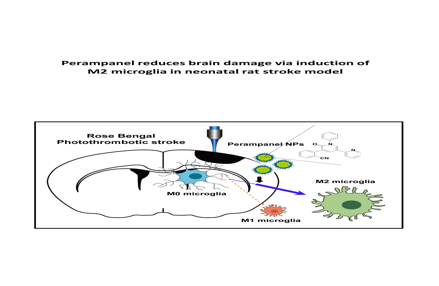
    Graphical Abstract:

    Keywords: ischemic stroke, neonate, poly lactic-co-glycolic acid, PLGA, nanoparticle, perampanel, microglial polarization

    Graphical Abstract:

    Introduction

    Perinatal ischemic stroke occurs in 1 per 4000 live births1 and causes significant morbidity and severe long-term neurologic sequelae in children. The pathophysiology of perinatal stroke may often relate to the unique environmental factors that surround the time of birth, with a relatively low recurrence rate, in contrast to adult cases.2 For the treatment of ischemic stroke, recanalization of blood vessels with thrombolytic or endovascular treatments can be helpful.3,4 However, this has a narrow therapeutic window of 4.5 hours from the onset of a stroke in certain eligible patients, and it is currently rarely performed in newborns.5 Thus, there is still no standard acute therapy available for perinatal stroke, and the focus is on supportive treatment and neuroprotection. It is important to investigate effective treatments for perinatal ischemic stroke.

    In response to ischemic stroke, microglia are activated and polarized to the pro-inflammatory M1 phenotype or the anti-inflammatory M2 phenotype. Also, pro-inflammatory M1 microglia, as the primary source of cytokines and free radicals, can induce secondary brain damage.6 Control of microglial polarization can be an important therapeutic strategy. The immunomodulatory molecules generated by these microglia, such as cytokines and chemokines, are closely associated with secondary brain damage or repair, respectively, following ischemic stroke. In the MCAO-induced ischemic stroke model, M1 phenotype microglia were increased, and ischemic lesions induced microglial inflammation in the ipsilateral cortex.7 Therefore, investigating microglial changes and their functions is crucial to understanding ischemic stroke pathophysiology.

    Nanoparticles have been widely applied in biomedical fields for use in diagnosis, biosensing, and drug delivery. Recently, many nanoparticles have been reported to be useful vehicles for delivery of drugs to inhibit the over-activation of microglia and neuro-inflammation.8,9 Biodegradable polymeric nanoparticles such as poly lactic-co-glycolic acid (PLGA) and poly lactic acid (PLA) have been well studied as stable drug carriers for drug delivery. Among them, PLGA has proven to be effective as a drug delivery method for gene regulators such as siRNAs, plasmids, and miRNA.10–12 PLGA nanoparticles (NPs) have also been approved for pharmaceutical application by the US Food and Drug Administration.13 Moreover, recent studies have shown that PLGA NPs are preferentially localized to microglia.14 Among the cells in CNS, microglia were shown to take up 63.41% of rhodamine-conjugated PLGA NPs at 24 hours.8,15

    Perampanel (PER) is a noncompetitive α-amino-3-hydroxyl-5-methyl-4-isoxazolepropionate (AMPA) receptor antagonist that is clinically used for seizure control. AMPA receptors are postsynaptic ionotropic excitatory receptors with binding sites for glutamate that are thought to participate in the induction of seizures by synchronizing excitatory glutamatergic signaling.16 In preclinical studies, PER was found to be effective in preventing seizures in a seizure model,17 and was studied in vitro as an inhibitor of the dose-dependent AMPA-mediated increase in intracellular calcium.18 Moreover, recent studies have reported that PER has neuroprotective effects in experimental models of stroke19,20 and traumatic brain injury.21 The AMPA receptor, the target of PER, is present not only in neurons but also in activated microglia.22 By examining the action on the AMPA receptor of PER contained in nanoparticles with high affinity for microglia, it may be possible to target this receptor for perinatal stroke treatment. There is still no research of the action of PER on microglia for the treatment of perinatal stroke. Based on the distribution of drug-loaded PLGA NPs, it is reasonable to hypothesize that PER-loaded NPs will influence microglia directly. In the present study, we investigated the effects of PER-loaded NPs on microglial polarization in a perinatal ischemic stroke rat model.

    More at link.