Changing stroke rehab and research worldwide now.Time is Brain! trillions and trillions of neurons that DIE each day because there are NO effective hyperacute therapies besides tPA(only 12% effective). I have 523 posts on hyperacute therapy, enough for researchers to spend decades proving them out. These are my personal ideas and blog on stroke rehabilitation and stroke research. Do not attempt any of these without checking with your medical provider. Unless you join me in agitating, when you need these therapies they won't be there.

What this blog is for:

My blog is not to help survivors recover, it is to have the 10 million yearly stroke survivors light fires underneath their doctors, stroke hospitals and stroke researchers to get stroke solved. 100% recovery. The stroke medical world is completely failing at that goal, they don't even have it as a goal. Shortly after getting out of the hospital and getting NO information on the process or protocols of stroke rehabilitation and recovery I started searching on the internet and found that no other survivor received useful information. This is an attempt to cover all stroke rehabilitation information that should be readily available to survivors so they can talk with informed knowledge to their medical staff. It lays out what needs to be done to get stroke survivors closer to 100% recovery. It's quite disgusting that this information is not available from every stroke association and doctors group.

Showing posts with label sarcasm. Show all posts

Wednesday, August 19, 2026

Seven mistakes a personal trainer says seniors should stop making at the gym

 You competent? doctor and therapist taught you all this already at the hospital gym, right? Sarcasm tag needed here!

Seven mistakes a personal trainer says seniors should stop making at the gym

Strength training is not solely the domain of bodybuilders; it is a crucial component of health and independence for everyone, particularly as we age.

When executed correctly, it plays a pivotal role in preserving bone density and building muscle mass, significantly enhancing overall quality of life.

However, the prospect of entering a gym can be daunting, and improper use of equipment poses risks, making safety paramount.

For seniors embarking on a strength training journey, understanding common pitfalls is essential to ensure a safe and effective start.

Here are seven common mistakes to watch out for when starting strength training as a senior.

1. Starting too heavy

“Our muscles, bones and connective tissues take time to adapt to resistance training, so if you start with weights that are too heavy, it creates unnecessary injury risk. It can also encourage poor movement patterns that are much harder to correct later,” explains Michael Betts, personal trainer and director of TRAINFITNESS.

Betts advises seniors looking to embark on their strength training journey to start off with body weight exercises or very light weights that almost feel too easy.

“Aim for 12-15 reps with perfect form before starting to think about increasing the weight,” recommends the personal trainer. “Approaching it like this builds strength progressively while at the same time teaching your nervous system the proper movement patterns to complete the exercises safely.”

2. Prioritising repetitions over technique

Sloppy technique doesn’t just stall progress; it increases injury risk.

“Prioritise slow, controlled movements over the amount of resistance or number of repetitions,” advises Leo Mathew, physiotherapist and rehabilitation service lead at Nellsar Care Homes. “Good technique helps prevent injuries and supports gradual progress.”

This is why seeking professional advice when starting out can be extremely beneficial.

“Invest in a few sessions with a qualified trainer who has experience working with older adults,” recommends Betts.

“They can assess your movement quality, design an appropriate programme and teach you proper form. This upfront investment prevents costly mistakes and gives you the confidence to train independently.”

3. Forgetting about a mobility warm-up

“If you’re strong but don’t have mobility, your movement patterns will be generally imbalanced and your exercise options will be limited,” says Betts.

“A lot of seniors jump straight into lifting before addressing issues like tight hips, rounded shoulders or limited mobility in their ankles which have developed from years of sitting.”

Betts recommends spending 10-15 minutes before a weights session working on dynamic stretches and mobility exercises to make your weight session more effective and reduce your risk of injury.

“The focus should be on your hips, shoulders and thoracic spine – the last of which is very commonly neglected,” says the personal trainer. “Preparing in this way prevents compensatory movements which are more likely to lead to injury and also ensures that the exercises are working the muscles they should be.”

4. Ignoring pain

Many people think pain is normal when it comes to strength training, but it’s important to listen to your body.

“Stop immediately if experiencing new pain, dizziness, or sharp discomfort – never force through these symptoms. Take advice if required.”

5. Neglecting balance and stability

“Traditional strength training often uses machines and supported positions that aren’t challenging for our balance systems,” notes Betts. “Balance is a fundamental part of functional strength that helps prevent falls and improves the quality of day-to-day movement – things that are incredibly important as we age.

“Include single-leg exercises, standing exercises and movements that challenge your stability. Simple progressions like standing on one foot, single-leg dead lifts or standing exercises with eyes closed help maintain the balance systems that naturally decline with age.”

6. Not allowing time for rest and recovery

Rest days from the gym are not lazy, they are crucial for your body’s recovery and for any long-term progress.

“Allow adequate rest during your exercise to improve strength, endurance and recovery,” advises Mathew. “Allow 2-3 rest days per week for muscle repair. Stay well-hydrated and eat nutritious foods to support and fuel recovery, performance and to maintain quality of life.”

7. Not eating enough protein

“Sufficient caloric and protein intake is required to build muscle and strength and repair from workouts and physical stress on the body,” explains Sam Quinn, personal training lead at Nuffield Health.

“We naturally maintain less muscle mass as we get older, and consuming the correct amount of protein to support our physical health and exercise goals is even more important. Between 1.1-2g of protein per kilogram of body weight is recommended to build strength and recover from your workouts.”

Good sources of protein include lean meats, poultry, fish, eggs, dairy products, beans, lentils, tofu, nuts and seeds.

The Independent is the world’s most free-thinking news brand, providing global news, commentary and analysis for the independently-minded. We have grown a huge, global readership of independently minded individuals, who value our trusted voice and commitment to positive change. Our mission, making change happen, has never been as important as it is today.

4 simple balance exercises you can do at home — no equipment needed

 You won't need these because your excellent doctors and therapists got you recovered so well, right! Sarcasm tag needed here! 

Videos at link.

4 simple balance exercises you can do at home — no equipment needed

If you’re just starting out with balance training, a routine of basic static and dynamic exercises is a great way to go, says Kimberly M. Burbank, a sports medicine physician at UCLA Health. The exercises are deceptively simple and easy to master. They require only a minute each — an entire routine is just five minutes a day. You can do the exercises at home, with little to no equipment, and you can easily progress with hand-held weights, if desired.

In fact, short investments of time, done consistently, is the best approach, Burbank says.

Good balance is beneficial for living a longer, healthier life — at any age. Check out our series featuring the latest research, a balance IQ test and simple at-home exercises.

“Generally, five minutes a day is better than 30 minutes all at once if you can do it daily or at least three times a week,” Burbank says. “Because you can train the neuromuscular system by frequently challenging it. And it’s easier to progress, or become better at it, the more frequently you do it.”

So what are static and dynamic exercises, exactly?

Static exercises mean you’re holding one position and staying still for the allotted amount of time. They’re especially helpful for postural control — you have to maintain good posture to stand on one leg — whereas dynamic exercises mean you’re maintaining control while moving through space. They hone coordination and proprioception skills (the awareness of where your body is in space) as well as joint positioning (like being cognizant of how your knee is bent in a lunge, for example, and adjusting that).


“So standing on one leg and holding it versus a lunge, where you’re moving while balancing,” Burbank says to illustrate the difference. “Static and dynamic exercises work in harmony together — you want control while standing and control while walking or moving through space.”

Consider static and dynamic exercises “the meat and potatoes” of balance training, Burbank says.

“They allow you to build a great balance foundation that you can then build upon with more complex balance training maneuvers or activities that require more balance — you can then go take that surf class or hop on a skateboard. Or, for older populations, safely walk through Disney World with more confidence knowing there’s a less likelihood of falling. It’s about confidence in day-to-day living.”

Here are four of Burbank’s favorite static and dynamic exercises to help you strengthen your balance. They’re demonstrated by Los Angeles Times staffer Christopher Buchanan.

1. Static airplane shallow squat (to build hip stability and balance control). Stand on one leg and slightly bend your standing knee. Lean your chest forward while extending the other leg behind you like an airplane. Hold for 5-10 seconds per side, for 2-3 sets per side, with 30-45 seconds rest between sets. (To progress the exercise, increase the depth of the squat and/or hold light weights.)


2. Static side lean balance hold (to challenge side-to-side stability and core strength). Stand tall on one leg and gently lean your upper body to one side while maintaining your balance. Hold for 15-20 seconds per side, for 2 reps per side, with 15-30 seconds rest between sets. (To progress the exercise, hold a small weight in one hand or add opposite arm reaches.)

3. Dynamic heel-to-toe walk (to train walking balance and coordination). Walk in a straight line placing the heel of one foot directly in front of the toes of the other foot. Take 15-20 steps, for 2-3 reps, with 30 seconds between reps. (To progress the exercise, walk backward or perform it on an uneven surface, such as grass or sand.)

4. Dynamic body weight lunges (to strengthen the legs, hips and core while improving balance during movement). Step one foot forward and lower your body until both knees are bent, ideally at a 90-degree angle. Push back up and switch sides. Do 8-12 reps per side, for 2-3 sets, with 45-60 seconds rest between sets. (To progress the exercise, hold light dumbbells or increase the depth of the lunge.)


Now that you are familiar with beginner balance exercises, try these powerful — and impactful — plyometric exercises for improving balance. 

Tuesday, May 19, 2026

It May Be Easier To Strengthen Your Brain Than Scientists Once Thought by mindbodygreen

 Your competent? doctor should already have all this information because they are studiously following brain research! (Should be a sarcasm tag here.)

It May Be Easier To Strengthen Your Brain Than Scientists Once Thought

For years, the prevailing belief was that cognitive decline is simply part of getting older: something to accept rather than actively address. But a growing body of research is challenging that assumption, suggesting the brain may remain far more adaptable throughout adulthood than scientists once believed.

New research adds compelling evidence1 to this shift, following nearly 4,000 adults over three years to measure changes in brain health—and the results suggest your daily habits may play a bigger role in long-term brain function than previously thought.

How the research was designed

The brain's capacity to change and adapt (known as neuroplasticity) has been well-documented in younger populations, but researchers have long debated how much this adaptability persists into middle age and beyond. This study set out to measure whether brain health could meaningfully improve across the adult lifespan with consistent engagement in cognitive training and lifestyle interventions.
Researchers at the Center for BrainHealth at The University of Texas at Dallas followed 3,966 adults ages 19 to 94 for three years. They developed a multidimensional assessment called the BrainHealth Index (BHI) to measure three core areas:

  • Clarity: cognitive function, including focus, reasoning, and memory
  • Connectedness: social engagement and sense of purpose
  • Emotional balance: mental well-being and stress regulation
Participants completed the BHI assessment every six months while engaging with an online platform that offered cognitive training, lifestyle modules, and one-on-one coaching.

Gains showed up at every age

Over three years, participants showed sustained improvements in overall brain health and all three component areas, regardless of where they started. Those who engaged more consistently with the training tools, strategy-based learning, and brain-healthy habits showed the greatest gains. Improvements were observed across age groups, genders, and education levels.

The concept of "brain health span" refers to how long a person maintains strong cognitive and emotional functioning throughout life: not just the absence of disease, but the presence of mental sharpness, emotional resilience, and purpose-driven engagement. The study authors describe extending brain health span as "critical to aligning health span with lifespan," essentially helping people live well for as long as they live.

Participants in this study were self-selected and likely highly motivated; they chose to sign up for a brain health program, which may not reflect the general population. Additionally, several study authors are inventors on a pending patent for the BrainHealth Index platform being studied, representing a potential conflict of interest.

Habits that support a sharper, more resilient brain

The study reinforces what neuroscience research has been pointing toward: consistent, everyday habits may help strengthen cognitive and emotional resilience over time.

Here are practical ways to put that into action:(I'm good at all of these)

Challenge your brain strategically: engage in mentally demanding activities that push you slightly beyond your comfort zone, such as learning a new skill, tackling complex problems, or having deep conversations that require synthesis and reasoning. Writing this blog
Prioritize social connection and purpose: the study's "Connectedness" factor highlights that relationships and a sense of meaning matter for brain health, so nurturing friendships, engaging in community, and pursuing activities that feel purposeful can all contribute. Many social connections.
Manage stress and protect your sleep: chronic stress and poor sleep are well-documented threats to cognitive function, so building in recovery time, practicing stress-regulation techniques, and prioritizing consistent sleep supports both emotional balance and mental clarity.NO stress once retired.
Move your body regularly: physical exercise increases blood flow to the brain and supports the growth of new neural connections, and even moderate, consistent movement counts. Regular walks in the 124 acre natural area close by.
Choose active engagement over passive consumption: the study found that higher engagement with training tools correlated with greater gains, and applying that principle more broadly, activities that require active participation (reading, problem-solving, creating) may benefit the brain more than passive scrolling. Writing this blog.
Focus on consistency over intensity: the participants who saw the greatest improvements were doing it regularly, so building sustainable brain-healthy habits over time appears to matter more than occasional bursts of effort. Yep.

The takeaway

This research adds to a growing body of evidence suggesting that brain health isn't fixed. The choices you make today may help shape your cognitive and emotional well-being for years to come.

Tuesday, March 11, 2025

Just one minute a week could slash your risk of stroke by almost 50% - flossing


Or your doctor could have competently years ago created a protocol on all this that reduces stroke risk by 307%.  Put all these together and you'll never have a stroke,(sarcasm tag here).

Like maybe a 307%  stroke risk reduction from these 11 possibilities?  

Just one minute a week could slash your risk of stroke by almost 50%

A new study has revealed that flossing once a week at a minimum can slash your risk of a stroke by 44%.


Vertigo dizzynes

Inflammation can contribute to stroke risk (Image: Getty )

Recent research has uncovered that adopting a straightforward weekly habit could dramatically reduce your stroke risk by nearly 50%. This revelation comes after a new study at the American Stroke Association's International Stroke Conference 2025 highlighted the 'lifesaving' advantages of regular teeth flossing.

The study revealed that flossing at least once a week, minimum, can significantly decrease the risk of stroke caused by a blood clot obstructing brain blood flow and irregular heart rhythms by up to 44%. The conference, held in Los Angeles, is a globally renowned gathering for researchers and clinicians committed to the science of stroke and brain health.

Despite the numerous benefits of flossing—not just for maintaining fresh breath or preventing cavities but also for overall wellness—reports indicate that around 33% of British adults never floss. Dental experts emphasise the importance of flossing as a key part of oral hygiene, alongside brushing twice daily and using mouthwash, even though many people neglect this step.

Poor dental health can significantly heighten your risk of stroke, heart disease, systemic inflammation, and even Alzheimer's disease. While flossing can be done as infrequently as once a week, it should ideally be a daily routine.

Flossing an adult set of teeth typically takes between one to two minutes, although the exact duration depends on how thorough you are, reports Surrey Live.

Thursday, May 30, 2024

Third Thumb: Public Quickly Adapts to Controllable Extra Thumb

 I'm absolutely positive your competent? doctor will see this and immediately implement protocols to use this for stroke rehab. I wish there was a sarcasm punctuation tag.

Third Thumb: Public Quickly Adapts to Controllable Extra Thumb

Summary: Researchers found that people easily learned to use a controllable, prosthetic “Third Thumb” for manipulating objects. The device, tested on a diverse group, enhances motor capabilities and shows promise for both productivity and aiding those with disabilities. The study highlights the importance of inclusive design in developing new technologies.

Key Facts:

  • 98% of participants successfully used the Third Thumb within a minute.
  • The device was tested on 596 participants aged 3 to 96.
  • Inclusive design is crucial for ensuring accessibility and functionality for all.

Source: University of Cambridge

Cambridge researchers have shown that members of the public have little trouble in learning very quickly how to use a third thumb – a controllable, prosthetic extra thumb – to pick up and manipulate objects.

The team tested the robotic device on a diverse range of participants, which they say is essential for ensuring new technologies are inclusive and can work for everyone.

An emerging area of future technology is motor augmentation – using motorised wearable devices such as exoskeletons or extra robotic body parts to advance our motor capabilities beyond current biological limitations.

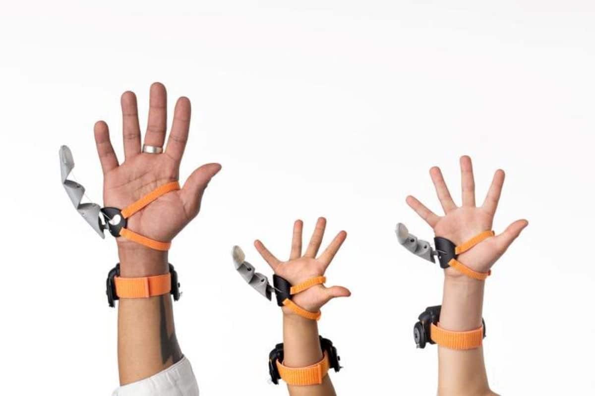
This shows people wearing the thumb.
The Third Thumb worn by different users. Credit: Dani Clode Design / The Plasticity Lab

While such devices could improve the quality of life for healthy individuals who want to enhance their productivity, the same technologies can also provide people with disabilities new ways to interact with their environment.

Professor Tamar Makin from the Medical Research Council (MRC) Cognition and Brain Sciences Unit at the University of Cambridge said: “Technology is changing our very definition of what it means to be human, with machines increasingly becoming a part of our everyday lives, and even our minds and bodies.

“These technologies open up exciting new opportunities that can benefit society, but it’s vital that we consider how they can help all people equally, especially marginalised communities who are often excluded from innovation research and development.

“To ensure everyone will have the opportunity to participate and benefit from these exciting advances, we need to explicitly integrate and measure inclusivity during the earliest possible stages of the research and development process.”

Dani Clode, a collaborator within Professor Makin’s lab, has developed the Third Thumb, an extra robotic thumb aimed at increasing the wearer’s range of movement, enhancing their grasping capability and expanding the carrying capacity of the hand.

This allows the user to perform tasks that might be otherwise challenging or impossible to complete with one hand or to perform complex multi-handed tasks without having to coordinate with other people.

The Third Thumb is worn on the opposite side of the palm to the biological thumb and controlled by a pressure sensor placed under each big toe or foot. Pressure from the right toe pulls the Thumb across the hand, while the pressure exerted with the left toe pulls the Thumb up toward the fingers.

The extent of the Thumb’s movement is proportional to the pressure applied, and releasing pressure moves it back to its original position.

In 2022, the team had the opportunity to test the Third Thumb at the annual Royal Society Summer Science Exhibition, where members of the public of all ages were able to use the device during different tasks.

The results are published today in Science Robotics.

Over the course of five days, the team tested 596 participants, ranging in age from three to 96 years old and from a wide range of demographic backgrounds. Of these, only four were unable to use the Third Thumb, either because it did not fit their hand securely, or because they were unable to control it with their feet (the pressure sensors developed specifically for the exhibition were not suitable for very lightweight children).

Participants were given up to a minute to familiarise themselves with the device, during which time the team explained how to perform one of two tasks.

The first task involved picking up pegs from a pegboard one at a time with just the Third Thumb and placing them in a basket. Participants were asked to move as many pegs as possible in 60 seconds. 333 participants completed this task.

The second task involved using the Third Thumb together with the wearer’s biological hand to manipulate and move five or six different foam objects. The objects were of various shapes that required different manipulations to be used, increasing the dexterity of the task.

Again, participants were asked to move as many objects as they could into the basket within a maximum of 60 seconds. 246 participants completed this task.

Almost everyone was able to use the device straightaway. 98% of participants were able to successfully manipulate objects using the Third Thumb during the first minute of use, with only 13 participants unable to perform the task.

Ability levels between participants were varied, but there were no differences in performance between genders, nor did handedness change performance – despite the Thumb always being worn on the right hand.

There was no definitive evidence that people who might be considered ‘good with their hands’ – for example, they were learning to play a musical instrument, or their jobs involved manual dexterity – were any better at the tasks.

Older and younger adults had a similar level of ability when using the new technology, though further investigation just within the older adults age bracket revealed a decline in performance with increasing age.

The researchers say this effect could be due to the general degradation in sensorimotor and cognitive abilities that are associated with ageing and may also reflect a generational relationship to technology.

Performance was generally poorer among younger children. Six out of the 13 participants that could not complete the task were below the age of 10 years old, and of those that did complete the task, the youngest children tended to perform worse compared to older children. But even older children (aged 12-16 years) struggled more than young adults.

Dani said: “Augmentation is about designing a new relationship with technology—creating something that extends beyond being merely a tool to becoming an extension of the body itself.

“Given the diversity of bodies, it’s crucial that the design stage of wearable technology is as inclusive as possible. It’s equally important that these devices are accessible and functional for a wide range of users. Additionally, they should be easy for people to learn and use quickly.”

Co-author Lucy Dowdall, also from the MRC Cognition and Brain Science Unit, added: “If motor augmentation – and even broader human-machine interactions – are to be successful, they’ll need to integrate seamlessly with the user’s motor and cognitive abilities.

“We’ll need to factor in different ages, genders, weight, lifestyles, disabilities – as well as people’s cultural, financial backgrounds, and even likes or dislikes of technology. Physical testing of large and diverse groups of individuals is essential to achieve this goal.”

There are countless examples of where a lack of inclusive design considerations has led to technological failure:

  • Automated speech recognition systems that convert spoken language to text have been shown to perform better listening to white voices over Black voices.
  • Some augmented reality technologies have been found to be less effective for users with darker skin tones.
  • Women face a higher health risk from car accidents, due to car seats and seatbelts being primarily designed to accommodate ‘average’ male-sized dummies during crash testing.
  • Hazardous power and industrial tools designed for a right-hand dominant use or grip have resulted in more accidents when operated by left-handers forced to use their non-dominant hand.

Funding: This research was funded by the European Research Council, Wellcome, the Medical Research Council and Engineering and Physical Sciences Research Council.

About this neurotech and neuroplasticity research news

Author: Craig Brierley
Source: University of Cambridge
Contact: Craig Brierley – University of Cambridge
Image: The image is credited to Dani Clode Design / The Plasticity Lab

Original Research: Closed access.
“Evaluating Initial Usability of a Hand Augmentation Device Across a Large and Diverse Sample” by Tamar Makin et al. Science Robotics


Sunday, September 20, 2020

Graphene-Based Nanoparticles as Potential Treatment Options for Parkinson’s Disease: A Molecular Dynamics Study

You might very well need this, so have your doctor and hospital follow this closely and create protocols for this.  Of course your doctors already know about graphene from this in February 2016, sorry there is no sarcasm marker that I can use for that last statement.

Graphene Shows Promise For Brain Implants February, 2016

Parkinson’s Disease May Have Link to Stroke March 2017 

The latest here:

Graphene-Based Nanoparticles as Potential Treatment Options for Parkinson’s Disease: A Molecular Dynamics Study

Authors Alimohammadi E, Khedri M, Miri Jahromi A, Maleki R, Rezaian M

Received 1 June 2020

Accepted for publication 8 September 2020

Published 18 September 2020 Volume 2020:15 Pages 6887—6903

DOI https://doi.org/10.2147/IJN.S265140

Checked for plagiarism Yes

Review by Single-blind

Peer reviewer comments 2

Editor who approved publication: Prof. Dr. Anderson Oliveira Lobo


Ehsan Alimohammadi1 *,* Mohammad Khedri2 *,* Ahmad Miri Jahromi,3 Reza Maleki,4 Milad Rezaian5

1Neurosurgery Department, Kermanshah University of Medical Sciences, Kermanshah, Iran; 2Department of Chemical Engineering, Amirkabir University of Technology (Tehran Polytechnic), Tehran 1591634311, Iran; 3Department of Petroleum Engineering, Amirkabir University of Technology (Tehran Polytechnic), Tehran 1591634311, Iran; 4Department of Chemical Engineering, Sharif University of Technology, Tehran, Iran; 5Department of Pharmacology, School of Medicine, Shahid Beheshti University of Medical Sciences, Tehran 19839-63113, Iran

*These authors contributed equally to this work

Correspondence: Reza Maleki
Department of Chemical Engineering, Sharif University of Technology, Tehran, Iran
Email Rezamaleki96@gmail.com

Introduction: The study of abnormal aggregation of proteins in different tissues of the body has recently earned great attention from researchers in various fields of science. Concerning neurological diseases, for instance, the accumulation of amyloid fibrils can contribute to Parkinson’s disease, a progressively severe neurodegenerative disorder. The most prominent features of this disease are the degeneration of neurons in the substantia nigra and accumulation of α-synuclein aggregates, especially in the brainstem, spinal cord, and cortical areas. Dopamine replacement therapies and other medications have reduced motor impairment and had positive consequences on patients’ quality of life. However, if these medications are stopped, symptoms of the disease will recur even more severely. Therefore, the improvement of therapies targeting more basic mechanisms like prevention of amyloid formation seems to be critical. It has been shown that the interactions between monolayers like graphene and amyloids could prevent their fibrillation.
Methods: For the first time, the impact of four types of last-generation graphene-based nanostructures on the prevention of α-synuclein amyloid fibrillation was investigated in this study by using molecular dynamics simulation tools.
Results: Although all monolayers were shown to prevent amyloid fibrillation, nitrogen-doped graphene (N-Graphene) caused the most instability in the secondary structure of α-synuclein amyloids. Moreover, among the four monolayers, N-Graphene was shown to present the highest absolute value of interaction energy, the lowest contact level of amyloid particles, the highest number of hydrogen bonds between water and amyloid molecules, the highest instability caused in α-synuclein particles, and the most significant decrease in the compactness of α-synuclein protein.
Discussion: Ultimately, it was concluded that N-Graphene could be the most effective monolayer to disrupt amyloid fibrillation, and consequently, prevent the progression of Parkinson’s disease.

Keywords: α-synuclein, amyloid, graphene, Parkinson’s disease, molecular dynamics

  This work is published and licensed by Dove Medical Press Limited. The full terms of this license are available at https://www.dovepress.com/terms.php and incorporate the Creative Commons Attribution - Non Commercial (unported, v3.0) License. By accessing the work you hereby accept the Terms. Non-commercial uses of the work are permitted without any further permission from Dove Medical Press Limited, provided the work is properly attributed. For permission for commercial use of this work, please see paragraphs 4.2 and 5 of our Terms.

Download Article [PDF]  View Full Text [HTML]

Saturday, September 28, 2019

Psychosocial working conditions, trajectories of disability, and the mediating role of cognitive decline and chronic diseases: A population-based cohort study

In my last 6 years working as a contractor there was zero social support at work.  My social support now that I'm retired is terrific.  I have no plans to get a chronic disease, that's what resveratrol in red wine and all my walking will do for me. I think my cognitive function is still pretty damn good, sarcasm still works. 

The highest form of intelligence: Sarcasm increases creativity for both expressers and recipients

The latest here:

Psychosocial working conditions, trajectories of disability, and the mediating role of cognitive decline and chronic diseases: A population-based cohort study 

PLoS Medicine — Pan KY, Xu W, Mangialasche F, et al. | September 20, 2019

In this study, researchers examined the association between job demand–control–support combinations and trajectories of disability in later life. In addition, they explored the role of cognitive decline and the co-occurrence of chronic diseases in mediating this association. They examined a cohort of 2,937 individuals aged 60 years and older for the association of psychosocial working conditions with the rate of disability progression over 12 years. In all, they noted a link between unfavorable psychosocial working conditions during working life and the progression of disability in later life. This association, they found, was partially related to the decrement in cognitive function and increase in chronic-disease burden, and especially the former. Findings emphasized the significance of social support at work in a high-strain work environment, given its capacity to attenuate the impact of high-strain jobs on disability accumulation.
Read the full article on PLoS Medicine

Thursday, November 23, 2017

Blue-Light Therapy following Mild Traumatic Brain Injury: Effects on White Matter Water Diffusion in the Brain

Would this help stroke patients? I don't think they are referring to the blue light of your smartphones or iPads. Anything would have been helpful to counteract the massive fatigue I had post-stroke.  Or you can ask your doctor about the effectiveness of near-infrared light since it is only two years old and your doctor and stroke hospital are so efficient they already have incorporated infrared into their stroke protocols. (sarcasm)

New Study Discovers Near-Infrared Light Therapy (NILT) Effectively Treats Traumatic Brain Injury (TBI) Patients  Aug. 2015

Blue-Light Therapy following Mild Traumatic Brain Injury: Effects on White Matter Water Diffusion in the Brain

  • Social, Cognitive and Affective Neuroscience Laboratory (SCAN Lab), Department of Psychiatry, College of Medicine, University of Arizona, Tucson, AZ, United States
Mild traumatic brain injury (mTBI) is a common and often inconspicuous wound that is frequently associated with chronic low-grade symptoms and cognitive dysfunction. Previous evidence suggests that daily blue wavelength light therapy may be effective at reducing fatigue and improving sleep in patients recovering from mTBI. However, the effects of light therapy on recovering brain structure remain unexplored. In this study, we analyzed white matter diffusion properties, including generalized fractional anisotropy, and the quantity of water diffusion in isotropic (i.e., isotropic diffusion) and anisotropic fashion (i.e., quantitative anisotropy, QA) for fibers crossing 11 brain areas known to be significantly affected following mTBI. Specifically, we investigated how 6 weeks of daily morning blue light exposure therapy (compared to an amber-light placebo condition) impacted changes in white matter diffusion in individuals with mTBI. We observed a significant impact of the blue light treatment (relative to the placebo) on the amount of water diffusion (QA) for multiple brain areas, including the corpus callosum, anterior corona radiata, and thalamus. Moreover, many of these changes were associated with improvements in sleep latency and delayed memory. These findings suggest that blue wavelength light exposure may serve as one of the potential non-pharmacological treatments for facilitating structural and functional recovery following mTBI; they also support the use of QA as a reliable neuro-biomarker for mTBI therapies.


Introduction

Mild traumatic brain injury (mTBI) is a common and often unobtrusive wound that occurs when kinetic energy is transferred to the brain through some form of traumatic event, such as a fall, blow to the head, or blast wave. While there are typically no exceptionally conspicuous physical or neuroimaging signs of mTBI, the mechanical trauma to the brain leads to a mild temporary disruption of consciousness or other alteration of ongoing cognition. Also commonly known as “concussion,” mTBI can further lead to persistent alterations in neuropsychological functions, including changes in mood (e.g., depression), poor attention and concentration, and memory problems (1, 2). Importantly, sleep deprivation is also known to produce many of these same symptoms (3, 4). It is therefore possible that sleep disturbances following mTBI may cause, or at least exacerbate, ongoing post-concussion symptoms. However, the nature of these complaints and their contribution to the experience of daytime sleepiness is not well understood (5). An objective measure of daytime sleepiness is the multiple sleep latency test (MSLT), which is used to determine the time it takes an individual to fall asleep (sleep onset latency) when given the opportunity to take a nap. Following a head trauma, symptoms are believed to result from neuronal damage in the form of diffuse axonal injury (6, 7), leading to the release of specific proteins that in turn promote maladaptive functional and structural changes within the brain (8). Identifying neuro-markers of these changes remains an important challenge in ongoing attempts to understand and treat mTBI and post-concussive symptoms.
A very limited number of treatment options for mTBI have been proposed and experimentally validated. Available treatments include cognitive behavior therapy (9), neuropsychological rehabilitation (10), educational intervention (11), and pharmacological intervention (12). Although the effects are small, some intervention studies report reliable reductions in post-concussion symptoms, including sleep problems, following successful treatment (13). Considering a range of post-concussion symptoms can also occur as the result of sleep loss, it is likely that improving sleep quality in particular would also lead to improvements of other post-concussion symptoms, such as attention, concentration, memory, and mood disturbances. While improving sleep makes sense, this is often easier said than done. A natural and potentially powerful method for regulating the sleep–wake cycle is through targeted exposure to bright light in the morning hours. Exposure to short wavelength light (~430–475 nm; blue wavelength light) has been demonstrated as an alternative to pharmacological treatment methods that focus on improving alertness, concentration, daytime sleepiness, as well as sleep quality (14, 15). Intrinsically photosensitive retinal ganglion cells are particularly responsive to light within the short wavelengths. These cells transmit signals to hypothalamic nuclei, which in turn regulate the production of melatonin (16, 17). Morning exposure to blue wavelength light leads to a suppression of melatonin production, which contributes to a phase delay and stabilization of the circadian rhythm (18), increases daytime alertness and vigilance, and earlier onset of evening sleep (19, 20). Interestingly, a recent clinical trial showed that 4 weeks of 45 min of morning blue-light therapy (BLT) in comparison to longer wavelength placebo light was effective at reducing self-rated fatigue and daytime sleepiness among individuals recovering from TBI (21). However, the extent to which these behavioral changes correspond to structural changes within the brain has not been explored.
When considering the potential influences of BLT on mTBI, it is important to consider that mTBI is associated with microscopic changes in brain structure, particularly within the white matter axonal tracts. Abnormalities in fractional anisotropy (FA) in the brain following an mTBI have been studied extensively using diffusion tensor imaging (DTI), a method that allows high-resolution imaging of the directional movement of water molecules along axonal fiber tracts (i.e., how fast water molecules move along fiber tracts). Abnormalities in FA in individuals with an mTBI are reported in areas such as uncinate fasciculus (UF) (22), superior longitudinal fasciculus (SLF) (23), anterior corona radiata (ACR) (22), corpus callosum (CC) (24), and thalamus (25). Alterations in FA within (a) UF are reported to be associated with changes in Mini-Mental State Examination (MMSE) scores (cognitive function) and specifically, memory performance (22, 26); (b) SLF and CC are reported to be associated with executive function (attention and memory) (27); (c) ACR changes are correlated with changes in attention (22); and (d) anterior thalamic nucleus changes are also linked to changes in executive function, memory, and attention (25). In addition, studies have found that individuals with mTBI show alterations in white matter within the frontal lobe (frontal cortex/dorsolateral prefrontal cortex, DLPFC), and that these alterations are correlated with lower executive control and related cognitive functions (28). Also, compared to healthy controls (HCs), there are multiple studies that have reported abnormally high FA values in individuals with mTBI within several areas, including the genu and splenium of CC, ACR (bilaterally), lUF, and internal capsule (IC; bilaterally) (29, 30). Recently, new diffusion measures—quantitative anisotropy (QA), isotropic diffusion (ISO), and generalized fractional anisotropy (GFA)—were introduced to the field of DTI for the analysis of diffusion properties of white matter (31). QA and ISO represent how much water diffuses (i.e., density) in a specific/restricted direction and in an isotropic fashion (i.e., total isotropic component), respectively. In contrast, GFA, which is calculated from an orientation distribution function, is a measure of how fast water diffuses (i.e., diffusivity) in an anisotropic fashion, i.e., it represents degree to which diffusion is anisotropic (31, 32). Highly significant correlations between FA and GFA were reported in the past (33). In addition, the difference between QA and GFA pertains to the fact that QA is a measure of water diffusion along each fiber orientation, whereas GFA/FA is defined for each voxel. Compared to GFA/FA, QA is also reported to have lower susceptibility to partial volume effects of crossing-fibers, free-water diffusion in ventricles, and non-diffusive particles (31). Moreover, normalization of QA helps to stabilize the spin-density measurement across subjects. In this study, we investigated multiple diffusion measures (i.e., diffusivity as well as density measures) simultaneously to better characterize the white matter properties; therefore, in conjunction with GFA, we also estimated normalized QA (NQA) and ISO measures. To the best of our knowledge, no study to date has used these metrics simultaneously to examine the effect of light exposure treatment on the brain following mTBI.
In individuals with mTBI, how changes in post-concussion symptoms following an exposure to BLT may correspond to structural changes within the brain has not yet been explored. Recent evidence suggests that sleep is important for clearing the neurotoxins that build up throughout the day (34) and increases the production of oligodendrocyte progenitor cells that contribute to myelin formation (35), which could conceivably facilitate repair of axonal damage. Based on this, we hypothesized that 6 weeks of daily morning BLT, compared to a placebo condition with an amber-light therapy (ALT) device, would improve sleep and, consequently, lead to changes in white matter water diffusion, improvements in cognitive abilities such as attention and memory, and daytime sleepiness. To this end, we investigated whether individuals in the BLT and ALT groups showed significant changes in diffusion (i.e., GFA, NQA, and ISO), cognitive, and sleep measures. Furthermore, we examined the correlations between changes in diffusion measures from pre- to post-treatment and changes in neuropsychological performance and sleep onset latency.