Changing stroke rehab and research worldwide now.Time is Brain! trillions and trillions of neurons that DIE each day because there are NO effective hyperacute therapies besides tPA(only 12% effective). I have 523 posts on hyperacute therapy, enough for researchers to spend decades proving them out. These are my personal ideas and blog on stroke rehabilitation and stroke research. Do not attempt any of these without checking with your medical provider. Unless you join me in agitating, when you need these therapies they won't be there.

What this blog is for:

My blog is not to help survivors recover, it is to have the 10 million yearly stroke survivors light fires underneath their doctors, stroke hospitals and stroke researchers to get stroke solved. 100% recovery. The stroke medical world is completely failing at that goal, they don't even have it as a goal. Shortly after getting out of the hospital and getting NO information on the process or protocols of stroke rehabilitation and recovery I started searching on the internet and found that no other survivor received useful information. This is an attempt to cover all stroke rehabilitation information that should be readily available to survivors so they can talk with informed knowledge to their medical staff. It lays out what needs to be done to get stroke survivors closer to 100% recovery. It's quite disgusting that this information is not available from every stroke association and doctors group.

Showing posts with label Proteomics. Show all posts

Thursday, March 19, 2026

Blood test predicts stroke 15 years before onset

 Will your doctor and hospital ensure further research that identifies the EXACT PREVENTION PROTOCOLS FOR THIS? 

Do you prefer your doctor, hospital and board of director's incompetence NOT KNOWING? OR NOT DOING? Your choice; let them be incompetent or demand action!

Blood test predicts stroke 15 years before onset

A blood test could predict stroke and other cardiovascular diseases up to 15 years before symptoms appear, researchers say.

The tool, called CardiOmicScore, uses a single blood sample to generate personalised risk scores for six major cardiovascular conditions: coronary artery disease, stroke, heart failure, atrial fibrillation, peripheral artery disease and venous thromboembolism.

The system was developed by a team at the University of Hong Kong’s LKS Faculty of Medicine using deep learning, a type of artificial intelligence that spots patterns in large amounts of data.

The study used large-scale population data from the UK Biobank, combining multiomics data, including genomics, metabolomics and proteomics, and analysing 2,920 circulating proteins and 168 metabolites from blood samples.

Unlike polygenic risk scores, which estimate inherited risk based on a person’s genes and are largely fixed at birth, the tool is designed to reflect a person’s current biological state and how it may be changing over time.

Professor Zhang Qingpeng, associate professor in the department of pharmacology and pharmacy at the university, said: “Genes determine where we start.

“They define our baseline health risk. However, proteins and metabolites reflect our current physical health.

“Our AI tool is designed to decode these complex molecular signals, enabling doctors and patients to identify risks much earlier, which can potentially change the trajectory of disease through timely lifestyle modifications and early prevention.”

The researchers said CardiOmicScore transformed complex multiomics measurements into personalised risk scores with substantially improved predictive performance compared with conventional polygenic risk scores.

When combined with clinical information such as age and gender, it significantly improved risk prediction accuracy and could flag elevated risk up to 15 years before symptoms appear.

Zhang added: “We aim to leverage technology to identify and prevent diseases before they develop.

“By shifting health management from reactive treatment to proactive prediction and intervention, we aim to create a lasting impact for both public health and individual patient care.”

Cardiovascular diseases remain the leading cause of death worldwide, accounting for about 19.8 million deaths in 2022.

Doctors usually assess risk using factors such as age, blood pressure and smoking, but those measures can miss subtle biological changes before disease becomes clinically apparent.

Wednesday, April 16, 2025

New biomarker tracks cognitive decline in Alzheimer’s disease

 With your risk of dementia post stroke your doctor and hospital (If competent) need to create this protocol and have dementia prevention protocols on hand. 

1. A documented 33% dementia chance post-stroke from an Australian study?   May 2012.

2. Then this study came out and seems to have a range from 17-66%. December 2013.`    

3. A 20% chance in this research.   July 2013.

4. Dementia Risk Doubled in Patients Following Stroke September 2018  

Do you prefer your doctor and hospital incompetence NOT KNOWING? OR NOT DOING?


New biomarker tracks cognitive decline in Alzheimer’s disease

At a Glance

  • Scientists uncovered a new biomarker that may help predict cognitive decline in people with Alzheimer’s disease.
  • The findings suggest measures of two proteins could improve early detection of Alzheimer’s disease and help predict or monitor cognitive decline.
Gloved hand holding a test tube with a blood sample. Researchers have been working to develop tests to help detect and track dementia early in the disease process. Westend61 on Offset / Shutterstock

In people with Alzheimer’s disease (AD), changes in the brain gradually erode the ability to think and remember. This cognitive decline involves an abnormal buildup of the proteins amyloid beta (Aβ) and tau. Measures of these protein biomarkers through brain scans or tests of cerebrospinal fluid (CSF) in the brain and spinal cord have improved AD diagnosis.

However, some people with high levels of Aβ and tau have no detectable cognitive problems. Existing biomarkers also can't fully account for the speed of progression from mild cognitive impairment to severe dementia, which can take from 2 to 20 years.

To learn more about the factors that affect cognitive decline, an NIH-funded team led by Drs. Hamilton Se-Hwee Oh and Tony Wyss-Coray at Stanford University analyzed CSF samples from about 3,400 people. The samples were from research studies in the U.S., Sweden, and Finland of people both with and without a diagnosis of AD who had volunteered to participate in the studies over many years.

The researchers used large-scale protein analysis, or proteomics, to measure levels of more than 7,000 proteins in each of the CSF samples. They searched for new proteins that might help explain differences in cognitive impairments, or thinking ability, among people with AD. To do so, they integrated their protein data with other data collected in the studies. Those included Aβ and tau measurements from CSF and brain scans, along with measures of cognitive ability, age, sex, and AD risk genes, including APOE. The results appeared in Nature Medicine on March 31, 2025.

The team found hundreds of proteins whose levels correlated with cognitive function. The most significant ones were related to synapse function. Synapses are the connections between neurons. In addition to the buildup of Aβ and tau, the loss of connections between neurons in the brain is a key feature of AD. Two synapse-related proteins, YWHAG and NPTX2, were the most closely related to measures of cognitive impairment.

The researchers used machine learning to search for patterns in the protein data that could reliably predict cognitive impairment. This analysis showed that a ratio of YWHAG:NPTX2 reflected a person’s cognitive impairment better than existing biomarkers for Aβ and tau.

YWHAG goes up in people with memory problems, while NPTX2 goes down. As a result, the YWHAG:NPTX2 ratio increases in people experiencing cognitive decline. It also rises in those at higher risk of advancing to full-blown dementia. These findings suggest that the YWHAG:NPTX2 ratio might be used to help predict the onset of AD symptoms and track disease progression. The researchers also found that this ratio rises somewhat as people age normally.

The team next used machine learning to try to develop a similar biomarker using less invasive proteomic blood tests. They were able to develop a set of protein measurements that correlated with the CSF YWHAG:NPTX2 ratio and could also help predict cognitive decline.

“More study is needed to understand the connection between these synaptic proteins and cognitive decline,” Wyss-Coray explains. “But our findings highlight the weakening and loss of neural connections as a driver of the decline.”

Further work will be needed to develop effective tests for use in the clinic. Such tests might one day be used to help detect memory loss sooner, perhaps even before it begins, to allow for early interventions. They could also help to select people for participation in clinical trials of promising new AD treatments and to measure treatment responses.

—by Kendall K. Morgan, Ph.D.

Thursday, December 12, 2024

Plasma proteomics identify biomarkers and undulating changes of brain aging

 Your competent? doctor can now verify and prevent your 5 lost years of brain cognition due to your stroke by creating EXACT PROTOCOLS TO PREVENT THIS PROBLEM!

Oh, your doctor can't do that? So, you don't have a functioning stroke doctor, do you? Just ask your doctor what their definition of competence in stroke rehab is. Anything less than 100% recovery is complete incompetence!

Plasma proteomics identify biomarkers and undulating changes of brain aging

Abstract

Proteomics enables the characterization of brain aging biomarkers and discernment of changes during brain aging. We leveraged multimodal brain imaging data from 10,949 healthy adults to estimate brain age gap (BAG), an indicator of brain aging. Proteome-wide association analysis across 4,696 participants of 2,922 proteins identified 13 significantly associated with BAG, implicating stress, regeneration and inflammation. Brevican (BCAN) (β = −0.838, P = 2.63 × 1010) and growth differentiation factor 15 (β = 0.825, P = 3.48 × 1011) showed the most significant, and multiple, associations with dementia, stroke and movement functions. Dysregulation of BCAN affected multiple cortical and subcortical structures. Mendelian randomization supported the causal association between BCAN and BAG. We revealed undulating changes in the plasma proteome across brain aging, and profiled brain age-related change peaks at 57, 70 and 78 years, implicating distinct biological pathways during brain aging. Our findings revealed the plasma proteomic landscape of brain aging and pinpointed biomarkers for brain disorders.

This is a preview of subscription content, access via your institution

Wednesday, June 26, 2024

Plasma proteomics identify biomarkers predicting Parkinson’s disease up to 7 years before symptom onset

Your doctor should be using this test on you because of your risk of Parkinsons post stroke.

Parkinson’s Disease May Have Link to Stroke March 2017

Your doctor is then required to have EXACT PROTOCOLS that prevent Parkinsons.  Your doctor has no excuses for not having EXACT PARKINSONS PREVENTION PROTOCOLS! 7 years is plenty of time for competent doctors to come up with those protocols, especially since thousands of doctors need those protocols! But since there is NO leadership in stroke, NOTHING ever gets done!

Plasma proteomics identify biomarkers predicting Parkinson’s disease up to 7 years before symptom onset

Abstract

Parkinson’s disease is increasingly prevalent. It progresses from the pre-motor stage (characterised by non-motor symptoms like REM sleep behaviour disorder), to the disabling motor stage. We need objective biomarkers for early/pre-motor disease stages to be able to intervene and slow the underlying neurodegenerative process. Here, we validate a targeted multiplexed mass spectrometry assay for blood samples from recently diagnosed motor Parkinson’s patients (n = 99), pre-motor individuals with isolated REM sleep behaviour disorder (two cohorts: n = 18 and n = 54 longitudinally), and healthy controls (n = 36). Our machine-learning model accurately identifies all Parkinson patients and classifies 79% of the pre-motor individuals up to 7 years before motor onset by analysing the expression of eight proteins—Granulin precursor, Mannan-binding-lectin-serine-peptidase-2, Endoplasmatic-reticulum-chaperone-BiP, Prostaglaindin-H2-D-isomaerase, Interceullular-adhesion-molecule-1, Complement C3, Dickkopf-WNT-signalling pathway-inhibitor-3, and Plasma-protease-C1-inhibitor. Many of these biomarkers correlate with symptom severity. This specific blood panel indicates molecular events in early stages and could help identify at-risk participants for clinical trials aimed at slowing/preventing motor Parkinson’s disease.

Introduction

Parkinson’s disease (PD) is a complex and increasingly prevalent neurodegenerative disease of the central nervous system (CNS). It is clinically characterised by progressive motor and non-motor symptoms that are caused by α-synuclein aggregation predominantly in dopaminergic cells, which leads to Lewy body (LB) formation1. The failure of neuroprotective strategies in preventing disease progression is due, in part, to the clinical heterogeneity of the disease—it has several phenotypes—and to the lack of objective biomarker readouts2. To facilitate the approval of neuroprotective strategies, governing agencies and pharmaceutical companies need regulatory pathways that use objectively measurable markers—potential therapeutical targets as well as state and rate biomarkers—directly associated with PD pathophysiology and clinical phenotypes3.

The recently emerged α-synuclein seed amplification assays (SAA) can identify α-synuclein pathology in vivo and support stratification purposes but still rely on cerebrospinal fluid (CSF) obtained through relatively invasive lumbar punctures4. Therefore, this test remains specialised and not readily suitable for large-scale clinical use. As peripheral fluid biomarkers are less invasive and easier to obtain, they could be used in repeated and long-term monitoring, which is necessary for population-based screenings for upcoming neuroprotective trials. While the only emerged serum biomarker in the last years, axonal marker neurofilament light chain (NfL), increases longitudinally and correlates with motor and cognitive PD progression5, it is non-specific to the disease process.

Growing data support evidence of PD pathology in the peripheral system, which increases the likelihood of finding a source of matrices for less invasive biomarkers. We know α-synuclein aggregation induces neurodegeneration, which is propagated throughout the CNS. Evidence indicates that additional inflammatory events are an early and potentially initial step in a pathophysiological cascade leading to downstream α-synuclein aggregation that activates the immune system6. Inflammatory risk factors in circulating blood (i.e. C-reactive-protein and Interleukin-6 and α-synuclein-specific T-cells) are associated with motor deterioration and cognitive decline in PD7,8. These inflammatory blood markers can even be identified in plasma/serum samples of individuals with isolated REM sleep behaviour disorder (iRBD), the early stage of a neuronal synuclein disease (NSD), and the most specific predictor for PD and dementia with Lewy bodies (DLB)6. NSD was recently proposed as a biologically defined term, for a spectrum of clinical syndromes, including iRBD, PD and DLB, that follow an integrated clinical staging system of progressing neuronal α-synuclein pathology (NSD-ISS)9.

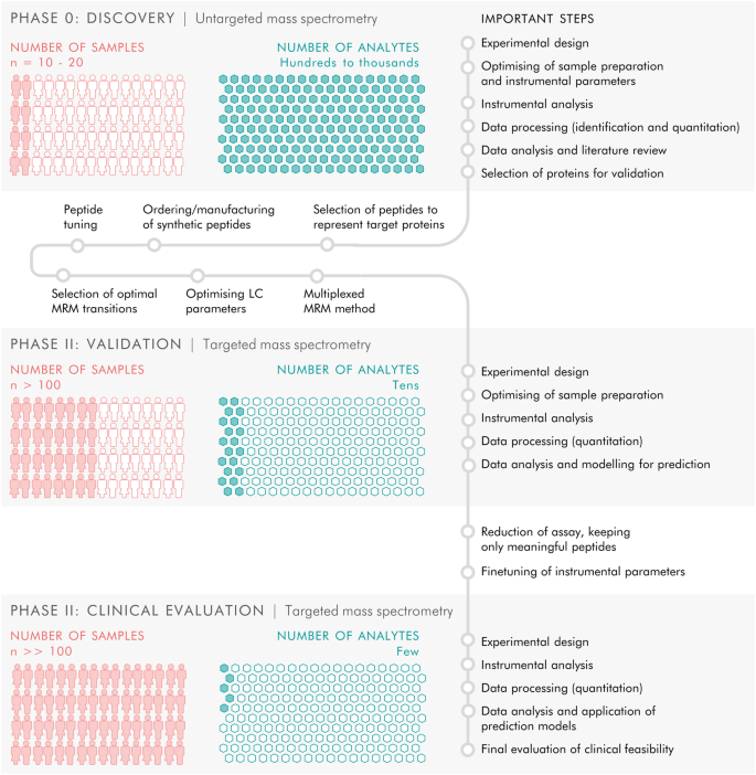
In this study, we used mass spectrometry-based proteomic phenotyping to identify a panel of blood biomarkers in early PD. In the initial discovery stage, we analysed samples from a well-characterised cohort of de novo PD patients and healthy controls (HC) who had been subjected to rigorous collection protocols10. Using unbiased state-of-the-art mass spectrometry, we identified putatively involved proteins, suggesting an early inflammatory profile in plasma. We thereafter moved on to the validation phase by creating a high-throughput and targeted proteomic assay that was applied to samples from an independent replication cohort, consisting of de novo PD, HC and iRBD patients. Finally, after refining the targeted proteomic panel to include a multiplex of only the biomarkers which were reliably measured, an independent analysis was performed on a larger and independent cohort of longitudinal, high-risk subjects who had been confirmed as iRBD by state-of-the-art video-recorded polysomnography (vPSG), including follow-up sampling of up to 7 years.

In summary, using a panel of eight blood biomarkers identified in a machine-learning approach, we were able to differentiate between PD and HC with a specificity of 100%, and to identify 79% of the iRBD subjects, up to 7 years before the development of either DLB or motor PD (NSD stage 3). Our identified panel of biomarkers significantly advances NSD research by providing potential screening and detection markers for use in the earliest stages of NSD for subject identification/stratification for the upcoming prevention trials.

Results

Proteomic discovery phase 0

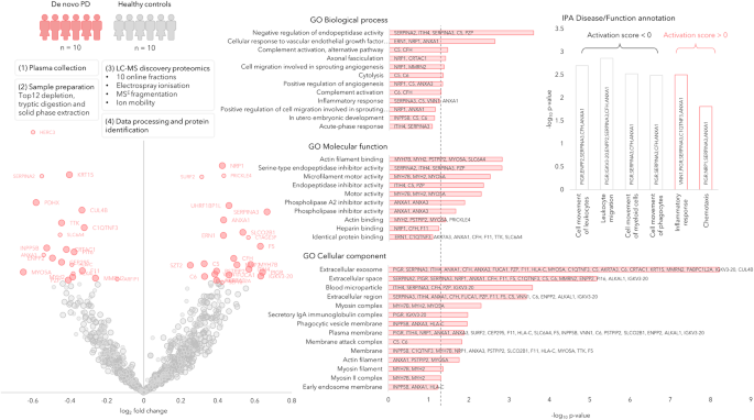
We performed a bottom-up proteomics analysis of plasma, which had been depleted of the major blood proteins, using two-dimensional in-line liquid chromatography fractionation into ten fractions and label-free mass spectrometric analysis by QTOF MSE. The discovery cohort consisted of ten randomly selected drug-naïve patients with PD and ten matched HC from the de novo Parkinson’s disease (DeNoPa10) cohort (details can be found in Supplementary Table 1). This analysis identified 1238 proteins when restricting identification to originate from at least one peptide per protein and at least two fragments per peptide. After excluding proteins with less than two unique peptides or with an identification score below a set threshold (see method section below), 895 distinct proteins remained. Of these proteins, 47 were differentially expressed between the de novo PD and control groups on a nominal significance level of 95%. Pathway analysis suggested enrichment in several inflammatory pathways. Workflow and Results are shown in Fig. 1, and 2 Supplementary Figs. 1, 2.

Fig. 1: All-over workflow of the study.
figure 1

The study included three phases. Phase 0 consisted of discovery proteomics by untargeted mass spectrometry to identify putative biomarkers, followed by phase I in which targets from the discovery phase were transferred to a targeted, mass spectrometric MRM method and applied to a new and larger cohort of samples, and finally phase II in which the targeted MRM method was refined and a larger number of samples were analysed to evaluate the clinical feasibility of the targeted protein panel.

Fig. 2: Discovery phase in plasma samples of de novo PD (n = 10) and healthy controls (n = 10) represented by a Volcano plot showing the protein expression differences between PD and controls (phase 0).
figure 2

The circle radii in the Volcano plot represent the identification certainty, where large radii represent proteins identified by at least two unique peptides and an identification score >15, smaller radii are given for proteins identified by two or more unique peptides or a confidence score >15. The horizontal axis shows log2 of the average fold-change and the vertical axis shows −log10 of the p values. The significantly different proteins are annotated by gene name and coloured in pink, while the non-significant proteins are coloured in grey. GO annotations for the significant proteins are shown, the dashed line represents p = 0.05. Disease and function annotations from IPA are shown, divided into annotations with a positive or negative activation score. Source data are provided as a Source Data file.


Wednesday, July 26, 2023

Proteomics analysis of plasma from middle-aged adults identifies protein markers of dementia risk in later life

I can't imagine any hospital using this to test for dementia risk; so useless.  And with NO protocols to prevent dementia, doubly useless.

What is the method of proteomics analysis?

The techniques that are most often used are electrospray ionization (ESI) and matrix-assisted laser desorption/ionization (MALDI). Both of them are representatives of so-called soft ionization techniques in which ions are created with low internal energies and thus undergo little fragmentation.

Proteomics analysis of plasma from middle-aged adults identifies protein markers of dementia risk in later life

Science Translational Medicine
19 Jul 2023
Vol 15, Issue 705

Abstract

A diverse set of biological processes have been implicated in the pathophysiology of Alzheimer’s disease (AD) and related dementias. However, there is limited understanding of the peripheral biological mechanisms relevant in the earliest phases of the disease. Here, we used a large-scale proteomics platform to examine the association of 4877 plasma proteins with 25-year dementia risk in 10,981 middle-aged adults. We found 32 dementia-associated plasma proteins that were involved in proteostasis, immunity, synaptic function, and extracellular matrix organization. We then replicated the association between 15 of these proteins and clinically relevant neurocognitive outcomes in two independent cohorts. We demonstrated that 12 of these 32 dementia-associated proteins were associated with cerebrospinal fluid (CSF) biomarkers of AD, neurodegeneration, or neuroinflammation. We found that eight of these candidate protein markers were abnormally expressed in human postmortem brain tissue from patients with AD, although some of the proteins that were most strongly associated with dementia risk, such as GDF15, were not detected in these brain tissue samples. Using network analyses, we found a protein signature for dementia risk that was characterized by dysregulation of specific immune and proteostasis/autophagy pathways in adults in midlife ~20 years before dementia onset, as well as abnormal coagulation and complement signaling ~10 years before dementia onset. Bidirectional two-sample Mendelian randomization genetically validated nine of our candidate proteins as markers of AD in midlife and inferred causality of SERPINA3 in AD pathogenesis. Last, we prioritized a set of candidate markers for AD and dementia risk prediction in midlife.

Editor’s summary

Pathological changes involved in Alzheimer’s disease (AD) occur decades before the onset of cognitive deficits but are not well understood. Here, Walker and colleagues used proteomics and genomics on a cohort of middle-aged adults followed longitudinally and identified pathway-specific plasma proteins that increased dementia risk up to 25 years later. The pathway signature of these proteins was characterized by dysregulated immune signaling and proteostasis in the earliest preclinical stages and abnormal coagulation and complement signaling around 10 years before dementia onset. The study indicates that distinct biological mechanisms may be relevant in earlier and later preclinical stages of AD. –Daniela Neuhofer

Get full access to this article

Friday, September 6, 2013

Quantitative Clinical Proteomic Study of Autopsied Human Infarcted Brain Specimens to Elucidate the Deregulated Pathways in Ischemic Stroke Pathology

Your doctor should be requesting you to allow your brain to be autopsied to help future stroke survivors. Why didn't they do this 30 years ago? You can tell your doctor not so politely where to go and hope they believe in a hell. 

Quantitative Clinical Proteomic Study of Autopsied Human Infarcted Brain Specimens to Elucidate the Deregulated Pathways in Ischemic Stroke Pathology


  • a School of Biological Sciences, Nanyang Technological University, Singapore 637551, Singapore
  • b Choju Medical Institute, Fukushimura Hospital, Toyohashi, Aichi 441-8124, Japan
  • c Graduate School of Biomedical Science and Engineering, Hanyang University, Seongdong-gu, Seoul 133-791, Republic of Korea

Highlights

Identification of human ischemic proteome (1520 proteins, FDR = 0.1%) by iTRAQ
Global failure of the cellular energy metabolism in the ischemic infarcts
Concomitant reduction of all participating proteins of the malate-aspartate shuttle
Iron-mediated oxidative imbalance as seen by the elevation of ferritin
Presence of reactive gliosis along with an increased anti-inflammatory response

Abstract

Ischemic stroke, still lacking an effective neuroprotective therapy is the third leading cause of global mortality and morbidity. Here, we have applied an 8-plex iTRAQ-based 2D-LC-MS/MS strategy to study the commonly regulated infarct proteome from three different brain regions (putamen, thalamus and the parietal lobe) of female Japanese patients. Infarcts were compared with age-, post-mortem interval- and location-matched control specimens.
The iTRAQ experiment confidently identified 1520 proteins with 0.1% false discovery rate. Bioinformatic data mining and immunochemical validation of pivotal perturbed proteins revealed a global failure of the cellular energy metabolism in the infarcted tissues as seen by the parallel down-regulation of proteins related to glycolysis, pyruvate dehydrogenase complex, TCA cycle and oxidative phosphorylation. The concomitant down-regulation of all participating proteins (SLC25A11, SLC25A12, GOT2 and MDH2) of malate-aspartate shuttle might be responsible for the metabolic in-coordination between the cytosol and mitochondria resulting in the failure of energy metabolism. The levels of proteins related to reactive gliosis (VIM, GFAP) and anti-inflammatory response (ANXA1, ANXA2) showed an increasing trend. The elevation of ferritin (FTL, FTH1) may indicate an iron-mediated oxidative imbalance aggravating the mitochondrial failure and neurotoxicity. The deregulated proteins could be useful as potential therapeutic targets or biomarkers for ischemic stroke.

Biological Significance

Clinical proteomics of stroke has been lagging behind other areas of clinical proteomics like Alzhemer’s Disease or Schizophrenia. Our study is the first quantitative clinical proteomics study where iTRAQ-2D-LC-MS/MS has been utilized in the area of ischemic stroke to obtain a comparative profile of human ischemic infarcts and age-, sex-, location- and post-mortem interval-matched control brain specimens. Different pathological attributes of ischemic stroke well-known through basic and pre-clinical research such as failure of cellular energy metabolism, reactive gliosis, activation of anti-inflammatory response and aberrant iron metabolism have been observed at the bedside. Our dataset could act as a reference for similar studies done in the future using ischemic brain samples from various brain banks across the world. Meta-analysis of these studies in the future could help to map the pathological proteome specific to ischemic stroke that will guide the scientific community to better evaluate the pros and cons of the pre-clinical models for efficacy and mechanistic studies.
Infarct being the core of injury should have the most intense regulation for several key proteins involved in the pathophysiology of ischemic stroke. Hence, a part of the up-regulated proteome could leak into the general circulation that may offer candidates of interest as potential biomarkers. In support of our proposed hypothesis, we report ferritin in the current study as one of the most elevated protein in the infarct, which has been documented as a biomarker in the context of ischemic stroke by an independent study. Overall, our approach has the potential to identify probable therapeutic targets and biomarkers in the area of ischemic stroke.
Full-size image (38 K)

Monday, October 29, 2012

Neuroproteomics approach & neurosystems biology analysis: ROCK inhibitors as promising therapeutic targets in neurodegeneration & neurotrauma

You'll have to talk to your researcher and doctor about what this could mean. 

Neuroproteomics approach & neurosystems biology analysis: ROCK inhibitors as promising therapeutic targets in neurodegeneration & neurotrauma


Several common degenerative mechanisms and mediators underlying the neuronal injury pathways characterize several neurodegenerative diseases including Alzheimer's Parkinson's, and Huntington's disease, as well as brain neurotrauma. Such common ground invites the emergence of new approaches and tools to study the altered pathways involved in neural injury alongside with neuritogenesis, an intricate process that commences with neuronal differentiation.
Achieving a greater understanding of the impaired pathways of neuritogenesis would significantly help in uncovering detailed mechanisms of axonal regeneration. Among the several agents involved in neuritogenesis are the Rho and Rho kinases (ROCKs) which constitute key integral points in the Rho/ROCK pathway that is known to be disrupted in multiple neuropathologies such as spinal cord injury, traumatic brain injury and Alzheimer's disease. This in turn renders ROCK inhibition as a promising candidate for therapeutic targets for treatment of neurodegenerative diseases.
Among the novel tools to investigate the mechanisms involved in a specific disorder is the use of neuroproteomics/systems biology approach, a growing subfield of bioinformatics aiming to study and establishing a global assessment of the entire neuronal proteome, addressing the dynamic protein changes and interactions. This review aims to examine recent updates regarding how neuroproteomics aids in the understanding of molecular mechanisms of activation and inhibition in the area of neurogenesis and how Rho/ROCK pathway/ROCK inhibitors, primarily Y-27632 and Fasudil compounds, are applied in biological settings, promoting neuronal survival and neuroprotection that has direct future implications in neurotrauma.

Wednesday, May 30, 2012

Urinary Proteomics to Support Diagnosis of Stroke

Rather than arguing with ER doctors about your belief you've had a stroke, just hand them a container of your pee. For guys they probably couldn't even stand up long enough, with the additional problem of only one hand to hold the pee cup and your penis. They could rule out drug interactions also. Something similar would need to be done for bleeds. How long after onset of stroke were samples taken? How much faster and accurate than a MRI? 

Urinary Proteomics to Support Diagnosis of Stroke


Accurate diagnosis in suspected ischaemic stroke can be difficult. We explored the urinary proteome in patients with stroke (n = 69), compared to controls (n = 33), and developed a biomarker model for the diagnosis of stroke. We performed capillary electrophoresis online coupled to micro-time-of-flight mass spectrometry. Potentially disease-specific peptides were identified and a classifier based on these was generated using support vector machine-based software. Candidate biomarkers were sequenced by liquid chromatography-tandem mass spectrometry. We developed two biomarker-based classifiers, employing 14 biomarkers (nominal p-value <0.004) or 35 biomarkers (nominal p-value <0.01). When tested on a blinded test set of 47 independent samples, the classification factor was significantly different between groups; for the 35 biomarker model, median value of the classifier was 0.49 (−0.30 to 1.25) in cases compared to −1.04 (IQR −1.86 to −0.09) in controls, p<0.001. The 35 biomarker classifier gave sensitivity of 56%, specificity was 93% and the AUC on ROC analysis was 0.86. This study supports the potential for urinary proteomic biomarker models to assist with the diagnosis of acute stroke in those with mild symptoms. We now plan to refine further and explore the clinical utility of such a test in large prospective clinical trials.