Interest in excitotoxicity expanded following its
implication in the pathogenesis of ischemic brain injury in the 1980s,
but waned subsequent to the failure of N-methyl-D-aspartate (NMDA)
antagonists in high profile clinical stroke trials. Nonetheless there
has been steady progress in elucidating underlying mechanisms. This
review will outline the historical path to current understandings of
excitotoxicity in the ischemic brain, and suggest that this knowledge
should be leveraged now to develop neuroprotective treatments for
stroke.
Introduction
It has now been 63 years since Lucas and Newhouse (1957) discovered the ability of parenterally administered glutamate to kill central neurons in the rodent retina, and 51 years since Olney (1969)
extended this observation to neurons in the hypothalamus and
hippocampus. Subsequently finding that a series of structurally related
neuroexcitatory amino acids exhibited similar neurotoxicity, with
potencies corresponding to known neuroexcitant potencies, Olney et al. (1974)
proposed that the “necrotizing effect is, in essence, an exaggeration
of the excitatory effect” and coined the term “excitotoxic amino acids”.
These seminal observations remained relatively fallow until the 1980s,
when advances in excitatory amino acid (EAA) pharmacology led to
widespread recognition of glutamate’s neurotransmitter function and
scientific interest in EAAs burgeoned. Over the next two decades, the
subfield of excitotoxicity likewise expanded, driven most prominently by
its implication in the pathogenesis of ischemic brain damage, and
several pharmaceutical companies developed glutamate receptor
antagonists as candidate treatments for stroke. By the early 2000s these
drugs had failed in clinical trials and excitotoxicity research lost
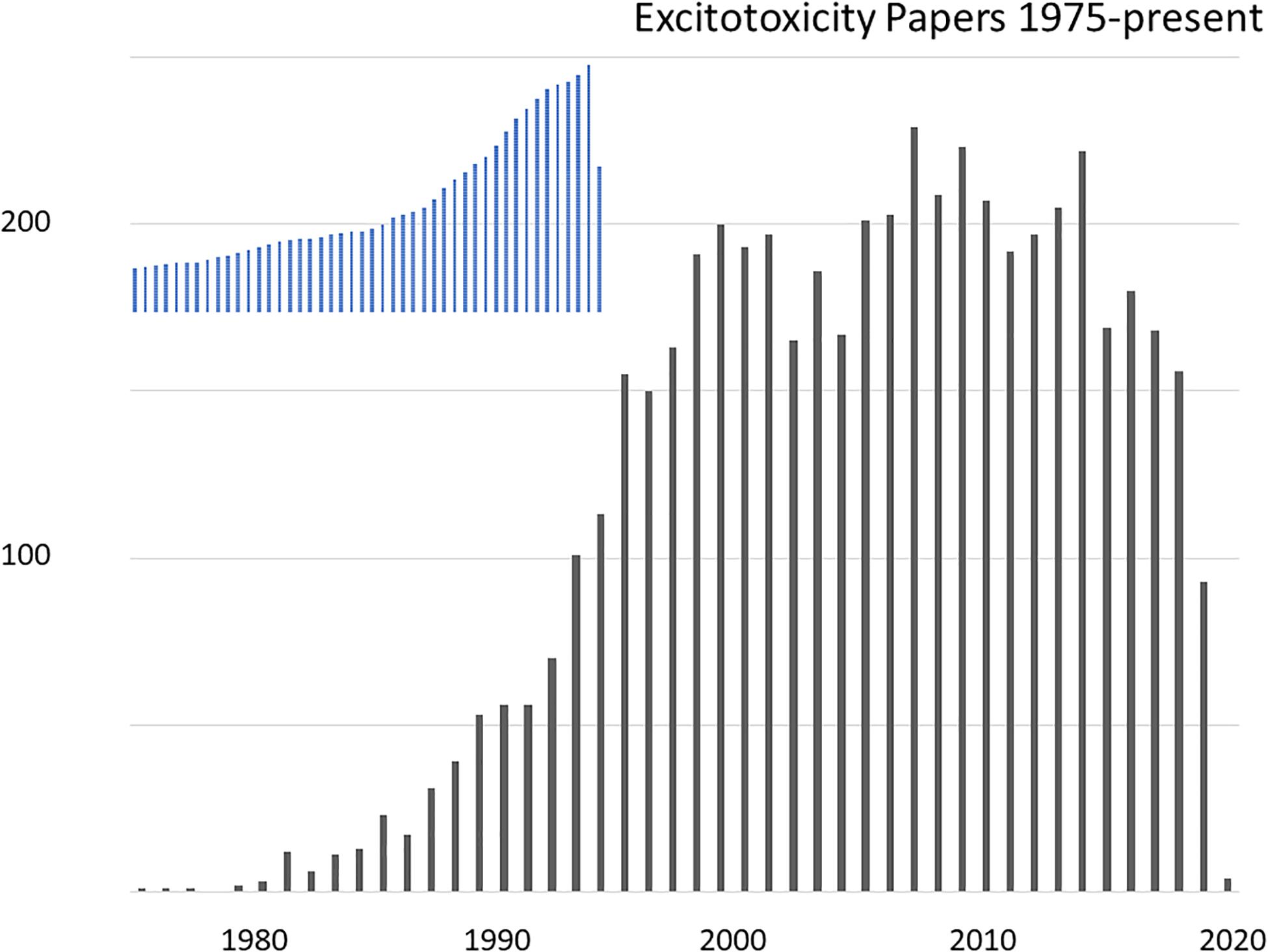
substantial momentum. A reflection of the excitotoxicity research boom
and subsequent slowdown can be found in the number of relevant
publications indexed by PubMed (Figure 1).
However, implication of excitotoxicity as an agent of human diseases has not gone away. The current issue of Frontiers in Neuroscience
is timely, marking a half-century of active excitotoxicity research. I
thank the editors for my invitation to contribute, and will provide here
a brief, semi-chronological, and at times personal, overview of this
research, focusing on the still extant path forward for developing an
anti-excitotoxic neuroprotective treatment for stroke. Recent reviews on
the topic have appeared (Wu and Tymianski, 2018; Fern and Matute, 2019; Hardingham, 2019; Ge et al., 2020).
Other articles in this issue will likely discuss the possible
contribution of excitotoxicity to neurodegeneration in chronic diseases
such as amyotrophic lateral sclerosis or Alzheimer’s disease.
Excitotoxicity – Early Days (Up to Early 1990s)
Brain tissue contains high concentrations of free glutamate, in the 5–15 mmol/kg range (Schousboe, 1981), but until the 1970s this glutamate was widely assumed to be serving purely metabolic functions. Hayashi (1954)
speculated that glutamate might be a neurotransmitter based on
convulsant activity on motor cortex, an idea that was strongly supported
by direct demonstration of neuroexcitatory properties (Curtis et al., 1960). Subsequent research on the topic of glutamate signaling proceeded gently for nearly two decades – a lull Watkins and Jane (2009) have dubbed “the dark ages”. But evidence for synaptic synthesis, Ca2+-dependent release and rapid cellular uptake accrued (Fagg and Lane, 1979),
and pharmacological tools were developed that enabled critical testing
of the transmitter hypothesis. Glutamate receptors were classified into
N-methyl-D-aspartate (NMDA) and non-NMDA types, the latter subsequently
further divided into kainite and quisqualate receptors (Watkins and Evans, 1981),
and then later, kainate, α-amino-3-methyl-4-isoxazoleproprionic acid
(AMPA), and G protein-coupled metabotropic receptors (mGluRs) (Krogsgaard-Larsen et al., 1980; Sugiyama et al., 1987; Hollmann and Heinemann, 1994); molecular subunit nomenclature is still evolving (Collingridge et al., 2009).
Glutamate antagonists blocked endogenous neural signaling in multiple
pathways, identifying glutamate as a neurotransmitter at insect and
crustacean neuromuscular junctions, and then throughout the mammalian
CNS (Nistri and Constanti, 1979; Cotman et al., 1981; McLennan, 1983).
Olney’s initial considerations of how excitotoxicity
might contribute to human disease focused on the possible dangers of
exogenous agonists, especially the monosodium glutamate added to food as
a flavor enhancer (Olney, 1982).
Recognition of ubiquitous glutamate neurotransmission elevated interest
in the possible pathophysiological importance of endogenous stores, and
excitotoxicity gained further attention as a laboratory tool, useful
for creating “axon-sparing” lesions in brain. These threads came
together when injection of kainate into the rat striatum was found to
reproduce some of the anatomical and biochemical features of
Huntington’s disease, raising speculation that progressive excitotoxic
damage might contribute to its pathogenesis (Coyle and Schwarcz, 1976; Mcgeer and Mcgeer, 1976). Further, the convulsant properties of kainate and the dependence of kainate neurotoxicity in vivo upon intact glutamatergic afferents supported a role for excitotoxicity in epileptic brain injury (Ben-Ari et al., 1980; Meldrum, 1985).
Thus, the stage was well set for three breakthrough studies that
leveraged newly available receptor antagonists to implicate endogenous
excitotoxicity in ischemic brain damage.
1. Rothman (1984) showed that the glutamate antagonist γ-D-glutamylglycine
(DGG) could block glutamate-induced depolarization and cell swelling,
as well as anoxic injury in cultured rat hippocampal neurons. This key
study followed up on his earlier demonstration that 10 mM MgCl2 could protect the neurons against damage induced by cyanide exposure (Rothman, 1983).
2. Simon R. et al. (1984)
found that direct hippocampal injection of the selective NMDA
antagonist, 2-amino-7-phosphonoheptanoic acid (APH), reduced
pathological changes in nearby neurons 2 h after transient global
ischemia (TGI) in rats. In retrospect, the study assessed neuronal
morphology too soon to determine lasting survival, as it was just
becoming recognized that selective neuronal death after TGI can occur
days later (Kirino, 1982).
Regardless, it demonstrated a neuroprotective effect of NMDA receptor
(NMDAR) blockade against a component of ischemic injury in vivo.
3. Wieloch (1985)
injected APH into the rat caudate, and observed markedly improved
survival of nearby neurons 1 week after 30 m exposure to hypoglycemia,
further implicating NMDARs in acute brain injury.
Additionally, microdialysis measurements revealed that
brain ischemia induced within minutes a large increase in extracellular
glutamate emanating from depolarized nerve terminals and astrocytes (Benveniste et al., 1984), indicating that neurons in the ischemic brain would inevitably be exposed to elevated glutamate concentrations.
It is worth pausing here. The entire cell death field
was then a shadow of what it would become. (PubMed papers retrieved by
“cell death” and published in 1985 are about 2% of the 14,000 such
papers published in 2019). Few people considered either acute or chronic
neurodegeneration a worthwhile research topic – cells die, right – and
the neurologists of the day managed patients presenting with Alzheimer’s
disease or stroke nihilistically, attending mainly to accompanying
medical conditions. Early excitotoxicity studies truly changed “normal
science” (Kuhn, 1962),
suggesting that some ischemic neuronal cell death occurred
consequential to specific events and pathways accessible to therapeutic
interdiction.
Inspired by Rothman’s in vitro studies, I set out
to investigate glutamate neurotoxicity in the mouse cortical cell
culture system I had going at the time. Having worked during the 1970s
as a graduate student on γ-aminobutyric acid (GABA) signaling and
benzodiazepines, I was emerging from clinical training as a neurologist
and setting up my own laboratory at Stanford, newly funded to study the
electrophysiology of glutamate receptors. Shifting research focus on the
fly was entirely feasible – thank you, NIH.
I found that bath exposure to 500 μM glutamate caused
cultured cortical neurons to swell immediately and then proceed to
disintegrate, similar to the “intracellular edema and neuronal necrosis”
observed by Olney (1969)
in mouse brains after glutamate injection. However, I wondered if
leaving glutamate in the bath, as Rothman had done, might not exaggerate
cell swelling and attendant damage over what would occur in vivo
within a three-dimensional brain and closed skull. I decided therefore
to terminate the bath exposure after 5 m, an exposure still widely
lethal by the next day. Replacing extracellular Na+ with an
impermeant cation eliminated acute neuronal swelling, but most neurons
still went on to degenerate over the next hours. In contrast, removing
extracellular Ca2+ increased early cell swelling, and yet most cells recovered and survived (Choi, 1985). This and other experiments (Choi, 1987; Choi et al., 1987) suggested that glutamate neurotoxicity at lower exposure levels was predominantly driven by delayed, Ca2+ -dependent processes rather than by the immediate entry of Na+ responsible for neuroexcitation and, together with Cl– and water, immediate excitotoxic swelling.
A Ca2+ -dependent death fit with observations of EAA-induced Ca2+ movement into brain tissue (Heinemann and Pumain, 1980; Berdichevsky et al., 1983), and aligned excitotoxic death with a larger theme of Ca2+ overload in other types of cell death, including the toxin-induced death of hepatocytes (Schanne et al., 1979) and agonist- or mechanical injury-induced muscle cell death (Bloom and Davis, 1972; Fleckenstein et al., 1975; Leonard and Salpeter, 1979). It also meshed with prior implications of Ca2+ overload in the neuronal death induced by ischemia (Siesjö, 1981; Simon R.P. et al., 1984) or prolonged seizures (Griffiths et al., 1982). Measurements with newly available Ca2+-sensitive microelectrodes revealed a rapid and large drop in brain extracellular Ca2+ after the onset of ischemia (Harris et al., 1981). The toxic EC50 for glutamate with 5 m exposure was 50–100 μM in mixed astrocyte + neuron cultures (Choi et al., 1987), dropping to 5 μM with 30 m exposure in astrocyte-poor cultures lacking protective cellular uptake (Rosenberg et al., 1992).
The exciting discovery that NMDA but not kainate gated Ca2+-permeable channels (MacDermott et al., 1986)
suggested that NMDARs would play a foreground role in glutamate
neurotoxicity. This was borne out. The selective NMDA antagonist
2-amino-5-phophonovalerate (APV) only modestly reduced the
neuroexcitation or acute neuronal cell swelling induced by brief
glutamate exposure, but markedly reduced later cell death (Choi et al., 1988). Selective NMDAR block was also effective in reducing the neuronal death induced by hypoxia (Weiss et al., 1986; Goldberg et al., 1987), glucose deprivation (Monyer et al., 1989) or mechanical trauma (Tecoma et al., 1989).
Non-NMDA agonists were also potently neurotoxic on
cortical neurons, but prolonged exposures of several hours was required
to produce widespread cell death. Consistent with the effects of
glutamate + APV, kainate induced immediate excitotoxic neuronal
swelling, but if exposure was terminated at 5 m, most cells recovered
and survived (Koh et al., 1990).
We considered it likely that this more slowly triggered neurotoxicity
mediated by kainate or AMPA receptors (KARs, AMPARs) was mediated by
slower Ca2+ overload secondary to excessive Na+ entry, involving voltage-gated Ca2+ channels and reverse operation of the electrogenic Na+/Ca2+ exchanger, NCX (Choi, 1988). In that mode, favored under conditions of membrane depolarization, high internal [Na+] and low external [Na+], NCXs provide high capacity transport for 3 Na+ ions out coupled to 1 Ca2+ ion in. The importance of NCXs to excitotoxicity was supported by implication in anoxic optic nerve injury (Stys et al., 1992). Later we showed that net cellular 45Ca2+ accumulation induced by NMDA or glutamate was much larger/faster than that induced by high K+, kainate, or AMPA (Hartley et al., 1993), and Hyrc et al. (1997) found a similar relationship for [Ca2+]i.
Identification of excessive Ca2+ entry as the primary mediator of excitotoxicity, likely augmented by Ca2+
release from endoplasmic reticulum (ER, triggered by mGluR activation;
see below), also bridged to prior studies implicating free radical
generation in the pathogenesis of ischemic brain injury (Siesjö, 1981). Multiple Ca2+-dependent enzymes including calpains, endonucleases and lipases, were known to be capable of damaging cells, and Ca2+ overload would impair mitochondrial energy production and perturb Ca2+-dependent signaling pathways (Cheung et al., 1986; Orrenius et al., 1989).
But the generation of free radicals seemed especially well positioned
to drive lethal cytodegeneration. Consequent to cellular Ca2+ overload, breakdown of lipid membranes into fatty acids mediated by phospholipase A2
and further metabolism via prostaglandin and leukotriene pathways,
damage to mitochondrial electron transport and conversion of xanthine
dehydrogenase to xanthine oxidase all generate free radicals, promoting
lipid peroxidation and membrane failure (Chan et al., 1985; Traystman et al., 1991; Halliwell, 1992).
Testing the hypothesis, 21-aminosteroid lipid peroxidation inhibitors
attenuated both glutamate neurotoxicity and oxygen-glucose deprivation
(OGD)-induced neuronal death (Monyer et al., 1990).
Toxic NMDA exposure increased superoxide formation in cultured
cerebellar neurons, and trapping this superoxide was neuroprotective (Lafon-Cazal et al., 1993).
Subsequent studies used electron paramagnetic resonance spectroscopy
and oxidation state-sensitive fluorescent dyes to detect mitochondrial
production of reactive oxygen species (ROS) after NMDAR-mediated Ca2+
overload, and demonstrated that this production could be substantially
reduced by inhibition of mitochondrial electron transport or dissipation
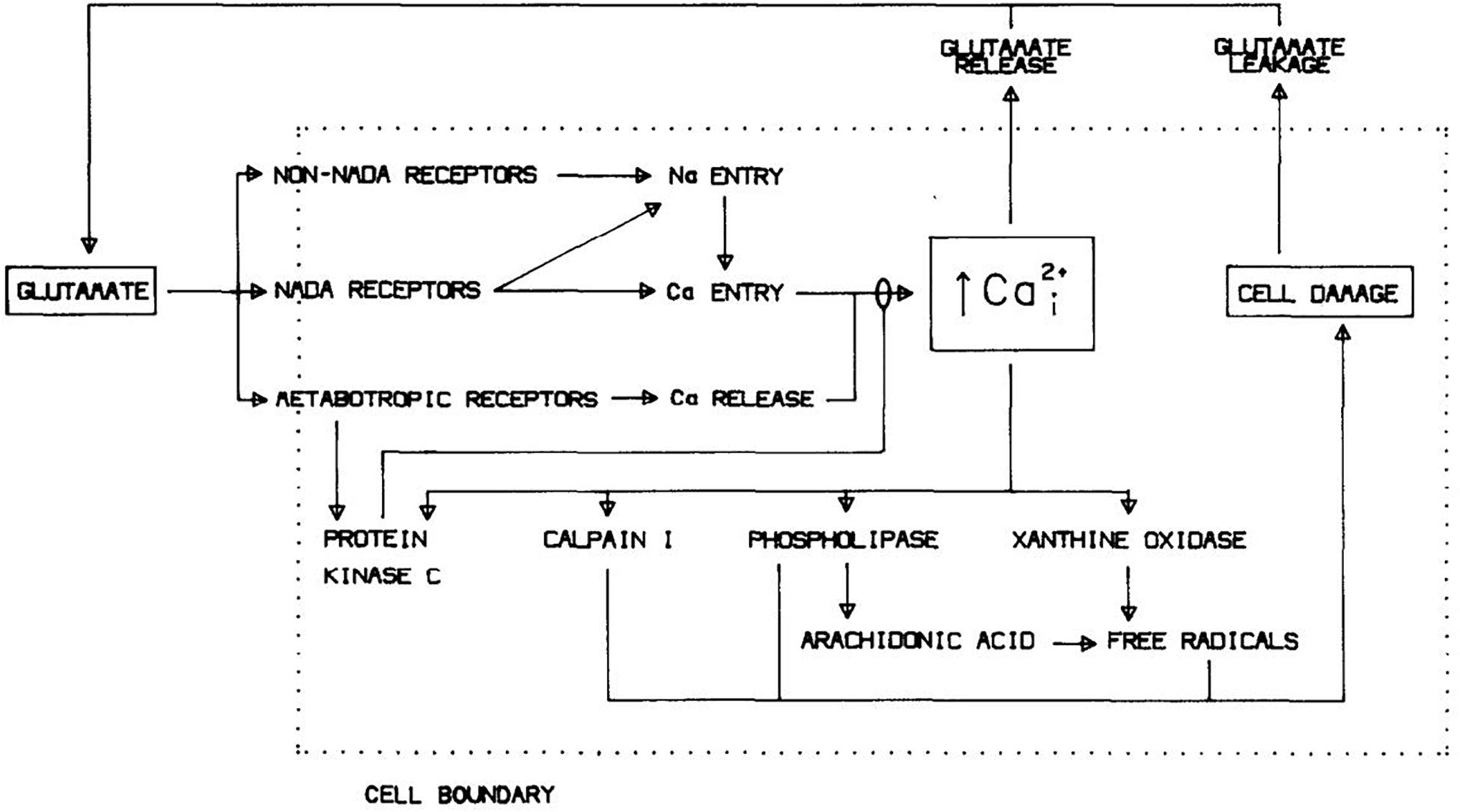
of the mitochondrial membrane potential (Dugan et al., 1995; Reynolds and Hastings, 1995). Figure 2 shows a diagram of excitotoxicity mechanisms as we saw it in 1988.
The discovery that NMDAR-mediated Ca2+
influx activated neuronal nitric oxide synthase (NOS) suggested that
nitric oxide (NO) might be both a normal neuronal signaling molecule and
a free radical mediator of glutamate neurotoxicity (Garthwaite et al., 1988; Bredt et al., 1990).
Freely diffusible through membranes, NO released from neurons activates
soluble guanylate cyclase in neighboring cells including smooth muscle.
Cytotoxicity at higher concentrations was harnessed by macrophages to
kill target cells, with NO reacting with superoxide to form the highly
reactive nitrogen species (RNS), peroxynitrite, and promoting
destructive hydroxyl radical formation (Halliwell and Gutteridge, 1984; Beckman et al., 1990). Pivotal experiments were carried out by Dawson et al. (1991),
demonstrating that inhibition of neuronal NOS (nNOS) selectively
attenuated NMDAR-mediated neurotoxicity but not kainate neurotoxicity.
Later, they would show that a major toxic consequence of NMDA-induced NO
formation was the hyperactivation of poly(ADP-ribose) polymerase
(PARP), likely consequential to peroxynitrite-induced DNA damage and
resulting in cellular energy depletion and nuclear translocation of
apoptosis-inducing factor (AIF), triggering a caspase-independent death
(“parthanatos”). Subsequent studies from other labs would raise a
possible role for oxidative intracellular Zn2+ release in connecting NO to PARP overactivation (see below).
As a sidebar, inhibiting nNOS did not reduce glutamate
neurotoxicity in the cortical cultures my lab was using in the 1990s. We
came to regard this as an idiosyncrasy, likely reflecting low baseline
nNOS expression and/or a large astrocyte presence sopping up NO. Raising
inducible NOS (iNOS) expression in the astrocyte layer did add a
NOS-dependent component to NMDAR-mediated toxicity in the cultures (Hewett et al., 1994).
In any case, this culture system difference illustrates that more than
one potentially lethal downstream cascade can be triggered in parallel
by glutamate receptor overactivation and robustly kill neurons. A messy “and”, not an “or”:
a race to death. Whether a specific cascade ends up directly
responsible for cell death such that its inhibition improves survival,
and the exact features of that death, may depend on where that cascade
lies in an injury hierarchy, as well as on the identity and closeness of
competitive injury pathways that may leave behind their own incomplete
molecular and morphological signatures.
NMDA Antagonists, Stroke, and Apoptosis
Demonstration of the neuroprotective effects of glutamate antagonists against hypoxia/ischemia/hypoglycemia in vivo and in vitro, together with identification of NMDAR overactivation and Ca2+
overload as primary mediators of acute glutamate toxicity, encouraged
global efforts to test NMDA antagonist drugs in various animal models of
brain ischemia and develop drugs suitable for human use. The first drug
candidate out of the gate was Merck’s MK-801, a potent and selective
NMDA antagonist with excellent brain penetrance. Gill et al. (1987)
reported that pretreatment with MK-801 reduced neuronal death in gerbil
brains subjected to either bilateral or unilateral forebrain ischemia.
Benefits of NMDA antagonists later proved variable in models of TGI, in
some cases perhaps confounded by hypothermia (Buchan and Pulsinelli, 1990),
but reduction of infarction or disability was robustly observed with
NMDA antagonists given up to 2 h after focal brain ischemia in rodents
or larger animals (Gotti et al., 1988; Kochhar et al., 1988; Ozyurt et al., 1988; Park et al., 1988; Steinberg et al., 1988).
By 1992 close to 20 published studies reported this finding.
AMPA/kainate antagonists also were neuroprotective in brain ischemia
studies, especially after TGI (Sheardown et al., 1990).
In 1994, Huang et al. reported that mice lacking nNOS exhibited reduced
infarcts after permanent occlusion of the middle cerebral artery
(pMCAO) (Huang et al., 1994).
However, the NMDA antagonist drug candidates brought
into development by several companies for use in stroke all failed in
clinical testing. Some were abandoned because of side effects; others
lacked efficacy. In two cases (CNS-1102, CGS 19755), there were
worrisome trends toward worse outcome in the treated group (Hoyte et al., 2004; Ginsberg, 2008; Lai et al., 2014).
As those reviewers noted, clinical studies did not match up well with
the earlier animal studies. Some drug dosing was too low (limited by
mechanism-associated behavioral side effects such as hallucinations) or
too late (most dosing was > 3 h post stroke onset, and in some trials
up to 48 h post onset, as enrolling patients at earlier time points was
not practical at that time). A smaller number of pilot studies with
AMPA antagonists were also disappointing, in one case (ZK20075)
unsurprisingly depressing consciousness (Ginsberg, 2008).
The first wave of NMDA antagonist stroke trials was
followed by two significant improvements, but these second-generation
efforts also failed. The first improvement, informed by insights into
the molecular biology and subunit composition of glutamate receptors,
was the use of antagonists selective for NR2B/GluN2B. The predominant
expression of NR2B in forebrain vs. cerebellum and limbic areas raised
hopes of achieving neuroprotection with less dose-limiting side effects.
Furthermore, as discussed below, consideration of Ca2+
source specificity and NMDAR signaling relationships suggested that NR2B
contributes more to excitotoxic death than the other major forebrain
subtype, NR2A. A older antagonist, ifenprodil, was discovered to be
conveniently NR2B selective, attractively use-dependent and
neuroprotective in multiple animal models of global and focal brain
ischemia (Wang and Shuaib, 2005; Gogas, 2006).
New molecular entity congeners of ifenprodil, SL-82.0715 and CP-101,606
were developed by Synthélabo and Pfizer, respectively, and exhibited
reduced side effects compared to pan NMDAR antagonists. But the former
failed to show efficacy in a stroke trial and the latter was felled by
electrocardiographic toxicity (QT prolongation) after showing some
promise against severe traumatic brain injury (Yurkewicz et al., 2005; Lai et al., 2014).
The second improvement was in stroke trial methodology.
Recognizing that treatment delay was problematic in prior stroke
neuroprotection trials, Saver et al. (2015)
completed a remarkable multicenter study (FAST-MAG) in which
intravenous magnesium sulfate was given to 1700 acute stroke patients by
paramedics within 2 h (and often within 1 h) of stroke onset. Still no
improvement in outcome measures was observed. An earlier study of Mg2+ treatment for stroke (IMAGES) with a 12 h treatment window was also negative (Muir et al., 2004).
While the methodological advance represented by FAST-MAG is clear,
direct relevance of FAST-MAG and IMAGES to the hypothesis of
NMDAR-mediated excitotoxicity in human stroke is less so. Mg2+
has multiple actions that might contribute to its modest
neuroprotective effects in animal stroke models, and it is likely that
its block of NMDA channels in the ischemic brain would be substantially
relieved by cellular depolarization (Mayer et al., 1984; Nowak et al., 1984).
Enthusiasm for developing anti-excitotoxic therapies for
stroke was also progressively dampened by the lack of efficacy in
stroke trials demonstrated by several other drugs targeting related
mechanisms, including voltage-gated Ca2+ channels
(nimodipine), free radicals (tirilazad mesylate, ebselen, NXY-059),
phospholipid hydrolysis (citicoline), and nitric oxide synthase
(lubeluzole). These trials also had shortcomings in concept or design,
commonly involving the use of doses and delay time windows that were not
supported by the enabling preclinical studies (Ginsberg, 2008; Sutherland et al., 2012). Administration of a lipophilic Ca2+/Zn2+ chelator, DP-b99, within 9 h of stroke onset also failed in Phase III testing (Lees et al., 2013).
The free radical drugs tested would be considered less
than compelling candidates today in updated comparisons. Neither
tirilazad (Hall, 1997) nor NXY-059 (Kuroda et al., 1999) penetrate well into brain parenchyma, and NYX-059 exhibited little antioxidant activity on cultured neurons (Antonic et al., 2018).
Ebselen is a mimic of glutathione peroxidase that would be expected to
decompose only a subset of harmful radicals (hydroperoxides); it has
little aqueous solubility and requires a thiol co-substrate like
glutathione to maintain activity. A fourth antioxidant drug, edaravone,
has a favorable chemical profile, being amphiphilic and able to directly
scavenge a range of harmful lipid- and water-soluble radicals (Watanabe et al., 2018).
It has protective effects in multiple animal models of brain ischemia,
and showed sufficient evidence of neuroprotective efficacy in clinical
testing to gain approval for use in stroke in Japan (Lapchak, 2010). Available published data documented a just significant (p
= 0.048) beneficial effect on 90 d clinical outcome in patients treated
within 72 h of stroke onset; clear benefit was suggested in an
exploratory analysis including only patients treated within 24 h (Edaravone Acute Infarction Study Group, 2003).
Subsequent clinical trials demonstrating ability to slow the
progression of amyotrophic lateral sclerosis supported the premise of
human neuroprotective activity, and led to US FDA approval in 2017 for
that indication (Radicava) (Cruz, 2018).
By 2001, I had moved to work in the pharmaceutical
industry and was in a position to champion an improved NMDA
antagonist/stroke trial, but I had become worried that there was
potentially a conceptual problem with hard blocking NMDARs in the
ischemic brain: apoptosis (Choi, 1995; Lee et al., 1999; Ikonomidou and Turski, 2002; Papadia and Hardingham, 2007). Programmed cell death had come to center stage in biological research, spearheaded by elegant genetic studies in C. elegans (Ellis and Horvitz, 1986; Hengartner and Horvitz, 1994)
and growing appreciation that apoptosis occurs in a wide range of
disease states outside of normal development. Hypoxic-ischemic cell
death was long considered to be a defining example of a non-apoptotic,
“accidental” death – necrosis – triggered by “violent and
non-physiological” environmental changes (Wyllie et al., 1980).
It, like classic excitotoxicity, was associated with prominent cellular
and organellar swelling and membrane rupture, in contrast to the
controlled cellular condensation characteristic of apoptosis. Yet
evidence was steadily emerging suggesting that some neurons underwent
apoptosis after ischemia, in particular selectively vulnerable neurons
dying in delayed fashion after TGI (Goto et al., 1990; Shigeno et al., 1990); but also after focal ischemia (Linnik et al., 1993; MacManus et al., 1994), especially threshold insults triggering “very delayed infarction” days later (Du et al., 1996; Endres et al., 1998).
As expected, intense excitotoxic death appeared typically not to be apoptotic (Ignatowicz et al., 1991; Dessi et al., 1993; Choi, 1996).
However, slowly triggered AMPAR/KAR-mediated neuronal death was
associated with internucleosomal DNA cleavage, consistent with
incomplete activation of apoptosis pathways (Gwag et al., 1997),
and neuronal apoptosis occurred after mild excitotoxic insults, either
in young cultures with limited EAA receptor expression (Kure et al., 1991) or with lower concentrations of NMDA (Bonfoco et al., 1995). These studies fit with findings that apoptosis could be induced by Ca2+ overload (Wyllie et al., 1984) or oxidative stress (Lennon et al., 1991), including NO (Albina et al., 1993). More recent studies have indicated that Ca2+
and oxidative surges can interact at the ER-mitochondrial signaling
interface to trigger mitochondrial membrane permeabilization, permitting
cytochrome c and AIF release, and the activation of caspase-dependent
or caspase-independent apoptosis pathways (Tajeddine, 2016; Hempel and Trebak, 2017; Humeau et al., 2018). Or resulting in necrosis, if mitochondrial and cellular failure is fulminant.
Together, these observations suggested that
excitotoxicity, like many other insults, has the potential to trigger
regulated cell death programs; but when intense, membrane, energy and
protein synthesis failure destroy neurons before these programs can
complete. Ca2+ ionophores likewise can induce neuronal apoptosis at low concentrations, and necrosis at high concentrations (Gwag et al., 1999).
A death race hierarchy was already apparent in cultured cortical
neurons deprived of oxygen and glucose: blockade of rapidly triggered
NMDAR-mediated death was necessary to reveal AMPAR/KAR-dependent death.
Blocking both NMDAR and AMPAR/KAR-mediated neurotoxicity rendered
neurons resistant to prolonged OGD, but then a further increase in OGD
duration (“blocked OGD”) drove neurons into apoptosis (Gwag et al., 1995; Choi, 1996).
Consideration of ischemic apoptosis suggested that a
sustained high degree of NMDAR blockade could be harmful. While
presumably beneficial initially in reducing acute excitotoxic necrosis
and the triggering of ischemic apoptosis, sustained blockade risks
keeping [Ca2+]i below a necessary survival “set-point” (Koike et al., 1989).
U-shaped curves are ubiquitous in cell and organismal biology, with
deleterious effects induced by both too little and too much of many
metabolites, messengers, drugs, etc. Apoptosis of young sympathetic
neurons induced by nerve growth factor (NGF) withdrawal can be
attenuated by raising [Ca2+]i from a basal
level of 90 nM to about 240 nM, the same higher basal level found in
older neurons capable of surviving without NGF (Koike and Tanaka, 1991). This survival-promoting level of [Ca2+]i is considerably lower than the levels associated with excitotoxic exposure to glutamate, which can exceed 10 μM (Stout and Reynolds, 1999)
and are associated with much larger amounts of net cellular calcium
loading than that induced by survival-promoting activation of
voltage-gated Ca2+ channels (Hartley et al., 1993). Lowering extracellular Ca2+ or reducing membrane Ca2+ channel opening induces apoptosis in a wide range of cell types [reviewed in Canzoniero et al. (2004)]. Many signaling pathways transduce the ability of moderate Ca2+
levels to promote cell survival, including the activation of
PI3K/Akt/mTOR, Ras/Raf/ERK, and AMPK pathways, as well as modulation of
gene expression by CREB and NFAT family transcription factors downstream
of Ca2+ / calmodulin (Pinto et al., 2015; Feldmann et al., 2018; Varghese et al., 2019).
Supporting the premise of Ca2+ starvation in neurons undergoing ischemic apoptosis, [Ca2+]i was abnormally low in neurons dying after exposure to blocked OGD, and normalizing [Ca2+]i with the voltage-gated Ca2+ channel opener, S)-(-)-BayK-8644, or even low concentrations of kainate improved neuronal survival (Canzoniero et al., 2004). 1–2 days NMDAR blockade alone reduced cultured neuronal [Ca2+]i below baseline levels and triggered / enhanced apoptosis (Takadera et al., 1999; Snider et al., 2002; Yoon et al., 2003); injection of a single dose of MK-801 induced neuronal apoptosis within hours in the developing rat brain (Ikonomidou et al., 1999). Consistent with a benefit of low level glutamate receptor activation in vivo,
delayed administration of the partial NMDA agonist, D-cycloserine,
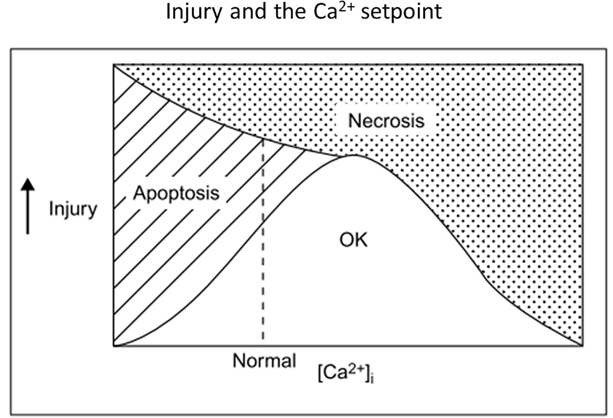
improved functional outcome in rats after traumatic brain injury (Adeleye et al., 2010) or focal ischemia (Dhawan et al., 2011). Figure 3 illustrates in broad brush strokes how neuronal [Ca2+]i, apoptosis and necrosis might interrelate after excitotoxic / ischemic insults.
In sum, whether NMDA antagonists
are beneficial or harmful in the ischemic brain may depend on multiple
insult and treatment specifics, with key issues being the relative
proportion of necrosis vs. apoptosis taking place; and the timing,
location and amount of [Ca2+]i lowering
induced by the drugs. A dynamic that might slow excitotoxic necrosis and
thus increase the overall contribution of apoptosis to ischemic brain
damage is a reduction of extracellular Na+ and Ca2+, as documented with ion-sensitive electrodes (Hansen, 1985);
also see below under Potassium). Furthermore, ischemic apoptosis might
be expected to be more prominent in human stroke, with its sometimes
stuttering onset and potentially large penumbral areas across complex
gyral anatomy, than after surgical artery occlusion in some
lissencephalic rat stroke models. Another reason why early
subtype-unspecific NMDA antagonists might have disappointed in human
stroke, despite their routine effectiveness in rat stroke.
Expansion and Refinement of Excitotoxicity Concepts
NMDARs and Ca2+ Source Specificity
Returning to the excitotoxicity story, the next major advance in the 1990s was appreciating that Ca2+ source makes a difference. Noting that elevations of [Ca2+]i mediated by voltage-gated Ca2+ channels lacked the toxicity associated with NMDAR activation, Tymianski et al. (1993)
proposed that NMDARs were preferentially linked to downstream mediators
of excitotoxic injury. The discovery that the PDZ domains of both NR2B (Moon et al., 1994) and nNOS (Brenman et al., 1996)
interacted with PDZ domains of the postsynaptic density protein,
PSD-95, fit the bill, and furthermore placed NR2B in the foreground of
excitotoxicity. Suppressing PSD-95 protein expression in cultured
neurons attenuated both NMDA-induced NO production and neuronal death,
without affecting NMDA currents or NMDA-induced Ca2+ loading (Sattler et al., 1999).
Extension of the Ca2+ source specificity
hypothesis was pursued in studies assessing the relative contributions
of extrasynaptic and synaptic subpopulations of NMDARs to excitotoxic
death. Synaptic and extrasynaptic NMDA receptors exhibit similar single
channel properties (Clark et al., 1997)
but have been reported to trigger different transcription factor and
gene expression changes; activation of extrasynaptic but not synaptic
NMDARs caused rapid loss of mitochondrial membrane potential and
neuronal death (Hardingham et al., 2002; Karpova et al., 2013). Mediation of excessive Ca2+ influx into mitochondria was postulated to be favored by extrasynaptic NMDAR activation, possibly due to spatial proximity (Bading, 2017).
However, the distinction between synaptic and extrasynaptic receptors
may not be sharp, as burst stimulation of inputs to hippocampal CA1
neurons activates both populations (Harris and Pettit, 2008). And arguing against synaptic localization per se
altering NMDAR contributions to excitotoxicity, delocalizing synaptic
NMDARs by depolymerizing F-actin did not alter NMDAR-induced current, Ca2+
loading, or cell death. The delocalization did attenuate death after
OGD, consistent with synaptic localization increasing exposure to the
glutamate released by nerve terminals (Sattler et al., 2000).
Comparing cell death after intense activation of
extrasynaptic vs synaptic NMDARs, some investigators have proposed that
these subpopulations have opposing effects on excitotoxic death:
extrasynaptic receptors promoting death, and synaptic receptors
promoting survival (Hardingham et al., 2002; Hardingham, 2009; Lai et al., 2011; Parsons and Raymond, 2014; Wu and Tymianski, 2018). Consistent with that proposal, synaptic activity repressed expression of the mitochondrial Ca2+ uniporter, Mcu and reduced neuronal vulnerability to excitotoxic death, a plausible neuroprotective adaptive mechanism (Qiu et al., 2013).
However, while supported by careful experiments, the receptor location
hypothesis is best considered to be still under test. It does not fit
easily with the prominence of PSD-95/nNOS signaling in NMDAR-mediated
excitotoxicity, since association with PSD-95 is a hallmark of synaptic
localization. Furthermore, as outlined above, the ability of NMDAR
activation to reduce apoptosis at lower levels and yet drive excitotoxic
apoptosis or necrosis at higher levels can be explained in terms of net
Ca2+ fluxes and Ca2+ setpoints, without invoking
this additional level of source specificity. The experimental necessity
of utilizing different paradigms to activate synaptic vs extrasynaptic
receptors (eg, bicuculline + 4-aminopyridine to stimulate the former,
bath glutamate + prior MK-801 to stimulate the latter) leaves open the
possibility that outcome differences primarily reflect differences in
ionic current envelopes or even just overall Ca2+ influx,
rather than differences in the fundamental properties of extrasynaptic
vs. synaptic receptors. The equivalence of cellular Ca2+ loading achieved by the extrasynaptic vs. synaptic activation paradigms is not assured by measuring peak [Ca2+]i, especially if high affinity indicators like Fluo-3 are used, as these may saturate below micromolar excitotoxic elevations (Hyrc et al., 1997; Stout and Reynolds, 1999). Additionally, modulatory influences present differentially at synapses vs elsewhere, e.g., pH changes or Zn2+
released by nerve terminals (see below), or differential levels of cell
injury may affect outcomes. The receptor location hypothesis might not
matter much in stroke anyway, as a large buildup of extracellular
glutamate would probably overstimulate both synaptic and extrasynaptic
receptors.
In the strongest formulation of Ca2+ source
specificity, NMDAR subtypes have been assigned both to distinct
locations and opposing roles, with NR2A assigned to synaptic locations
and pro-survival roles, and NR2B assigned to extrasynaptic locations and
pro-death roles (Liu et al., 2007).
However, electrophysiological examination of ifenprodil sensitivity in
cultured hippocampal neurons suggested that NR2A and NR2B can both be
located in either synaptic or extrasynaptic compartments (Thomas et al., 2006).
This formulation will also need adjustment to accommodate other subunit
compositions of NMDARs, as many are likely triheteromeric, containing
both NR2A and NR2B subunits (Tovar et al., 2013).
Additional to activation of nNOS, other NR2B-linked
signaling pathways have been proposed to be mediators of excitotoxic
death, including death-associated protein kinase 1 (DAPK1) (Nair et al., 2013), phosphatase and tensin homolog deleted on chromosome 10 (PTEN) (Ning et al., 2004)
and NOX2 (see below). DAPK1 was recruited to the NR2B protein complex
after transient middle cerebral artery occlusion (tMCAO), binding to a
unique region of the NR2B C-terminal region, phosphorylating NR2B and
upregulating channel current; administration of a peptide uncoupling
DAPK1 from NR2B or genetic deletion of DAPK1 reduced infarction after
tMCAO and selective neuronal death after TGI (Tu et al., 2010).
However, another study did not find evidence of DAPK1 modifying NR2B
after excitotoxic insults, or reduced neuronal death after TGI in Dapk1–/– mice (McQueen et al., 2017).
Downregulating PTEN expression with antisense oligodeoxynucleotides
enhanced Akt signaling and reduced the death of vulnerable hippocampal
CA1 neurons after TGI (Zhang et al., 2007).
Likely many signaling pathways affecting cell death are
triggered by glutamate receptor overactivation, not only via direct
signaling linkages, but also unspecifically consequent to cellular
damage. Example of the latter are ROS/RNS-induced DNA single strand
breaks activating PARP, or mitochondrial damage releasing ROS and
cytochrome c. Another example of an unspecific link would be damage to
ER and accumulation of misfolded proteins, activating the PERK/eIF2
pathway, increasing levels of ATF4 and CHOP, and promoting apoptosis (Sokka et al., 2007; Almanza et al., 2019).
Furthermore, whether a given downstream signaling pathway or event is
ultimately responsible for excitotoxic death, or even whether it
promotes death vs survival, may depend on quantitative specifics and
cellular state. As discussed above, raising [Ca2+]i
can be either survival-promoting or death-promoting, depending.
PI3K/Akt signaling is often survival promoting, but in some settings can
enhance apoptosis (Los et al., 2009),
and cytoplasmic PI3K activation enhances superoxide production by NOX2
(see below). JNK signaling is usually pro-apoptotic but anti-apoptotic
functions have been described (Liu and Lin, 2005); and Jnk3 –/– mice exhibited reduced kainate-induced and ischemic apoptosis (Yang et al., 1997; Kuan et al., 2003).
Yet another example of context-dependent pathway effects on cell
survival would be upregulation of BDNF expression by glutamate
receptor-induced Ca2+ influx or ischemia (Lindvall et al., 1992; Zafra et al., 1992; Favaron et al., 1993; Hardingham et al., 2002). While BDNF is classically anti-apoptotic, it accentuated excitotoxic neuronal necrosis (Koh et al., 1995) and glutathione depletion-induced death of immature neurons (Ratan et al., 1996).
NADPH Oxidase
NADPH oxidase (NOX) comprises a family of 7
membrane-active protein complexes, NOX1 through NOX5, DUOX1, and DUOX2,
that catalyze the transfer of electrons from NADPH to molecular oxygen,
primarily generating superoxide (Bedard and Krause, 2007).
While long recognized as a key component of neutrophil antimicrobial
defenses, NOX was later found in many tissues, including brain, and
implicated in acute and chronic neurodegeneration (Infanger et al., 2006; Sorce and Krause, 2009). NOX1, NOX2 and NOX4 are the main brain isoforms, and mice with genetic deletion of the gp91phox
catalytic unit of NOX2 exhibited reduced brain infarction after tMCAO,
even if neutrophil NOX2 was rescued with a bone marrow transplant (Walder et al., 1997; Tang et al., 2011).
Subsequent studies showed that ischemia raised the expression of NOX2
and NOX4 in multiple brain cell types over ensuing hours-days, and that
amelioration of ischemic infarction could also be achieved by genetic
disruption of NOX4 (Radermacher et al., 2013; Ma et al., 2017).
Increased expression of endothelial and neuronal NOX4, which is
constitutively active, likely contributes to blood-brain barrier (BBB)
breakdown and neuronal death, respectively (Casas et al., 2017).
Brennan et al. (2009)
identified NOX2 as a specific source of ROS production after NMDAR
overactivation, demonstrating that both NMDA-induced ROS production and
neuronal death in cortical cultures were blocked by the NOX2 inhibitor,
apocynin, the hexose monophosphate pathway inhibitor,
6-aminonicotinamide (reducing NADPH), or deletion of the NOX2 subunit
gene, p47phox, as well as by removal of extracellular Ca2+ or inhibition of PKCζ, the latter responsible for phosphorylating and activating p47phox (Heinecke et al., 1990; Bedard and Krause, 2007). Subsequent study implicated NR2B and phophoinositide-3 kinase (PI3K) upstream of PKCζ (Brennan-Minnella et al., 2013). PI3K activation is likely mediated by Ca2+ / calmodulin (Joyal et al., 1997), as well as possibly through the NR2B C-terminal / PSD-95 signalosome (Chen et al., 2015; Minnella et al., 2018). Another way this signalosome might facilitate NOX2 activation is via Src and Panx1 channel opening; loss of Zn2+
homeostasis may also contribute (see below). Importantly, the NR2B/NOX2
pathway is strongly inhibited by small drops in intracellular pH (Lam et al., 2013), so its contribution to ischemic injury may be limited to onset, the outer penumbra and reperfusion (see Protons, below).
AMPA Receptors
As noted above, AMPA receptors (AMPARs) mediate Na+ influx and hence can contribute to excitotoxic Ca2+
overload and neuronal death. Although these receptors lack direct
linkage to NOS/NOX, they participate substantially in brain damage after
focal and global ischemia, in the latter setting typically contributing
more than NMDARs to the delayed death of selectively vulnerable neurons
(Sheardown et al., 1990; Buchan et al., 1991; Hicks et al., 1999). This prominent contribution to delayed selective neuronal death may be largely due to upregulated expression of Ca2+-permeable AMPA receptors.
The Ca2+ (and Zn2+, see below)
permeability of central AMPARs during development and in response to
synaptic activity is regulated by the expression of an RNA-edited form
of AMPA subunit GluR2 that contains an arginine residue instead of the
genetically-coded glutamine in a key position within the second
transmembrane domain (Burnashev et al., 1992; Liu and Zukin, 2007). While most central AMPARs contain such edited GluR2 subunits and exhibit low Ca2+
permeability, certain telencephalic and cerebellar neurons, including
aspiny interneurons and neurons with high levels of nNOS, express Ca2+ -permeable AMPARs and are highly vulnerable to AMPA-induced neuronal death (Hope et al., 1991; Weiss et al., 1994). Ischemia enhances the expression of Ca2+ -permeable AMPARs in vulnerable neuronal populations (Pellegrini-Giampietro et al., 1992), which then constitute a dominant route for toxic Ca2+ /Zn2+
entry, limiting the protective reach of NMDA antagonists. This
enhancement is mediated by turning on expression of the gene silencer
REST, reducing GluR2 promoter activity (Calderone et al., 2003).
Kainate Receptors
While less studied than other glutamate receptor due to a
historical paucity of selective pharmacological tools, kainate
receptors (KARs) are expressed postsynaptically at glutamatergic
synapses and presynaptically on glutamatergic and GABAergic nerve
terminals, modifying transmitter release (Jane et al., 2009; Traynelis et al., 2010).
Postsynaptic KARs contribute to synaptic excitation, more at some
synapses, such as mossy fiber inputs to hippocampal CA3 neurons, than
others; and likewise participate variably in synaptic plasticity.
Analogous to AMPARs, a minority of KARs containing unedited GluK1 or
GluK2 subunits can be Ca2+ permeable and convey heightened
vulnerability to glutamate excitotoxicity where heavily expressed, for
example on somatostatin-containing interneurons (Sun et al., 2009).
Administration of the GluK1R antagonist, LY377770, 2 h after
endothelin-1-induced focal ischemia in rats reduced infarction and
produced a surprisingly large reduction in extracellular glutamate
levels in ipsilateral striatum (O’Neill et al., 2000).
Metabotropic Glutamate Receptors
Glutamate released from nerve terminals and astrocytes
will activate G-protein coupled mGluRs on neurons and glia concurrently
with ionotropic glutamate receptors. There are 8 major mGluRs and
several splice variants, divided into 3 groups based on structure,
function, and pharmacology: Group I (mGluR1, mGluR5) linked to
activation of phospholipase C (PLC); and Groups II (mGluR2, mGluR3) and
III (mGluR4, mGluR6, mGluR7, mGluR8) linked to inhibition of adenylate
cyclase (Schoepp et al., 1990; Pin and Duvoisin, 1995).
mGluRs modulate synaptic transmission and plasticity; although they
probably do not play a primary role in mediating acute excitotoxic brain
injury, they influence this injury and are thus worth keeping in view
for possible secondary therapeutic targeting in stroke (Sun et al., 2019).
Group I mGluR-activated PLC generates inositol 1,4,5-trisphosphate, triggering Ca2+ release from ER, and diacylglycerol, which together through PKC can enhance excitotoxic Ca2+ entry through NMDARs, NCXs and NHEs (see below) (Benquet et al., 2002; Lipp and Reither, 2011); and activate phospholipase A2, promoting ROS formation and lipid peroxidation (Bonventre, 1997; van Rossum and Patterson, 2009; Guemez-Gamboa et al., 2011). Possibly through PLC / diacylglycerol signaling, mGluR1 also opens plasmalemmal TRP (TRPC3) cation channels (see below) (Kim et al., 2003; Hartmann et al., 2011).
As expected from these actions, and the typically net excitatory
effects of Group I agonists on neural circuits, activation of Group I
mGluRs enhanced NMDA-induced neuronal death; and inhibition, especially
of mGluR1, reduced that death in vitro (Bruno et al., 1995b, 1999; Strasser et al., 1998) as well as ischemic injury in vivo (Pellegrini-Giampietro et al., 1999; Li et al., 2013).
Besides canonically activating PLC, Group I mGluRs could potentially
have neuroprotective effects through G-protein mediated downmodulation
of NMDARs (Yu et al., 1997; Bertaso et al., 2010) and neuroprotective Akt signaling via Homer, PIKE-L and nuclear PI3K (Rong et al., 2003; Xu et al., 2007). However at least the latter action may be quickly abrogated by calpain (Xu et al., 2007).
Surprisingly though, interfering with the C-terminal interactions
between mGluR1 and NR2A with cell-permeable peptides was recently
reported to attenuate NMDA-induced neuronal death in vitro and infarction after tMCAO (Lai et al., 2019).
In contrast, Group II and III mGluRs most often have
inhibitory effects on neural circuits. and anti-excitotoxic effects of
Group II or Group III agonists have been observed in vitro (Bruno et al., 1995a). The Group III agonist ACPT-1 reduced infarction in rats after tMCAO (Domin et al., 2018).
Within Group II however, neuronal mGluR2 activation has been proposed
to enhance excitotoxicity, perhaps by limiting GABA release (Corti et al., 2007). A novel selective mGluR2 negative allosteric modulator, ADX92639, reduced selective neuronal death after TGI (Motolese et al., 2015).
Protons
In addition to Na+ and Ca2+, another cation participating in excitotoxicity is H+.
Rapid local changes in extracellular pH accompany physiological
neuronal activity and modulate the behavior of many receptors, channels
and transporters (Chesler, 1990), including NMDARs, which are inhibited by H+ around physiological pH (Tang et al., 1990).
In the ischemic brain, tissue and extracellular pH typically drops
within minutes toward 6.5 or lower, due to reliance on anerobic
glycolysis for ATP resynthesis (Ljunggren et al., 1974; Kraig et al., 1985; Dennis et al., 1991; Katsura et al., 1991), an increase in extracellular H+ sufficient to attenuate NMDAR channel openings (Tang et al., 1990) and NOX2 activity (see above). Expectedly NMDAR-mediated excitotoxicity is also reduced (Giffard et al., 1990b; Tombaugh and Sapolsky, 1990).
Despite reducing NMDAR and NOX activation, acidosis is
still likely a net contributor to ischemic brain damage as long
postulated, as movements of H+ or H+ equivalents amplify excitotoxicity. Ischemic acidosis enhances neurotoxic Ca2+
overload via the gating of acid-sensing ion channels (ASICs – see
below). Furthermore, several studies have shown that glutamate- or
NMDA-induced Ca2+ influx into hippocampal neurons is accompanied by rapid intracellular acidification, in part due to Ca2+ displacement of H+ from intracellular binding sites (Hartley and Dubinsky, 1993; Irwin et al., 1994; Koch and Barish, 1994). NOX2 activity (Lam et al., 2013) and electroneutral operation of the plasma membrane calcium ATPase (PMCA) may also contribute, the latter importing 2 H+ ions for every Ca2+ ion exported (Stawarski et al., 2020).
Intracellular pH dropped initially to around 6.5 during excitotoxic
glutamate exposure (500 μM × 5 m) before progressively recovering and
overshooting to pH 7.5–8.0 as a result of acid-extrusion mechanisms,
especially membrane Na+/H+ exchangers (NHEs) (Raley-Susman et al., 1991; Rathje et al., 2013). Sustained operation of NHEs loads intracellular Na+, which then raises intracellular Ca2+
via NCXs. Consistent with this sequence contributing to excitotoxicity,
inhibition of NHE-1 with cariporide reduced glutamate-induced cortical
neuronal death, as well as three predicted intermediate steps: a later
increase in [Ca2+]i, loss of mitochondrial membrane potential, and intracellular production of ROS (Lee and Jung, 2012). NHE–/– mice exhibited reduced infarction after tMCAO (Luo et al., 2005).
In addition, ischemic acidosis is likely itself cytotoxic. Exposure to pH 6.5 for 6 h is lethal to both neurons and glia (Nedergaard et al., 1991). Astrocytes are especially vulnerable to proton-induced death (Goldman et al., 1989; Giffard et al., 1990a),
and can be killed after only 15–20 m exposure to pH 6.6 if other
extracellular ions are altered to levels found in ischemic brain (low Ca2+ and Na+, high K+, hypoxia), conditions likely to impair cellular pH homeostasis (Swanson et al., 1997; Bondarenko and Chesler, 2001).
Sustained intracellular acidity likely becomes lethal due to myriad
disturbances in protein conformation and essential cellular processes (Nedergaard et al., 1991), including energy failure (Swanson et al., 1997). If cellular H+ favors release of ferrous iron (Kraig et al., 1987), the Fenton reaction (Jung et al., 2009) may facilitate hydroxy radical formation, lipid peroxidation, and a “ferroptosis” form of regulated cell death (Xie et al., 2016; Ratan, 2020) if necrosis does not supervene. Proton-induced death of cerebellar neurons is accompanied by an increase in [Zn2+]i and reduced by Zn2+ chelation, raising the possibility that disturbance in Zn2+ storage or other homeostatic mechanisms may also contribute to H+ cytotoxicity (Isaev et al., 2010).
Potassium
A core property of the cation channels gated by ionotropic glutamate receptors, besides permeability to Na+, and in certain cases Ca2+/Zn2+, is high permeability to K+ (Nowak et al., 1984). Although glutamate receptor-mediated K+ movement has received less attention than Na+ or Ca2+
movement, it may contribute significantly to excitotoxic apoptosis
after mild insults or under other conditions where excitotoxic necrosis
is blunted.
Noting that the hallmark cell volume loss occurring during apoptosis would require K+ to exit, my laboratory examined K+
currents in cortical neurons undergoing serum deprivation-induced
apoptosis and observed early enhancement of the delayed rectifier
current, IK. Attenuating this current with tetraethylammonium (TEA) or raising extracellular K+ inhibited apoptosis, even if [Ca2+]i was kept at resting levels with gadolinium (Yu et al., 1997). This implication of K+ efflux in neuronal apoptosis converged with observations implicating that efflux in other cell types (Bortner and Cidlowski, 2007), and in particular two findings: 1) lymphocyte apoptosis required enough K+ efflux to lower [K+]i to less than 50 mM (Bortner et al., 1997); and 2) in those cells, activation of pro-caspase-3 by cytochrome c and apoptotic nuclease activity was [K+]i -dependent, inhibited by normal [K+]i and released by lowered [K+]i (Hughes et al., 1997).
Supporting the notion that K+ efflux may
promote apoptosis after mild excitotoxic insults, in a cortical culture
model of NMDA-induced neuronal apoptosis (achieved by using a low
concentration of NMDA, lowering extracellular Na+ and Ca2+ to levels found in the ischemic brain and extending exposure time), NMDAR activation markedly reduced cellular K+ content (Yu et al., 1999).
Furthermore, intraventricular injection of TEA reduced infarction in
rats after tMCAO, a finding contrary to prediction from a classic
excitation / excitotoxicity standpoint, but consistent with
participation of K+ efflux in ischemic apoptosis (Wei et al., 2003).
Zinc
Yet another less-studied cation that participates in excitotoxicity is zinc (Choi and Koh, 1998; Frederickson et al., 2004; Sensi and Jeng, 2004; Granzotto and Sensi, 2015). Zn2+ is a dietary requirement and has many essential roles in cell biology (Parisi and Vallee, 1969; Maret, 2017), including service as a central neurotransmitter/neuromodulator regulating circuit behavior and synaptic plasticity (Frederickson, 1989; Smart et al., 1994; Frederickson et al., 2005). Reactive Zn2+
is stored in presynaptic vesicles within a subset of glutamatergic
nerve terminals throughout CNS and co-released with glutamate in a Ca2+-dependent fashion upon nerve terminal firing. Synaptically released Zn2+
modifies the behavior of multiple moieties on postsynaptic membranes,
including voltage- and agonist-gated channels, with primary signaling
actions likely on NMDA and AMPA receptors (Peters et al., 1987; Westbrook and Mayer, 1987; Kalappa et al., 2015). It can also enter (and likely leave) postsynaptic target neurons though Ca2+ routes that might equally well be called Ca2+/ Zn2+ routes, including voltage-gated Ca2+ channels, NMDARs, Ca2+-permeable AMPARs, TRP channels, ASIC1a and in depolarized cells, reverse operation of NCXs (Sensi et al., 1997; Kerchner et al., 2000; Inoue et al., 2015), the last augmented by intracellular H+ and NHE operation (Kang et al., 2020). Ischemic acidosis may also favor Zn2+ entry over Ca2+ entry through voltage-gated Ca2+ channels (Sheline et al., 2002).
Gating of unselective Panx1 channels, on neurons by NMDAR-Src
signaling, and on astrocytes and oligodendrocytes by P2X7 signaling,
might be another route for cellular Zn2+ entry or egress (see below).
Once “translocated” from afferent terminals into postsynaptic target cells, Zn2+ participates in myriad intracellular signaling pathways including the activation of a metabotropic Zn2+ receptor, mZnR/GPR39, and inducing intracellular Ca2+ release (Besser et al., 2009). Additional to translocated Zn2+, a major source of intracellular signaling Zn2+ is mobilization from intracellular stores, especially metallothioneins, by oxidation reactions (Maret, 1994).
Cellular [Zn2+]i is
normally tightly regulated, but in ischemia this homeostasis is
disrupted. Release from depolarized nerve terminals and oxidized
intracellular stores, together with failure of energy dependent
transport, produces increases in extracellular and intracellular Zn2+ that can be cytotoxic, especially to neurons (Yokoyama et al., 1986; Lobner et al., 2000; Bozym et al., 2010). Zn2+ like glutamate induces apoptosis at lower toxic exposures and necrosis at higher exposures (Manev et al., 1997; Lobner et al., 2000). The parallel between glutamate and Zn2+ neurotransmission (Frederickson, 1989) thus extends to excitotoxicity: both are potential transmitter-killers in the ischemic brain.
Evidence that Zn2+ dysregulation contributes to acute brain injury was provided by observations of apparent Zn2+
translocation from afferent terminals to the cytoplasm of neurons
degenerating after sustained seizures or transient global ischemia (Sloviter, 1985; Tønder et al., 1990), and supported by the finding that intraventricular CaEDTA, which chelates extracellular Zn2+, blocked both cytoplasmic Zn2+ accumulation and degenerative changes in vulnerable neurons after TGI (Koh et al., 1996). At the time we thought CaEDTA was only chelating Zn2+ released from nerve terminals, but later studies indicated that CaEDTA can also pull Zn2+ from intracellular stores (Frederickson et al., 2002; Lavoie et al., 2007). Calderone et al. (2004) determined that the major accumulation of Zn2+
in vulnerable hippocampal CA1 neurons occurred more than 48 h after
TGI, and that early injection of CaEDTA attenuated later appearance of
Ca2+-permeable AMPARs, consistent with the possibility that loss of cellular Zn2+ homeostasis constituted a trigger for reducing GluR2 expression. Contribution of Zn2+
to infarction after short duration tMCAO was similarly detected:
intraventricular CaEDTA reduced infarct volume 3 days later, but this
protective effect was lost after full maturation of the infarct 11 days
later, suggesting that an early Zn2+-triggered component of ischemic infarction was eventually overtaken by other injuries (Lee et al., 2002).
Zn2+ like Ca2+ also serves as a
downstream mediator of excitotoxicity – the latter’s shadow, unleashed
and dysregulated by glutamate receptor overactivation. The relationship
between Zn2+ and Ca2+ in mediating excitotoxicity
is complex, with target overlap and reciprocal influences; and
delineating roles has been further challenged by lack of specificity in
historical Ca2+ assay tools such as fura-2 (Cheng and Reynolds, 1998). Studies with Zn2+ selective dyes indicated that OGD induced an early increase in [Zn2+]i within hippocampal slice CA1 neurons; a Zn2+ chelator blocked this increase and attenuated neuronal death (Stork and Li, 2006). Further examination revealed that an OGD-induced increase in neuronal [Zn2+]i preceded mitochondrial depolarization, Ca2+ deregulation and membrane failure, with Zn2+ likely entering mitochondria and contributing to loss of mitochondrial membrane potential (Medvedeva et al., 2009). This meshed with earlier demonstration that elevated intracellular Zn2+, like Ca2+, can enter and damage mitochondria, leading to their swelling, loss of membrane potential and, at high levels of Zn2+, increased ROS generation (Sensi et al., 2003; Clausen et al., 2013), the last likely a consequence of disturbances in mitochondrial electron transport as well as Zn2+ induction of p47PHOX and increased NOX activity (Noh and Koh, 2000; Slepchenko et al., 2017). Downstream ROS/RNS-induced release of Zn2+ from intracellular stores (Berendji et al., 1997; Cuajungco and Lees, 1998; Aizenman et al., 2000) thus drives further oxidative stress. Other injury-promoting events linked to Zn2+ overload include activation of nNOS, PARP (Kim and Koh, 2002) and cyclin-dependent kinase 5 (Cdk5 – Tuo et al., 2018; see Wang et al., 2003; Ko et al., 2019) for links to excitotoxicity). Neuronal Zn2+ overload together with Ca2+-activated CaMKII increase insertion of delayed rectifier Kv2.1 channels into the plasma membrane, increasing K+ efflux and facilitating apoptosis (Aras et al., 2009; McCord and Aizenman, 2013). Zn2+ may also upregulate NMDAR activity through a Src-dependent mechanism (Manzerra et al., 2001).
In addition to promoting neuronal death, excitotoxic Zn2+ dysregulation may contribute to the death of adjacent non-neuronal cells in the ischemic brain. Zn2+-induced death of astrocytes was potentiated by concurrent hypoxia and reduced by PARP knockdown (Pan et al., 2013). Intracellular Zn2+
release was implicated in mediating peroxynitrite-induced death of
oligodendrocytes, through activation of ERK42/44, 12-lipoxygenase and
further ROS generation, rather than immediately through hydroxyl radical
formation (Zhang et al., 2006). OGD-induced death of cultured oligodendrocytes was attenuated by Zn2+ chelation or PARP inhibition (Domercq et al., 2013). Hydrogen peroxide-induced death of endothelial cells (Wiseman et al., 2007) and astrocytes (Furuta et al., 2019) was also attenuated by Zn2+ chelators At sublethal levels, Zn2+
can upregulate ICAM-1 expression in vascular endothelial cells, thereby
promoting leukocyte attraction and microvascular leakage (Sumagin et al., 2008; Yeh et al., 2011).
Together, these observations on glia and endothelial cells suggest that reduction of downstream Zn2+
toxicity may help account for the observed ability of glutamate
antagonists to reduce brain infarction, not just neuronal death, in
animal models of stroke.
Other Cation Channels
Several other membrane channels, likely activated in
part consequential to glutamate receptor overstimulation, can contribute
to toxic Ca2+/ Zn2+ overload and other ionic derangements in the ischemic brain:
1.
Transient receptor potential channels. These are variably selective
cation channels, subdivided into 7 groups and including receptor-,
second messenger- and store-operated members (Venkatachalam and Montell, 2007).
Two members of the melastatin group, TRPM7 and TRPM2 are plasma
membrane channels highly expressed in brain and implicated in the
pathogenesis of ischemic brain damage. TRPM7 is activated in the
ischemic brain by ROS/RNS and augments Ca2+ overload and oxidative stress (Aarts et al., 2003). Suppressing TRPM7 expression in cortical cultures subjected to either OGD or blocked OGD attenuated Ca2+ uptake and neuronal death, leading Aarts et al. (2003) to suggest that TRPM7 might be a foreground pathway for neuronal Ca2+ influx into anoxic neurons. Notably, the divalent cation most permeable through TRPM7 is Zn2+ (Monteilh-Zoller et al., 2003).
TRPM2 is opened by ADP-ribose accumulating secondary to PARP activity downstream of oxidative DNA damage (Kaneko et al., 2006; Li et al., 2015).
Knockdown of TRPM2 expression or drug inhibitors protected male but not
female cortical neurons in culture from OGD-induced death, and
similarly reduced infarction after focal ischemia in male but not female
mice (Jia et al., 2011).
2.
Acid-sensing ion channels are part of the Epithelial Na
channel/degenerin (ENaC/DEG) family of monovalent plasma membrane cation
channels widely expressed throughout the nervous system and gated by
extracellular protons (Vullo and Kellenberger, 2020). Xiong et al. (2004) found that pH 6.0 activation of ASIC1a, which is permeable to divalent cations, mediated Ca2+-dependent
neuronal injury and augmented the neuronal death induced in cortical
cultures by blocked OGD. The ability of ischemic acidosis to activate
ASICs is potentiated by arachidonic acid liberated by phospholipase A2 (Allen and Attwell, 2002), as well as by NO, extracellular Zn2+, and Ca2+ signaling through CaMKII (Gao et al., 2004; Leng et al., 2014).
Mice lacking ASIC1a or pretreated with blocking PcTX venom developed
smaller infarcts after tMCAO, and the protective effect of PcTX added to
that of the NMDA antagonist, memantine (see below). In a combined pH
6.0 + blocked OGD paradigm, reducing TRP7 activation by adding an
antioxidant or NOS inhibitor was not neuroprotective (Xiong et al., 2004) – perhaps another example of one pathway masking another in a parallel race to death.
3.
Pannexin ion channels constitute 3 new members of the gap junction
superfamily, first cloned from mammalian tissue 20 years ago (Barbe et al., 2006; Yeung et al., 2020).
Panx1 is widely expressed on the plasma membrane of central neurons,
glia, and endothelial cells. While constitutively permeable to Cl–,
when activated Panx1 becomes a large conductance pore unspecifically
permeable to ions as well as to some larger molecules such as ATP and
dyes. It can be activated downstream of NMDARs, primarily through a Src
kinase signalosome, but possibly additionally through Ca2+ or NO signaling (Zhang et al., 2008; Weilinger et al., 2012).
Panx1 can also be activated by P2X7 purinergic receptors and
irreversibly activated by C-terminal cleavage via caspases, an event
that likely contributes to the execution of apoptosis.
NMDAR-Src activation of Panx1 contributes heavily to anoxic depolarization (Thompson et al., 2006; Weilinger et al., 2012).
This linkage positions Panx1 as an excitotoxicity amplifier, operating
in parallel with the NMDA channel to augment disturbances in Ca2+, Zn2+, Na+, and K+. Administration of a novel peptide interfering with NMDAR-Src activation of Panx1, TAT-Panx308, reduced OGD-induced elevation in neuronal [Ca2+]i,
mitochondrial membrane permeabilization and neuronal death in
hippocampal cultures, as well as infarction after tMCAO in rats (Weilinger et al., 2016).
4. Store-operated Ca2+ entry provides Ca2+ entry and replenishment of ER Ca2+
stores in both non-excitable cells and excitable cells, and has been
implicated in the pathogenesis of excitotoxicity and ischemic brain
damage (Li et al., 2015; Serwach and Gruszczynska-Biegala, 2019). The system consists of Orai1-3, plasma membrane channels selective for Ca2+ [blocked by Zn2+ (Gore et al., 2004)] and gated by the ER Ca2+ sensors, stomal interaction molecule isoforms, STIM1-2. Release of ER Ca2+ induces STIM oligomerization and migration to ER-plasma membrane junctions, where these open Orai to permit cytoplasmic Ca2+
entry, which is then taken up by ER via SERCA ATP pumps. As noted
above, Group I mGluR activation likely contributes to excitotoxic Ca2+ overload by triggering ER Ca2+ release, placing SOCE in an enabling role. STIM2 is prominently expressed in brain, and stim2–/– mice exhibited reduced neuronal vulnerability to hypoxia in vitro and infarction after tMCAO in vivo (Berna-Erro et al., 2009).
Excitotoxic Glial Cell Death
Although excitotoxicity was originally described as
specific to neurons, oligodendrocytes express a full array of glutamate
receptors (Káradóttir and Attwell, 2007)
and are highly vulnerable to excitotoxic injury and death. Young
cultured oligodendrocytes can be killed by 24 h exposure to kainate or
glutamate; this toxicity was blocked by an AMPAR/KAR antagonist and
enhanced by cyclothiazide, which reduces AMPAR desensitization (Yoshioka et al., 1995).
Vulnerability to a non-glutamate receptor-mediated mechanism involving
inhibition of cystine update by high concentrations of glutamate was
also demonstrated (Murphy et al., 1989; Oka et al., 1993). More prominent AMPAR/KAR-mediated, extracellular Ca2+-dependent
excitotoxicity was demonstrated on cultured optic nerve
oligodendrocytes, and a 5–7 days infusion of kainate destroyed optic
nerve oligodendrocytes in vivo (Matute et al., 1997).
Similarly, after maturing 3–5 weeks on a monolayer of astrocytes,
oligodendrocyte vulnerability to AMPAR/KAR-mediated excitotoxicity was
comparable to that of neurons, with death induced by exposure to as
little as 30 μM AMPA for 3 h (McDonald et al., 1998). OGD triggered AMPAR/KAR-mediated oligodendrocyte death in cultures (McDonald et al., 1998) or in adult brain slices (Tekkök and Goldberg, 2001). Ca2+ -permeable AMPA receptors are expressed on oligodendrocytes (Matute et al., 1997; Barron and Kim, 2019) and may substantially mediate this excitotoxic vulnerability. Loss of cellular Zn2+ homeostasis may contribute, as oligodendrocytes exposed to OGD developed increased [Zn2+]i and subsequent death was reduced by a Zn2+ chelator (Domercq et al., 2013).
In the presence of AMPAR/KAR blockade, OGD induced extracellular Ca2+-dependent damage to oligodendrocyte myelinating processes (Salter and Fern, 2005), likely mediated by process-specific expression of an unusual NR3-containing NMDAR relatively insensitive to Mg2+ block (Káradóttir et al., 2005).
Another contributor to ischemic oligodendrocyte damage may be the their
Panx1 channels, possibly activated by P2X7 receptors and ATP released
from nearby dying or permeabilized cells (Domercq et al., 2010).
Most astrocytes express AMPARs and mGluRs, but NMDARs and Ca2+-permeable AMPARs are generally not abundant (Bradley and Challiss, 2012; Ceprian and Fulton, 2019; Skowrońska et al., 2019).
In contrast to oligodendrocytes, astrocytes appear to be relatively
insensitive to excitotoxicity, although as noted above they are
vulnerable to Zn2+ or H+-induced damage, and they express Panx1 channels (Iglesias et al., 2009). Hence their death could be enhanced secondary to excitotoxicity occurring in nearby neurons or oligodendrocytes.
Blocking Excitotoxicity in the Ischemic Human Brain
It is time to get back in the water.
Failure of the first wave of NMDA antagonists in stroke
trials, viewed with the easy clarity of informed hindsight, does not
come close to excluding excitotoxicity as a major driver of ischemic
brain damage or the feasibility of reducing that damage with
anti-excitotoxic approaches. Those drugs needed to be in the brain at
sufficient levels to ameliorate excitotoxicity at stroke onset, not >
3 h later when efficacy can no longer be demonstrated in animal models.
Patients with slowly progressive vascular occlusions might still have
benefited, but in many patients the drugs would have entered brain after
cells were largely en route to death in a glutamate
receptor-independent fashion. Neurons in the outer stroke penumbra with
less severe NMDAR overactivation would have been the slowest to die, but
there pan NMDA receptor blockade may have sometimes lowered [Ca2+]i too far and exacerbated regulated cell deaths (Fricker et al., 2018).
Over the last 25 years much has been added to the basic NMDAR-Ca2+
overload scenario that served as the basis for initial NMDA
antagonist/stroke trials. Returning to an earlier process framework for
excitotoxicity (induction, amplification, expression - (Choi, 1992), I would highlight the following events in an updated four-stage working scenario:
1.
Induction. Excitotoxicity in the ischemic brain is predominantly
triggered by overactivation of neuronal NR2B receptors at stroke onset,
and in the ischemic penumbra later, raising [Ca2+]i,
activating nNOS and NOX2, and generating ROS/RNS. Concurrent
overactivation of other NMDARs, AMPARs, KARs, and mGluR1 augments
neuronal Ca2+ overload. Ca2+-permeable AMPARs, KARs, and NR3-containing NMDARs mediate Ca2+ overload in oligodendrocytes; reverse operation of NCXs convert Na+ entry to Ca2+ overload in axons. Zn2+ overload shadows Ca2+ overload everywhere. Intracellular pH falls and intracellular K+ begins to leave.
2. Amplification. Excitotoxic Ca2+/Zn2+
overload is promoted by the activation of other plasmalemmal channels,
including TRPM7, TRPM2, ASIC1, Panx1 and Orai; NCXs enhanced by Na+ import via NHEs; oxidative Zn2+ release from intracellular metallothioneins; and in some neurons, increasing expression of Ca2+/Zn2+-permeable
AMPARs. Astrocytic swelling and oxidative stress activate the
volume-regulated anion channel (VRAC – see below), facilitating
glutamate release and intensifying excitotoxic induction.
3. Expression. An expanding array of concurrent injury and signaling pathways, activated by Ca2+/Zn2+ overload, K+ efflux, intracellular H+
and oxidative stress head toward regulated cell deaths over the next
hours to days. DNA damage and PARP activation, membrane ATPases, and
mitochondrial failure deplete energy stores. Catabolic enzymes,
especially phospholipase A2 and calpains (Markgraf et al., 1998), damage cellular structures and promote ROS/RNS formation. Increasing intracellular H+ synergizes with elevated [Zn2+]i
to damage all cells. In severely ischemic cells (core, inner penumbra),
fulminant energy and structural failure culminate in necrosis before a
regulated cell death can complete. Oxidative damage is enhanced if
reperfusion occurs (Traystman et al., 1991), which it does with increasing frequency in human stroke today due to interventions.
4. Later events. Excitotoxicity per se
triggers and augments subsequent inflammatory processes to continue
destroying brain tissue. The neurovascular unit (neurons, glia, vascular
elements) releases cytokines and chemokines, recruiting leukocytes to
the evolving infarct over hours to days, advancing microvascular damage
and oxidative stress (Zoppo et al., 2000; Anrather and Iadecola, 2016; Jayaraj et al., 2019).
In particular, iNOS is expressed in infiltrating neutrophils and
endothelial cells 6–96 h after MCAO in rats and in human cerebral
infarcts (Forster et al., 1999), adding NO fuel to the fire and synergizing oxidatively with superoxide emanating from neutrophil NOX2 (Tang et al., 2011)
and endothelial NOX4. Remarkably, inhibition of iNOS with
aminoguanidine reduced infarct volume after pMCAO in rats when given 24 h
later (Iadecola et al., 1995). Cellular release of lysosomal cathepsin B may also contribute to remote cell damage and later secondary degeneration (Zuo et al., 2018).
There have also been significant advances in relevant
drug development methodologies and clinical trial capabilities. In the
wake of the first NMDA antagonist failures, academic and industry
investigators met to devise guidelines for improving the testing of
candidate neuroprotective treatments in animal models and human trials (Stroke Therapy Academic Industry Roundtable, 1999; Fisher et al., 2009).
Vitally, in the interim stroke-is-untreatable nihilism has departed
from medicine, as both medical (tissue plasminogen activator, tPA)
thrombolysis and, more recently, endovascular thrombectomy improve
outcomes in selected patients, even if the latter is delayed up to 24 h
after stroke onset (Malik et al., 2020).
Major hospitals now deploy specialized stroke teams, capable of
completing brain imaging and delivering drugs within minutes of a
patient’s arrival, and emergency medical services are tuned to getting
patients to stroke centers as quickly as possible. In some communities,
mobile stroke vans can image and treat patients in remote locations.
Neuroprotective treatment aimed at reducing ischemic
excitotoxicity could be in theory directed at points anywhere along the
causality chain from excessive extracellular accumulation of glutamate
to the downstream activation of regulated cell death pathways and
destruction of cellular structures. Upstream targeting has the general
advantage of gaining leverage on multiple divergent downstream pathways
but requires early intervention and could be problematic in terms of
side effects or interference with beneficial downstream events.
Downstream targeting has the advantage of a potentially longer
therapeutic time window, greater specificity and less side effects, but
risks lower efficacy if unblocked pathways still reach cell death.
Safely reducing glutamate release from
depolarized/de-energized neurons seems a stretch goal, although
observations noted above with a GluK1R antagonist are worth following
up, and reverse operation of neuronal transporters has been proposed to
be a major mediator (Rossi et al., 2000).
Ischemic glutamate release from astrocytes may be a more promising
target, as this appears substantially mediated by druggable
volume-regulated anion channels (VRACs). VRACs are activated by cell
swelling and oxidative stress, and permeable to certain organic anions
including glutamate (Haskew-Layton et al., 2005). Astrocyte-specific deletion of the obligate VRAC subunit gene, Swell1, reduced brain infarction in mice subjected to tMCAO (Yang et al., 2019).
This excitotoxicity amplification mechanism might be specifically
targeted by VRAC inhibitors, or unspecifically and collaterally targeted
by antioxidant approaches (Kimelberg, 2005; Dohare et al., 2014).
Blocking all glutamate receptors at stroke onset would
likely have great anti-excitotoxic efficacy even against initial
excitotoxic necrosis, but widespread loss of fast synaptic excitation
would likely be poorly tolerated. Pan block of NMDARs could be tried
again, achieved with a short-acting drug that could be withdrawn in time
to avoid later Ca2+ starvation. However, as cells in an
evolving stroke are unlikely to be synchronously in the same state,
getting this timing right might be challenging. Future advances in
methods for staging the progression of stroke pathophysiology or for
location-specific drug delivery might bring this approach back into
active consideration.
Block of NR2B is an appealing upstream approach,
especially given experience suggesting that this is doable with an
acceptable level of side effects. If the strong Ca2+ source specificity hypothesis turns out correct, then full block should be the goal to interdict all death signaling. The Ca2+
setpoint perspective (and the complication of triheteromeric NR2A +
NR2B receptors) would favor a more conservative goal of partial block
with a low affinity antagonist that would spare some potentially
anti-apoptotic Ca2+ signaling. A South Korean biotechnology
company, GNT Pharma Co., has developed such a new molecular entity NR2B
antagonist, Neu2000 / Nelonemdaz, which also has potent antioxidant
properties (Cho et al., 2010; Visavadiya et al., 2013).
Nelonemdaz has a favorable profile in animal stroke model paradigms
including delayed treatment of tMCAO and pMCAO, and is currently in
phase II clinical trials in patients undergoing endovascular
thrombectomy (SONIC) (Hong et al., 2018)
or presenting with acute ischemic stroke (ENIS I). Another way to
improve the therapeutic index of NR2B antagonists may be to dial in pH
sensitivity, so that greater block would occur in brain regions with
severe ischemia and low pH, and lesser block in brain regions with more
normal pH (Yuan et al., 2015).
Moving one step downstream, one might seek to decouple
NR2B from signalosome interactions mediating toxicity, including
PSD-95-nNOS, Src-Panx1 and possibly PI3K-NOX2, while leaving NR2B
channel gating intact. Tymianski and colleagues have explored this
latter approach, using a novel eicosapeptide, Tat-NR2B9c that disrupts
the interaction of the NR2B C-terminal region with PSD-95 (Ballarin and Tymianski, 2018). Tat-NR2B9c did not block NMDAR-mediated Ca2+ influx, but reduced NMDA-induced neuronal death even if applied 1 h after NMDA (Aarts et al., 2002), and also reduced infarction when applied 3 h after tMCAO or 1 h after pMCAO (Sun et al., 2008).
Interestingly, another group has proposed that the Tat targeting
sequence of Tat-NR2B9c, pulled from a human immunodeficiency virus
protein, may have unexpected anti-excitotoxic properties independent of
its NR2B9c payload, perhaps mediated by inducing endocytic
internalization of membrane ion channels (Meloni et al., 2015).
A Toronto-based biotech company, NoNO Inc. has taken
Tat-NR9B2c (NA-1 / Nerinetide) into clinical development. In a phase II
trial (ENACT), 185 patients undergoing endovascular repair of
intracranial aneurysms received NA-1 or placebo at the end of their
procedure, and the NA-1 treated group sustained fewer ischemic infarcts (Hill et al., 2012).
The results of a just-completed phase III trial of Nerinetide
(ESCAPE-NA1) in 1105 patients with acute ischemic stroke were less
encouraging, with no difference in clinical outcome 3 m after stroke.
However an exploratory analysis suggested that drug treatment within a
12 h window was associated with improved outcome in the subset of
patients that did not receive tPA (Hill et al., 2020). The investigators have hypothesized that plasmin generated by tPA may have cleaved Nerinetide.
Inhibiting many individual steps implicated in
augmenting excitotoxic induction (e.g., blocking mGluR1, AMPAR, or KAR
activation), or downstream in amplifying or expressing excitotoxicity
has been demonstrated to be neuroprotective in animal stroke models, but
taking such selective targeting forward into the clinic bears
significant risk of being bypassed by concurrent unblocked pathways,
especially given the huge variability of human stroke presentations. To
achieve a robust protective effect in the human wild, it may prove
necessary to inhibit multiple excitotoxic mechanisms. The idea of
combination therapies has been endorsed for a long time by many workers
in the stroke neuroprotection field. My personal view, after time spent
working in the pharmaceutical industry, is that a historical focus on
single drug, single mechanism therapies is a central reason why the
development of innovative drugs has had a lower success rate in
neuroscience than in other areas. Several important drugs, especially in
the infectious disease or oncology areas, would disappoint if tested as
monotherapies.
An anti-excitotoxic drug combination approach would
ideally be configured to ameliorate multiple processes and pathways at
both early and late time points. The development and approval process
for combination drugs is more complicated than for monotherapies, but
not insurmountably so; indeed the number of combination drugs approved
by the US FDA has increased each decade since the 1950s, with over 400
combination drugs approved to date (Das et al., 2019).
Development of an innovative combination drug can be eased if only one
constituent is a new molecular entity and other(s) are previously
developed drugs that are well-understood, well-tolerated, and easily
available. A growing number of such drugs have useful anti-excitotoxic
properties that might boost the efficacy or improve the side effect
profile (by permitting lower dosing) of a co-administered new molecular
entity anti-excitotoxic therapy. Some examples of candidates for service
as a background “partner” (or two) in anti-excitotoxic drug
combinations:
1.
Memantine. The prototype low-affinity NMDA antagonist, approved since
2003 to treat cognitive deficiency in patients with Alzheimer’s disease
and now off-patent. Although NMDA receptor subtype non-selective (Bresink et al., 1996), it may preferentially block extrasynaptic receptors (Xia et al., 2010).
It is a low-affinity blocker of the NMDA channel with a fast off-time
and use-dependence, properties that likely explain its low side effect
profile (Lipton, 2004). It reduces NMDAR-mediated excitotoxicity in cultures and brain infarction after focal ischemia (Chen et al., 2017).
2.
Perampanel. Non-competitive AMPA antagonist approved since 2012 as an
anticonvulsant. It is still under US patent protection, but this begins
to lapse in 2021. The basis for its favorable side effect profile is
unclear but perhaps reflects partial antagonism of central AMPA
receptors, analogous to memantine on NMDA receptors. It reduces
infarction in rats after focal ischemia (Niu et al., 2018),
and even if dosed too low to achieve neuroprotection on its own, it
might aid the survival of neurons and oligodendrocytes expressing Ca2+/Zn2+-permeable AMPA receptors.
3.
Minocycline. Minocycline is a second generation, brain-penetrant
tetracycline antibiotic in clinical use since 1971. Aside from
antibiotic properties, it has unexpected neuroprotective effects in a
wide range of acute and chronic injury models (Garrido-Mesa et al., 2013), including infarct reduction after focal ischemia in nearly 20 animal studies (Naderi et al., 2019).
This ischemic neuroprotection has been largely attributed to inhibition
of matrix metalloproteinases (MMPs) at standard antimicrobial doses (Koistinaho et al., 2005).
Brain MMP-9 cellular expression and release to the extracellular space
is increased after ischemia, likely contributing to inflammation and BBB
breakdown (Chaturvedi and Kaczmarek, 2014). However minocycline also inhibits PARP at lower concentrations (10–100 nM) than needed for MMP-9 inhibition (Alano et al., 2006). It has been tested as a monotherapy in human stroke in several small pilot studies, and shown suggestions of benefit (Lampl et al., 2007; Padma Srivastava et al., 2012); a third study was negative, cooling enthusiasm (Kohler et al., 2013).
4.
Pyruvate. The glycolytic metabolite, pyruvate, is sold as a human
dietary supplement, typically with suggestions that it may aid weight
loss or improve athletic performance. High (mM) concentrations of
pyruvate reduce Zn2+ neurotoxicity in vitro (Sheline et al., 2000), a protective effect that may be due to intracellular Zn2+ chelation by accumulating citrate and isocitrate (Sul et al., 2016), additional to benefits on cellular NAD+ and ATP stores (Sheline et al., 2000). Anti-inflammatory effects have also been described (Wang et al., 2009). Large doses of pyruvate (500 mg/kg) powerfully blocked selective neuronal death in rat hippocampus after TGI (Lee et al., 2001) and 30 m-1 h delayed treatment with lower doses (62.5–125 mg/kg) reduced infarction after tMCAO or pMCAO (Yi et al., 2007). Chronic doses of oral pyruvate (22–44 g/d × 4 weeks) were well tolerated in a human dietary trial, except for some diarrhea (Stanko et al., 1994).
5. Hydrogen. While the physicochemical properties of H2 have been long known, recent studies have highlighted how these position H2 favorably for use as a biological protectant. H2
efficiently quenches toxic hydroxyl and peroxynitrite radicals, but has
little effect on the superoxide or hydrogen peroxide molecules that
participate in normal cellular signaling (Ohsawa et al., 2007), a profile partially shared with edaravone. H2 readily diffuses through tissues and penetrates cell membranes, quenching hydroxyl radicals generated even in cell nuclei. H2
exposure is not associated with any known toxicity and hence
environmental concerns focus on explosivity (occurring at 41,000 ppm
with ambient O2). Humans are routinely exposed to H2
as a trace gas in air at about 0.5 ppm, and this can reach 7,500 ppm
(0.75%) in closed environments such as within submerged submarines (National Research Council, 2008). Inhalation of 49% H2 (with 50.2% helium and 0.8% O2) in a gas mixture called Hydreliox has been used by human divers.
Administration of H2
has been reported to produce protective and anti-inflammatory effects
in multiple experimental injury settings, including cardiac ischemia,
organ transplantation, sepsis, and
1-methl-4-phenyl-1,2,3,6-tetrahydropyridine (MPTP)-induced dopaminergic
neuronal loss (Ohta, 2014). Inhalation of 2% H2 gas reduced infarction in rats after tMCAO (Ohsawa et al., 2007) and brain injury in swine after TGI (Cole et al., 2019).
Besides radical scavenging, indirect mechanisms, e.g., activation of
transcription factor Nrf2, have been suggested to contribute to
protective effects (Kawamura et al., 2013). A small randomized controlled trial of H2 gas inhalation in stroke patients reported good safety and a hint of benefit (Cole et al., 2019). My colleagues at Stony Brook University have initiated a pilot therapeutic trial of H2 in patients with acute ischemic stroke in combination with a 5 days course of minocycline (H2M treatment – ClinicalTrials.gov Identifier NCT03320018).
Another potential partner drug is the endogenous purine metabolite and plasma antioxidant, uric acid (Ames et al., 1981). Uric acid reduces excitotoxicity in culture and infarction after tMCAO in rats (Yu et al., 1998); further reviewed in Llull et al. (2016),
and a Phase IIb clinical trial (URICO-ICTUS) examining the effect of a 1
g dose administered together with tPA showed a trend toward better
outcomes at 90 days (Chamorro et al., 2014).
However uric acid can have pro-oxidant effects in membrane and
intracellular compartments, promoting NOX activity and itself forming
radicals (Sautin and Johnson, 2008).
Non-pharmacological partner approaches should also be
considered. Mild hypothermia attenuates a broad swath of injury
cascades, including early and late excitotoxicity pathways, and protects
the human brain after TGI (cardiac arrest/resuscitation) (Ginsberg, 2008; Yenari and Han, 2012).
Implementation challenges and side effects have slowed efforts to bring
hypothermia forward as a monotherapy for stroke, but these issues would
recede if it were implemented leniently without need to reach
monotherapy effectiveness (Yenari and Han, 2012). As noted by Ginsberg (2008), ischemic brain injury lies on a temperature-dependence continuum: even mild hyperthermia is clearly bad for the injured brain.
There are of course a daunting – factorial, if order
matters – number of possible rational drug and dosage combinations. If
only a highly optimized combination will succeed, the discovery and
development road ahead could end up lengthy, but everything we know at
present leaves open the more favorable possibility that many combination
approaches now on the table would end up working well enough to be
clinically useful. Perhaps Radicava or Nerinetide are already there as
monotherapies.
To systematically identify the most promising and robust
combinations for clinical testing, the animal efficacy bar might be
further raised to the point that most monotherapies would fail. This has
been done historically by increasing treatment delay after tMCAO or
pMCAO, but it may be that commitment points to various deaths will
constitute a hard assay ceiling, limiting ability to differentiate among
multiple promising approaches. And a long treatment window per se
is no longer a requirement for clinical value, as treatment delays are
becoming progressively shorter in today’s stroke centers. Other ways to
raise the laboratory animal efficacy bar might be to raise core
temperature, raise pre-stroke glucose (increasing ischemic acidosis),
add concurrent organ stressors, or utilize specific genetic backgrounds
prone to severe strokes. Adequate survival times to permit delayed
infarction to complete, efficacy in both male and female animals, and
demonstration of white matter protection in animal models larger than
rodents should also be sought (Stroke Therapy Academic Industry Roundtable, 1999; Fisher et al., 2009).
Stroke remains today a leading cause of death and disability throughout the world (Katan and Luft, 2018),
and hypoxic-ischemic CNS injury occurs additionally in other common
settings such as head trauma, spinal cord injury, cardiac arrest and
surgery. The unabridged strength of the central hypothesis that
excitotoxicity damages brain in human stroke, expanded understandings of
how excitotoxicity works and where it might be interdicted, readiness
of the health care system to manage stroke patients with alacrity
(finally), and the accessibility of several promising partner treatments
for combination therapies, all suggest that it is time to recharge the
global effort to develop anti-excitotoxic neuroprotective drugs for
stroke. Unfortunately, in recent years large pharmaceutical companies
have substantially shifted investment away from discovering and
developing innovative neuroscience drugs, based on return-on-investment
assessments (Choi et al., 2014).
Alzheimer’s disease R&D may still be on the table, but stroke
R&D is certainly not - the failure of earlier stroke neuroprotection
trials contributed palpably to corporate de-investment decisions. Thus
the recharge will need to be led, at least initially, by academic
investigators and biotechnology companies.


![author['full_name']](https://clf1.medpagetoday.com/media/images/author/johnGever_188.jpg)
 Dennis W. Choi