Changing stroke rehab and research worldwide now.Time is Brain! trillions and trillions of neurons that DIE each day because there are NO effective hyperacute therapies besides tPA(only 12% effective). I have 523 posts on hyperacute therapy, enough for researchers to spend decades proving them out. These are my personal ideas and blog on stroke rehabilitation and stroke research. Do not attempt any of these without checking with your medical provider. Unless you join me in agitating, when you need these therapies they won't be there.

What this blog is for:

My blog is not to help survivors recover, it is to have the 10 million yearly stroke survivors light fires underneath their doctors, stroke hospitals and stroke researchers to get stroke solved. 100% recovery. The stroke medical world is completely failing at that goal, they don't even have it as a goal. Shortly after getting out of the hospital and getting NO information on the process or protocols of stroke rehabilitation and recovery I started searching on the internet and found that no other survivor received useful information. This is an attempt to cover all stroke rehabilitation information that should be readily available to survivors so they can talk with informed knowledge to their medical staff. It lays out what needs to be done to get stroke survivors closer to 100% recovery. It's quite disgusting that this information is not available from every stroke association and doctors group.

Showing posts with label axonal regeneration. Show all posts

Wednesday, August 5, 2026

New Stroke Treatment Turns Brain Cavities Into Repair Hubs

 After your competent? doctor gets human testing going then s/he can create the protocols that fill those cavities with axon pathfinding and dendritic branching! Not understanding any of this IS PURE INCOMPETENCE from your doctor!

New Stroke Treatment Turns Brain Cavities Into Repair Hubs

An injectable biomaterial turned stroke-damaged areas into hubs of repair, helping mouse brains grow new blood vessels and nerve fibers while restoring near-normal movement.

A stroke can leave behind more than damaged brain cells. In severe cases, it creates an empty cavity where living tissue once carried signals, supplied blood, and controlled movement. Duke University researchers are now testing an injectable material designed to turn that biological void into a place where repair can begin.

In mice, the treatment drew immune cells into the stroke cavity and helped organize them into a coordinated healing response. New blood vessels spread through the injured area, nerve fibers became more abundant, and the animals regained motor abilities that approached those of healthy mice.

The findings were published in Cell Biomaterials. The material was injected directly into the damaged region five days after the stroke, meaning it was tested as a repair strategy rather than an emergency treatment.

Why Stroke Damage Is So Hard to Repair

Most strokes occur when a clot cuts off blood flow to part of the brain. Clot-dissolving drugs and procedures that physically remove the blockage can save threatened tissue when delivered quickly. Once brain cells have died, however, restoring circulation cannot bring them back.

A major ischemic stroke may destroy enough tissue to leave a fluid-filled cavity. Rehabilitation can train surviving brain networks to take on new roles, but medicine currently has no established way to reconstruct the missing region itself.

“Once brain tissue has been lost, restoring blood flow is no longer enough,” said Tatiana Segura, the Robert Plonsey Distinguished Professor of Biomedical Engineering at Duke. “Our goal is to engineer the injured space so that immune, vascular, and neural repair processes can begin to work together.”

Tatiana Segura
Tatiana Segura. Credit: Duke University

An Injectable Scaffold for Brain Repair

Rather than trying to manufacture replacement brain tissue, Segura’s team developed a temporary framework that encourages the body to do more of the rebuilding itself.

The treatment is based on MAPS, or microporous annealed particle scaffolds. These injectable materials are assembled from tiny hydrogel particles that connect after delivery while leaving open spaces between them. Unlike a solid gel, the porous structure gives cells room to enter, move, and form new tissue. Microporous scaffolds can support cellular infiltration and blood vessel growth without waiting for the entire material to break down first.

The Duke team had previously investigated similar materials for stroke repair. In the new work, the researchers added biological instructions intended to shape the immune response inside the scaffold.

Astrocyte Signals Guide the Healing Response

Those instructions came from astrocytes, star-shaped cells that support neurons, help regulate the brain’s environment, and react rapidly to injury. Astrocytes communicate partly by releasing extracellular vesicles, or EVs. These nanoscale packages transport proteins, lipids, and genetic material between cells.

Researchers grew astrocytes in the laboratory and exposed them to different signaling molecules. They then collected the EVs produced under those conditions and tested whether the packages could attract immune cells and encourage tissue repair.

Simply releasing EVs into the damaged brain would allow many of them to disperse. To keep the signals where they were needed, the researchers chemically attached the vesicles to the hydrogel particles.

Turning the Scaffold Into a Signaling Hub

This design transformed the scaffold into more than a physical support. It became a localized signaling hub where incoming cells could repeatedly encounter molecular instructions.

“We are not simply placing a material into the brain,” Segura said. “We are engineering a local environment that can coordinate several parts of the repair response.”

EVs produced after astrocytes were exposed to IL-4 and C1q generated the strongest results. The combination attracted macrophages and a surprisingly persistent population of neutrophils into the stroke cavity.
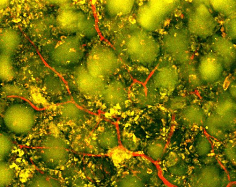
Neutrophil Infiltration
Two-photon imaging at day 27 using Ly6G-green fluorescent protein (GFP) reporter mice to visualize neutrophils. IL-4/C1q-EV + MAPS implants exhibited dense vascularization and focal accumulation of GFP+ cells within scaffold pores. Credit: Duke University

Immune Cells Take on a Surprising Role

Neutrophils are among the immune system’s fastest responders. After a stroke, they are often associated with inflammation and additional tissue damage, especially during the early phase of injury. Yet immune cells do not always have a single fixed role. Their behavior can change depending on timing, location, and the molecular signals surrounding them.

Inside the engineered scaffold, neutrophils appeared to become part of the repair process rather than merely contributing to destruction.

The researchers tested that possibility by depleting the immune-cell population rich in neutrophils. Blood vessel formation dropped sharply, and the scaffold underwent far less remodeling. The experiment showed that these cells were not simply present at the injury site. They were helping drive the response.

“This result changes how we think about neutrophils after stroke,” said Shangjing Xin, lead scientist of the study and a postdoctoral fellow in the Segura Laboratory. “Their role appears to depend on when they arrive, where they are located, and the signals they receive from their surroundings. Our study demonstrates a potential engineering strategy to recruit and retain these cells at the right time.”

New Blood Vessels and Nerve Fibers Emerge

The treatment produced visible changes throughout the damaged region. Blood vessels grew across the cavity, potentially creating the circulation needed to support living tissue. Researchers also detected more axonal fibers within and around the injury. Axons are the long projections neurons use to carry electrical signals to other cells.

Those biological changes were accompanied by improved movement.

During a grid-walking test, scientists measured how often the mice misplaced a front paw while crossing an uneven surface. Animals treated with the optimized scaffold made fewer errors over time. By eight weeks, their performance could not be statistically distinguished from that of healthy control mice, and the improvement continued through the end of the study.

The scaffold itself proved essential. When researchers delivered the EVs without MAPS, they did not observe comparable blood vessel growth. The result suggests that the treatment depended on both components: the biological messages carried by the vesicles and the porous structure that concentrated those messages while giving cells space to organize.

Toward a Scalable Human Stroke Therapy

The study relied on EVs collected from primary rat astrocytes, which would not be a practical source for a widely available human therapy.

Segura’s laboratory is now exploring astrocytes made from human-induced pluripotent stem cells. These cells can be produced from reprogrammed adult cells and expanded in the laboratory, potentially offering a more scalable and clinically relevant source of EVs. Researchers may also be able to adjust the conditions under which the astrocytes grow to better control the messages their vesicles carry.

“You do not restore an ecosystem simply by containing the initial damage,” Segura said. “You have to create the conditions that allow life to return. That is how we think about the stroke cavity. The material is not intended to reproduce the brain itself but to create an environment where the body’s own cells can enter, communicate, and participate in rebuilding vascularized tissue.”

Reference: “IL-4/C1q activated astrocyte-derived extracellular vesicles promote stroke infarct recovery by recruiting peripheral leukocytes” by Shangjing Xin, Lucy Zhang, Nhi V. Phan, Mengying An, Ligen Shi, S. Thomas Carmichael and Tatiana Segura, 21 July 2026, Cell Biomaterials.
DOI: 10.1016/j.celbio.2026.100543


Saturday, July 11, 2026

Study Overturns Decades of External Axon Growth Theory

 How will your competent? doctor use this to get your needed axon pathfinding working properly AND have your doctor proves it is working!

Study Overturns Decades of External Axon Growth Theory

Summary: Researchers discovered that axon generation is actually an autonomous, internally driven process. The team proved that young nerve cells utilize an internal protein complex to methodically “unzip” their own structural scaffolding from the inside out, establishing early brain wiring via an intrinsic genetic protocol.

Key Facts

  • The Intrinsic Control Shift: Rather than being passively shaped by external biological chemical trails, embryonic neurons actively remodel their own cytoskeleton (the cell’s internal structural scaffold) to trigger axon outgrowth, originating from the cell body (soma).
  • The Dance of the Neurites: Early symmetric neurons exhibit small, bud-like extensions called neurites. These extensions display a highly rhythmic “two steps forward, one step back” movement behavior, constantly stretching outward and shrinking backward on a minute-by-minute basis.
  • The Arp2/3 Molecular Zipper: The key to breaking this loop is a specialized protein complex called Arp2/3. Acting like a microscopic molecular zipper, Arp2/3 locally opens the cell’s tight, tension-bearing structural “corset,” allowing individual neurites to bulge outward.
  • Wave-Like Propagation: This internal unzipping action travels outward through a single neurite at a time like a physical wave. The wave continues until it meets the mechanical resistance of the remaining cellular corset, which forces the neurite back into a brief rest state.
  • Microtubule Lock-In: While these random, wave-driven expansions alternate between different neurites by chance, a parallel internal process is underway: rigid structural proteins (microtubules) grow outward from within. Eventually, one lucky neurite accumulates enough rigid microtubule scaffolding to resist pulling back.
  • Symmetry Shattered: Once this stable tipping point is reached within roughly 48 hours, that specific neurite transitions into independent, rapid growth to become the official axon. The overall wave-driven shape-shifting stops, and the remaining neurites are locked into becoming input-receiving dendrites.
  • Neurons in the brain and spinal cord form a vast network in which each cell receives many inputs but sends output through only a single, long extension: the “axon”.

“If our neurons had multiple axons, this would cause chaos in the brain,” says Professor Frank Bradke, a neurobiologist and research group leader at DZNE. 

“Nature has therefore found a clever way to make sure that neurons generate only one axon. This applies not only to humans, but across the entire animal kingdom. So, we’re dealing with very fundamental processes that shape the wiring of the brain and nervous system.”

Breaking symmetry

During early embryonic development, neurons are initially largely symmetric, exhibiting small projections known as neurites. E

ventually one of these develops into the axon, thereby breaking the symmetry. Until now, it was largely assumed that this process is determined by biological growth factors that act on a neuron from the outside – and that, much like attractants, lead to the development of the axon. The team led by Frank Bradke reaches a different conclusion.

“According to our observations, the axon forms as a result of a remodeling of the cytoskeleton initiated by the young neuron. The process originates in the cell body, the so-called soma – the very center of the neuron,” says Dr. Tien-chen Lin, first author of the current publication and a scientist at DZNE.

Young neurons display a rhythmic behavior: Their neurites stretch out somewhat, and then shrink back slightly.

“This happens on a minute-by-minute basis. In a sense, the process follows the principle of two steps forward and one step back. This sequence repeats again and again,” says Tien-chen Lin.

Within typically 48 hours, however, one of the neurites grows into an axon. The remaining neurites later develop into receptors for inputs.

“Actually, this recurring process was already known. But it was unclear what lies behind it. We have now been able to shed considerable light on the underlying mechanisms.”

The key lies in the neuron’s cytoskeleton, a tension‑bearing, molecular scaffold that acts like a corset around the cell.

“This is where a protein complex called Arp2/3 enters the picture. Our findings show that it works like a zipper, locally opening the cell’s corset,” says Tien-chen Lin. “By doing so, Arp2/3 drives the cell’s rhythmic shape-shifting, repeatedly loosening a network that would otherwise tighten up again.”

Wave-like propagation

The researchers found that Arp2/3 always acts on only one neurite at a time, temporarily enabling its growth.

“This comes with a locally confined restructuring of the cytoskeleton that spreads like a wave,” says Tien-chen Lin. “These events continue until the wave subsides because, although the cellular corset has been loosened, it still offers a certain degree of resistance. Then the process begins again. The same neurite may be affected once more, or a different one may be involved. Which one seems to be a matter of chance.”

Parallel to this outward extension, relatively rigid structural proteins grow into the neurites from within.

“Eventually, one of the neurites becomes stable enough to resist being pulled back. It can then continue growing independently of Arp2/3 and ultimately develops into the axon. Meanwhile, the overall ’wave-driven’ outgrowth comes to a stop,” says Tien-chen Lin.

Open questions

“We cannot exclude the possibility that external factors play a certain role. However, given our data, we are convinced that the basic process that drives axon growth originates within the cell itself,” says Frank Bradke.

Open questions remain: What initiates the remodeling? Why does it proceed rhythmically and one neurite at a time? And, why does remodeling stop as soon as one of the neurites has grown large enough?

“The young nerve cell presumably follows a protocol encoded in its genome. However, we do not yet know the relevant genetic program, and our understanding of the associated regulatory processes remains limited. Thus, there is still plenty of research ahead, which motivates us to continue pursuing this topic.”

Key Questions Answered:

Q: Why would it cause “chaos in the brain” if a single nerve cell accidentally developed two or three axons?

A: Think of a neuron like an ultra-precise telephone line. It is biologically designed to collect information from thousands of neighbors through its many input roots (dendrites), but it must broadcast its finalized message through a single, dedicated output wire (the axon). If a neuron sprouted multiple axons, it would blast its electrical signals into multiple unintended circuits simultaneously, causing widespread cross-talk, short-circuiting sensory loops, and destroying the brain’s delicate computational symmetry.

Q: How does the Arp2/3 protein complex function like a “zipper” to change the shape of a cell?

A: Every young neuron is wrapped in a highly tense, rigid mesh network of structural proteins that acts like a cellular corset, keeping the cell tight and round. Dr. Tien-chen Lin discovered that the Arp2/3 complex acts as a localized release valve. When it activates at the base of a neurite, it unzips the interlocking threads of that corset, temporarily loosening the tension. This allows the internal contents of the cell to push outward in a wave, extending that specific branch further into space.

Q: If this entire process is managed from inside the cell, how does the neuron decide which branch becomes the final axon?

A: According to the DZNE data, the initial selection process is actually driven by chance. The Arp2/3 complex randomly shifts from one neurite to another, causing them all to rhythmically stretch and contract. However, as these branches take turns expanding, rigid rod-like structural proteins called microtubules are continuously growing outward from the center. Eventually, purely by coincidence, one neurite remains expanded just long enough for these rigid rods to pack inside and lock its skeleton in place, permanently stabilizing it so it can no longer shrink back.

Editorial Notes:

  • This article was edited by a Neuroscience News editor.
  • Journal paper reviewed in full.
  • Additional context added by our staff.

About this neuroscience research news

Author: Marcus Neitzert
Source: DZNE
Contact: Marcus Neitzert – DZNE
Image: The image is credited to Neuroscience News

Original Research: Open access.
“An intrinsic cytoskeletal oscillator establishes neuronal polarity” by Tien-chen Lin (林天正), Charlotte H. Coles, Eissa Alfadil, Florian Fäßler, Andreas Husch, Sebastian Dupraz, Thorben Pietralla, Akihiro Narita, Max Schelski, Kevin C. Flynn, Sina Stern, Christoph Möhl, Brett J. Hilton, Franz Vauti, Hans-Henning Arnold, Florian K. M. Schur & Frank Bradke. Nature
DOI:10.1038/s41586-026-10755-6

Wednesday, November 26, 2025

Axonal Regeneration, Growth Cone, and the FIGN Gene Family: A Comprehensive Review

 Your competent? doctor created dendritic branching and axon pathfinding and regeneration protocols years ago, right? Oh, your incompetent doctor hasn't even started creating protocols! WOW!

Send me hate mail on this: oc1dean@gmail.com. I'll print your complete statement with your name and my response in my blog. Or are you afraid to engage with my stroke-addled mind? No excuses are allowed! You're medically trained; it should be simple to precisely state EXACTLY WHY you haven't created those protocols in the last decade with NO EXCUSES! Your definition of competence in stroke is obviously much lower than stroke survivors' definition of your competence! Swearing at me is allowed, I'll return the favor. Don't even attempt to use the excuse that brain research is hard.

Axonal Regeneration, Growth Cone, and the FIGN Gene Family: A Comprehensive Review


Affiliations 

Abstract

Mature mammalian central nervous system (CNS) neurons exhibit limited regenerative capacity, resulting in severe and often irreversible functional deficits following injuries such as spinal cord injury or stroke. Both inhibitory extrinsic environments and intrinsic neuronal constraints govern this regenerative failure. Growth cones-cytoskeleton-rich structures at axonal tips-are critical for axon extension and pathfinding during neural repair, with their structural integrity and motility heavily dependent on microtubule (MT) stability and dynamics. Pharmacological stabilization of MTs has been shown to prevent growth cone collapse and support regeneration, highlighting MTs as a promising therapeutic target. The FIGN gene family-FIGN, FIGNL1, and FIGNL2-encodes microtubule-severing enzymes (MSEs) that regulate MT remodeling, thereby directing axonal growth and regeneration. FIGN and FIGNL2 act as key negative regulators: FIGN destabilizes dynamic tyrosinated MTs, whereas FIGNL2 selectively severs stable acetylated MTs, both leading to excessive microtubule fragmentation, growth cone collapse, and impaired regeneration. Inhibition of FIGN or FIGNL2, particularly via siRNA-mediated knockdown, enhances MT integrity, promotes robust axonal regrowth, and improves functional recovery in preclinical models. In contrast, FIGNL1 modulates MT dynamics in a context-dependent manner, primarily contributing to developmental neurite outgrowth. These proteins intersect with critical signaling pathways, including RhoA/ROCK and PI3K/Akt, linking cytoskeletal regulation to regenerative signaling. Targeting FIGN or FIGNL2 represents a promising neuropharmacological strategy to overcome intrinsic barriers to CNS repair. This review elucidates the regulatory mechanisms and functional roles of the FIGN gene family in neural regeneration and proposes novel therapeutic approaches for CNS injury and neurodegenerative diseases.

Keywords: Axonal regeneration; FIGN gene family; cytoskeleton.; gene therapy; microtubule stability.

Thursday, September 19, 2024

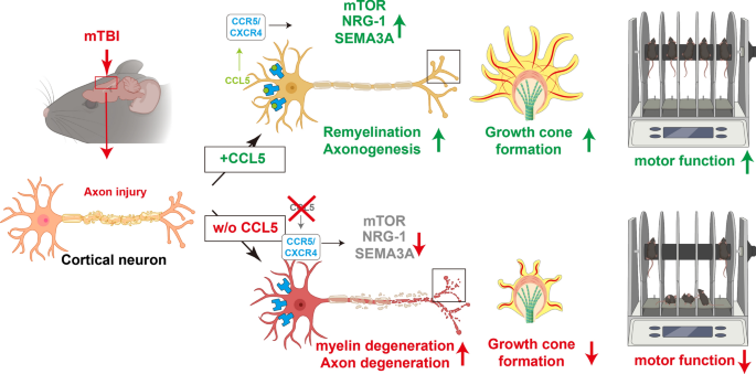
CCL5 is essential for axonogenesis and neuronal restoration after brain injury

 Does your competent? doctor and hospital have enough functioning brain cells to see that this might help in stroke and get further research done in humans? Oh, you don't have a functioning stroke doctor or hospital, do you?

CCL5 is essential for axonogenesis and neuronal restoration after brain injury

Abstract

Background

Traumatic brain injury (TBI) causes axon tearing and synapse degradation, resulting in multiple neurological dysfunctions and exacerbation of early neurodegeneration; the repair of axonal and synaptic structures is critical for restoring neuronal function(Sounds exactly like the needs in stroke). C-C Motif Chemokine Ligand 5 (CCL5) shows many neuroprotective activities.

Method

A close-head weight-drop system was used to induce mild brain trauma in C57BL/6 (wild-type, WT) and CCL5 knockout (CCL5-KO) mice. The mNSS score, rotarod, beam walking, and sticker removal tests were used to assay neurological function after mTBI in different groups of mice. The restoration of motor and sensory functions was impaired in CCL5-KO mice after one month of injury, with swelling of axons and synapses from Golgi staining and reduced synaptic proteins-synaptophysin and PSD95. Administration of recombinant CCL5 (Pre-treatment: 300 pg/g once before injury; or post-treatment: 30 pg/g every 2 days, since 3 days after injury for 1 month) through intranasal delivery into mouse brain improved the motor and sensory neurological dysfunctions in CCL5-KO TBI mice.

Results

Proteomic analysis using LC-MS/MS identified that the “Nervous system development and function”-related proteins, including axonogenesis, synaptogenesis, and myelination signaling pathways, were reduced in injured cortex of CCL5-KO mice; both pre-treatment and post-treatment with CCL5 augmented those pathways. Immunostaining and western blot analysis confirmed axonogenesis and synaptogenesis related Semaphorin, Ephrin, p70S6/mTOR signaling, and myelination-related Neuregulin/ErbB and FGF/FAK signaling pathways were up-regulated in the cortical tissue by CCL5 after brain injury. We also noticed cortex redevelopment after long-term administration of CCL5 after brain injury with increased Reelin positive Cajal-Rerzius Cells and CXCR4 expression. CCL5 enhanced the growth of cone filopodia in a primary neuron culture system; blocking CCL5’s receptor CCR5 by Maraviroc reduced the intensity of filopodia in growth cone and also CCL5 mediated mTOR and Rho signalling activation. Inhibiting mTOR and Rho signaling abolished CCL5 induced growth cone formation.

Conclusions

CCL5 plays a critical role in starting the intrinsic neuronal regeneration system following TBI, which includes growth cone formation, axonogenesis and synaptogensis, remyelination, and the subsequent proper wiring of cortical circuits. Our study underscores the potential of CCL5 as a robust therapeutic stratagem in treating axonal injury and degeneration during the chronic phase after mild brain injury.

Graphical Abstract

Introduction

Traumatic brain injury (TBI) is a complex disorder caused by external forces and is also the most significant cause of death and disability for people under the age of 40 [23, 31]. According to the Glasgow Coma Scale (GCS), TBI can be clinically divided into mild (GCS:14–15), moderate (GCS:9–13) and severe (GCS:3–8). Mild TBI is the most common brain injury caused by contact sports (i.e., hockey, football), motor vehicle accidents, and falls. Evidence from long-term studies on soldiers and athletes suggest that mild and repeated mild brain injuries are associated with the development of Chronic Traumatic Encephalopathy (CTE) [19, 37, 44], and also increase the risk of early onset Parkinsonism [18], dementia, and Alzheimer’s disease (AD) [25, 48].

Axonal shredding and tearing upon the impact, also called traumatic axonal injury (TAI) or diffuse axonal idiopathic injury (DAI), are the initial direct damages from mechanical impact on brain tissue. TAI is characterized by impaired axoplasmic transport, axonal swelling, disconnection with small hemorrhagic and/or non-hemorrhagic lesions, and brain swelling [7]. Unfortunately, much evidence suggests that neurons within the adult mammalian CNS cannot regenerate their axons after injury [13, 43]. Several deleterious cascades will be activated, leading to axon degeneration after injury. First, damaged glial cells/oligodendrocytes may not be able to provide sufficient energy to support neurons and cause neuron degeneration after injury [33, 49]. Second, increased calcium (Ca2+) influx leads to neuron apoptosis and degeneration [33, 49]. Third, glia scar and myelin-associated inhibitory proteins, such as Nogo, myelin-associated glycoprotein (Mag), and oligodendrocyte myelin glycoprotein (OMgp), can inhibit axonal regeneration after injury [15].

Recent evidence suggests that CNS neurons can revert to an embryonic-like growth state in the chronic/remodeling phase of injury to facilitate axon regeneration [46]. This “redevelopment” state provides a permissive microenvironment and the intracellular machinery for axon regrowth [9]. The endogenous repair systems activate and repair damaged axons within the chronic phase after axonal injury. The molecular machinery underlying axon regeneration is very similar to axon growth, such as Rho-GTPases Cdc42, Rac-1, and RhoA, phosphoinositide 3-kinase (PI3K)/AKT signaling pathways, and mitogen-activated protein kinase (MAPK) signaling [38]. Importantly, Rho is also the target molecule for myelin-associated inhibitory proteins [15]. PI3K/Akt activates its downstream molecules - the mammalian target of rapamycin (mTOR) and promotes axonal regeneration, synaptic plasticity, and neuronal survival after injury [35, 45]. Modulating the mTOR pathway is a novel strategy to promote axon regeneration [10]. Among three types of MAP kinases, the Erk cascade is a major pro-survival intracellular signaling pathway that can be activated by GDNF (glial cell-line derived neurotrophic factor) to promote neurite outgrowth in the spinal cord [29].

Chemokine CCL5; C-C motif ligand 5, also known as RANTES (regulated on activation, normal T cell expressed and secreted), shows many protective roles after neuronal damage, such as in stroke [51] and AD [24, 28, 32, 53]. In brain trauma, the plasma level of CCL5 increases in both human patients and animals after injury [2, 20]. Interestingly, CCL5 was found to be raised around the axonal transection site in mice immediately after injury [3]. The increased CCL5 around the axon transection site induces leukocyte infiltration to the injury site, but its function on neurons is still unclear. Our previous study identified that CCL5 is an essential factor in activating glutathione peroxidase 1 (GPX1) after brain injury, consequently reducing oxidative stress and protecting hippocampal neurons from oxidative stress-induced death; this effect facilitated memory-cognition recovery in mice after mild brain injury [22]. We also showed that CCL5 contributes to hippocampal neurons’ ATP generation and synaptic complex formation [1]. In addition, CCL5 expression after spinal cord injury is associated with axon regeneration and immune suppression [57]. The plasma level of CCL5 is highly correlated with BDNF, EGF (epidermal growth factor), and VEGF (vascular endothelial growth factor) after stroke [51]; those trophic factors contribute to neuron growth and proper brain function. Taken together, these findings suggest that CCL5 can help neurons survive from energy shortage and promote axon and synapse regrowth after injury.

In the present study, we identified that this chemokine has a robust and attractive effect on neuron growth cone formation, axon growth and myelination during post-injury repair. We specifically demonstrate for the first time that these effects are related to the ability of neuronal CCL5 to promote axon growth cone formation, axonogenesis and myelination through P70S6/mTOR signaling and the NRG1/ErbB and FGF pathways, ultimately augmenting neuronal axon and synapse regrowth after injury. Together, CCL5 promotes recovery of axogenesis and neurogenesis after brain injury.

Saturday, June 24, 2023

Insight into protein synthesis in axon regeneration

You need this, what is your doctor doing to ensure this occurs? 

Do you prefer your  doctor incompetence NOT KNOWING? OR NOT DOING?

Insight into protein synthesis in axon regeneration


, , , , ,
https://doi.org/10.1016/j.expneurol.2023.114454Get rights and content

Abstract

Successful axon regeneration is crucial for the treatment of numerous nerve injuries and neurodegenerative diseases, which requires adequate and accurate protein synthesis, including mRNA translation, both in the neuron somas and locally in the axons. Recent studies have shed light on novel functions and mechanisms of protein synthesis that are relevant for axon regeneration, with a particular focus on local translation. Here, we review the new developed technologies and approaches for investigating local translation, discuss the roles of local translation in axon regeneration, and summarize the key signaling molecules and pathways that regulate local translation during axon regeneration. Additionally, we give an overview of local translation in the peripheral and central nervous systems neurons and the latest progress in protein synthesis in neuron somas respectively. Finally, we consider the potential directions for future research in this field to provide insights into protein synthesis in axon regeneration.

Introduction

Numerous disorders involving axon damage can benefit from successful axon regeneration, including stroke, traumatic brain injury, spinal cord injury, peripheral nerve injury, and a range of neurodegenerative diseases (Li et al., 2020; Qian and Zhou, 2020). As a complex and synergistic process involving multiple cellular events, axon regeneration requires the sustained activity of structural and regulatory proteins in both neuronal somas and axons (Rozenbaum et al., 2018; Terenzio et al., 2018; Zheng et al., 2001), implying that de novo protein synthesis in somas and axons is critical for axon regeneration in adult mammalian nervous systems (Belin et al., 2015; Donnelly et al., 2013; Weng et al., 2018). Due to technical constraints, it has long been believed that transcripts and their translational machinery are found in the dendrites of neurons but not in axons. Although polysomes have been found at the base of dendritic spines (Steward and Levy, 1982), mature axons typically exhibit low levels of ribosomes, which are primarily localized in periaxoplasmic ribosomal plaques (Calliari et al., 2014; Koenig et al., 2000; Sotelo-Silveira et al., 2008; Sotelo-Silveira et al., 2004). Thus, the prevalent view has been that axons are incapable of locally synthesizing proteins.

However, with methodological advances, it was eventually discovered that axon compartments contain a variety of RNA molecules, including ribosomal RNA, transfer RNA, and messenger RNA (Koenig, 1979; Koenig et al., 2000; Van Minnen, 1994; Weiner et al., 1996), suggesting protein synthesis in axons. The earliest direct evidence of local translation in mature mammalian axons came from sensory neurons in the peripheral nervous system (PNS). Zhang et al. separated pure regenerating axons from the dorsal root ganglion (DRG) cell bodies, and found that the axons can actively synthesize proteins (Zheng et al., 2001). Then numerous studies demonstrated that mature neurons in the central nervous system (CNS) can also synthesize proteins in their axons (Baleriola et al., 2014; Kalinski et al., 2015). And the local protein synthesis dynamically control the neuronal function in the CNS (Akins et al., 2017; Monday et al., 2022). In addition, studies have revealed that translation and ribosome biogenesis are rate-limiting processes for axon and dendrite growth (Slomnicki et al., 2016; Williams et al., 2016) and that the equilibrium of protein synthesis in the soma and axon influences axonal growth rates (Perry et al., 2016). These observations suggest that protein synthesis regulation at the levels of both axons and somas plays an important role in axon regeneration.

However, compared with transcriptional regulation, translational regulation in axon repair after injury is less understood. This emerging field is working toward the goal of revealing novel functions of protein synthesis in axon regeneration and identifying potential pharmacological targets in nerve injury or neurodegenerative diseases. Here, we will discuss these functions and provide a brief overview of the current understanding of the underlying mechanisms, including local translation and protein synthesis in the soma. Furthermore, we discuss what advancements in this field can be expected in the future.

More at link.

Insight into protein synthesis in axon regeneration

How is your doctor using this to successfully reconnect neurons for your recovery? 

Your doctor should have intuited that this could work for stroke rehab way back in September 2015. If not you don't have a functioning stroke doctor.

Protein synthesis in regenerating spinal cord axons September 2015 

The latest here:

Insight into protein synthesis in axon regeneration

Abstract

Successful axon regeneration is crucial for the treatment of numerous nerve injuries and neurodegenerative diseases, which requires adequate and accurate protein synthesis, including mRNA translation, both in the neuron somas and locally in the axons. Recent studies have shed light on novel functions and mechanisms of protein synthesis that are relevant for axon regeneration, with a particular focus on local translation. Here, we review the new developed technologies and approaches for investigating local translation, discuss the roles of local translation in axon regeneration, and summarize the key signaling molecules and pathways that regulate local translation during axon regeneration. Additionally, we give an overview of local translation in the peripheral and central nervous systems neurons and the latest progress in protein synthesis in neuron somas respectively. Finally, we consider the potential directions for future research in this field to provide insights into protein synthesis in axon regeneration.

Introduction

Numerous disorders involving axon damage can benefit from successful axon regeneration, including stroke, traumatic brain injury, spinal cord injury, peripheral nerve injury, and a range of neurodegenerative diseases (Li et al., 2020; Qian and Zhou, 2020). As a complex and synergistic process involving multiple cellular events, axon regeneration requires the sustained activity of structural and regulatory proteins in both neuronal somas and axons (Rozenbaum et al., 2018; Terenzio et al., 2018; Zheng et al., 2001), implying that de novo protein synthesis in somas and axons is critical for axon regeneration in adult mammalian nervous systems (Belin et al., 2015; Donnelly et al., 2013; Weng et al., 2018). Due to technical constraints, it has long been believed that transcripts and their translational machinery are found in the dendrites of neurons but not in axons. Although polysomes have been found at the base of dendritic spines (Steward and Levy, 1982), mature axons typically exhibit low levels of ribosomes, which are primarily localized in periaxoplasmic ribosomal plaques (Calliari et al., 2014; Koenig et al., 2000; Sotelo-Silveira et al., 2008; Sotelo-Silveira et al., 2004). Thus, the prevalent view has been that axons are incapable of locally synthesizing proteins.

However, with methodological advances, it was eventually discovered that axon compartments contain a variety of RNA molecules, including ribosomal RNA, transfer RNA, and messenger RNA (Koenig, 1979; Koenig et al., 2000; Van Minnen, 1994; Weiner et al., 1996), suggesting protein synthesis in axons. The earliest direct evidence of local translation in mature mammalian axons came from sensory neurons in the peripheral nervous system (PNS). Zhang et al. separated pure regenerating axons from the dorsal root ganglion (DRG) cell bodies, and found that the axons can actively synthesize proteins (Zheng et al., 2001). Then numerous studies demonstrated that mature neurons in the central nervous system (CNS) can also synthesize proteins in their axons (Baleriola et al., 2014; Kalinski et al., 2015). And the local protein synthesis dynamically control the neuronal function in the CNS (Akins et al., 2017; Monday et al., 2022). In addition, studies have revealed that translation and ribosome biogenesis are rate-limiting processes for axon and dendrite growth (Slomnicki et al., 2016; Williams et al., 2016) and that the equilibrium of protein synthesis in the soma and axon influences axonal growth rates (Perry et al., 2016). These observations suggest that protein synthesis regulation at the levels of both axons and somas plays an important role in axon regeneration.

However, compared with transcriptional regulation, translational regulation in axon repair after injury is less understood. This emerging field is working toward the goal of revealing novel functions of protein synthesis in axon regeneration and identifying potential pharmacological targets in nerve injury or neurodegenerative diseases. Here, we will discuss these functions and provide a brief overview of the current understanding of the underlying mechanisms, including local translation and protein synthesis in the soma. Furthermore, we discuss what advancements in this field can be expected in the future.

Friday, June 23, 2023

H2S-RhoA/ROCK Pathway and Glial Cells in Axonal Remyelination After Ischemic Stroke

 Didn't your doctor give you hydrogen sulfide gas immediately post stroke?

 Well, for your fart smelling needs there are still sales on eBay. What is your doctor's recommendation on this? 

Among the top gas-producing foods are beans and other legumes as well as cruciferous vegetables, such as:

  • Cabbages.

  • Turnips.

  • Kale.

  • Broccoli.

  • Brussels sprouts.

  • Arugula.

  • Cauliflower.


Does this explain all these benefits?

12 posts on hydrogen sulfide helping with stroke prevention, reduction in stroke damage, protects stem cells and helps neurogenesis.

Or this?

Smelling farts could be the best thing you do today

The latest here:

H2S-RhoA/ROCK Pathway and Glial Cells in Axonal Remyelination After Ischemic Stroke

Abstract

Ischemic stroke is one of the main reasons of disability and death. Stroke-induced functional deficits are mainly due to the secondary degeneration of the white matter characterized by axonal demyelination and injury of axon-glial integrity. Enhancement of the axonal regeneration and remyelination could promote the neural functional recovery. However, cerebral ischemia-induced activation of RhoA/Rho kinase (ROCK) pathway plays a crucial and harmful role in the process of axonal recovery and regeneration. Inhibition of this pathway could promote the axonal regeneration and remyelination. In addition, hydrogen sulfide (H2S) has the significant neuroprotective role during the recovery of ischemic stroke via inhibiting the inflammatory response and oxidative stress, regulating astrocyte function, promoting the differentiation of endogenous oligodendrocyte precursor cells (OPCs) to mature oligodendrocyte. Among all of these effects, promoting the formation of mature oligodendrocyte is a crucial part of axonal regeneration and remyelination. Furthermore, numerous studies have uncovered the crosstalk between astrocytes and oligodendrocyte, microglial cells and oligodendrocyte in the axonal remyelination following ischemic stroke. The purpose of this review was to discuss the relationship among H2S, RhoA/ROCK pathway, astrocytes, and microglial cells in the axonal remyelination following ischemic stroke to reveal new strategies for preventing and treating this devastating disease.

This is a preview of subscription content, access via your institution.