Changing stroke rehab and research worldwide now.Time is Brain! trillions and trillions of neurons that DIE each day because there are NO effective hyperacute therapies besides tPA(only 12% effective). I have 523 posts on hyperacute therapy, enough for researchers to spend decades proving them out. These are my personal ideas and blog on stroke rehabilitation and stroke research. Do not attempt any of these without checking with your medical provider. Unless you join me in agitating, when you need these therapies they won't be there.

What this blog is for:

My blog is not to help survivors recover, it is to have the 10 million yearly stroke survivors light fires underneath their doctors, stroke hospitals and stroke researchers to get stroke solved. 100% recovery. The stroke medical world is completely failing at that goal, they don't even have it as a goal. Shortly after getting out of the hospital and getting NO information on the process or protocols of stroke rehabilitation and recovery I started searching on the internet and found that no other survivor received useful information. This is an attempt to cover all stroke rehabilitation information that should be readily available to survivors so they can talk with informed knowledge to their medical staff. It lays out what needs to be done to get stroke survivors closer to 100% recovery. It's quite disgusting that this information is not available from every stroke association and doctors group.

Showing posts with label quantification. Show all posts

Thursday, October 12, 2023

A robot-based interception task to quantify upper limb impairments in proprioceptive and visual feedback after stroke

 Quantification DOES NOTHING to get survivors recovered! I'd fire you all for absolute stupidity! I can do no reaching with left arm until spasticity is cured.

A robot-based interception task to quantify upper limb impairments in proprioceptive and visual feedback after stroke

Abstract

Background

A key motor skill is the ability to rapidly interact with our dynamic environment. Humans can generate goal-directed motor actions in response to sensory stimulus within ~ 60-200ms. This ability can be impaired after stroke, but most clinical tools lack any measures of rapid feedback processing. Reaching tasks have been used as a framework to quantify impairments in generating motor corrections for individuals with stroke. However, reaching may be inadequate as an assessment tool as repeated reaching can be fatiguing for individuals with stroke. Further, reaching requires many trials to be completed including trials with and without disturbances, and thus, exacerbate fatigue. Here, we describe a novel robotic task to quantify rapid feedback processing in healthy controls and compare this performance with individuals with stroke to (more) efficiently identify impairments in rapid feedback processing.

Methods

We assessed a cohort of healthy controls (n = 135) and individuals with stroke (n = 40; Mean 41 days from stroke) in the Fast Feedback Interception Task (FFIT) using the Kinarm Exoskeleton robot. Participants were instructed to intercept a circular white target moving towards them with their hand represented as a virtual paddle. On some trials, the arm could be physically perturbed, the target or paddle could abruptly change location, or the target could change colour requiring the individual to now avoid the target.

Results

Most participants with stroke were impaired in reaction time (85%) and end-point accuracy (83%) in at least one of the task conditions, most commonly with target or paddle shifts. Of note, this impairment was also evident in most individuals with stroke when performing the task using their unaffected arm (75%). Comparison with upper limb clinical measures identified moderate correlations with the FFIT.

Conclusion

The FFIT was able to identify a high proportion of individuals with stroke as impaired in rapid feedback processing using either the affected or unaffected arms. The task allows many different types of feedback responses to be efficiently assessed in a short amount of time.

Introduction

A key feature of our motor system is the ability to use sensory feedback for the online control of movement, such as responding when your arm is accidentally bumped when reaching for an object [1]. A broad range of studies highlight how proprioceptive and visual feedback can be used to generate goal-directed motor corrections and initiate new motor actions within 100ms [2, 3]. Impressively, even responses requiring cognitive functions such as aborting an on-going motor action can be generated within 200ms [4]. This use of proprioceptive and visual feedback for goal-directed motor actions is supported by highly distributed circuits including both cortical and subcortical structures [1, 5].

Previous studies highlight how stroke can impair an individual’s ability to generate rapid and accurate motor corrections [6,7,8,9,10,11]. Marsden et al. studied individuals with brain damage and identified delays in generating responses to mechanical perturbations applied during thumb movements [9]. Schaefer and colleagues examined responses to visual perturbations while participants reached towards a goal. They found individuals with stroke had delayed response initiation when compared to healthy controls [7]. Of note, ~ 30% of individuals with stroke display these impairments with their ‘unaffected’ arm [8, 12]. Identifying impaired corrective responses after stroke is crucial as reaction time impairments have been associated with reduced quality of life and an increased risk of falls [10, 13,14,15,16]. Importantly, the ability to use sensory feedback for motor function is not adequately considered in common clinical tools such as Fugl-Meyer Assessment (FMA) and Chedoke McMaster Stroke Assessment (CMSA) [17, 18].

The present study and associated behavioural task address several aspects of goal-directed feedback control. First, neural circuits associated with feedback processing involve a broad range of cortical and subcortical circuits. Thus, we hypothesize that impairments in using sensory feedback for goal-directed motor actions will be commonly observed in individuals following stroke. Second, neural pathways for proprioceptive and visual feedback overlap, but also involve distinct brain regions [19]. For example, primary somatosensory cortex and parietal area 5 are associated with somatosensory feedback, whereas occipital and posterior parietal regions are associated with visual feedback [20]. Thus, we hypothesize that individuals with stroke may display impairments that are limited to one sensory modality, proprioceptive or visual feedback. Finally, sensory feedback for motor and cognitive functions are also somewhat distinct, such as the involvement of medial regions in the frontal lobe engaged for inhibitory control [21]. Thus, we hypothesize that individuals with stroke may display selective impairment for either motor corrections or cognitive control.

We examined these hypotheses by developing a novel interception task that requires individuals to maintain their hand at a spatial location and were instructed to intercept a target moving rapidly towards them. On random trials, we induce mechanical or visual perturbations that require rapid corrective responses to intercept the moving target, or when instructed by a color cue, to avoid the target. The results highlight that most individuals with stroke are impaired in at least one form of feedback processing, and in some cases, selectively for one form of feedback processing.

More at link.

Friday, May 26, 2023

System identification: a feasible, reliable and valid way to quantify upper limb motor impairments

No clue how quantifying any of this helps survivors recover.  I don't understand how mentors and senior researchers can allow such useless research. The actual goal of stroke research is survivor recovery, if you can't link your research to that in some way it was useless.

System identification: a feasible, reliable and valid way to quantify upper limb motor impairments

Abstract

Background

Upper limb impairments in a hemiparetic arm are clinically quantified by well-established clinical scales, known to suffer poor validity, reliability, and sensitivity. Alternatively, robotics can assess motor impairments by characterizing joint dynamics through system identification. In this study, we establish the merits of quantifying abnormal synergy, spasticity, and changes in joint viscoelasticity using system identification, evaluating (1) feasibility and quality of parametric estimates, (2) test–retest reliability, (3) differences between healthy controls and patients with upper limb impairments, and (4) construct validity.

Methods

Forty-five healthy controls, twenty-nine stroke patients, and twenty cerebral palsy patients participated. Participants were seated with the affected arm immobilized in the Shoulder-Elbow-Perturbator (SEP). The SEP is a one-degree-of-freedom perturbator that enables applying torque perturbations to the elbow while providing varying amounts of weight support to the human arm. Participants performed either a ‘do not intervene’ or a resist task. Elbow joint admittance was quantified and used to extract elbow viscosity and stiffness. Fifty-four of the participants performed two sessions to establish the test–retest reliability of the parameters. Construct validity was assessed by correlating system identification parameters to parameters extracted using a SEP protocol that objectifies current clinical scales (Re-Arm protocol).

Results

Feasibility was confirmed by all participants successfully completing the study protocol within ~ 25 min without reporting pain or burden. The parametric estimates were good with a variance-accounted-for of ~ 80%. A fair to excellent test–retest reliability was found (

) for patients, except for elbow stiffness with full weight support (). Compared to healthy controls, patients had a higher elbow viscosity and stiffness during the ‘do not intervene’ task and lower viscosity and stiffness during the resist task. Construct validity was confirmed by a significant (all ) but weak to moderate (

) correlation with parameters from the Re-Arm protocol.

Conclusions

This work demonstrates that system identification is feasible and reliable for quantifying upper limb motor impairments. Validity was confirmed by differences between patients and controls and correlations with other measurements, but further work is required to optimize the experimental protocol and establish clinical value.

Introduction

Upper limb motor impairments in hemiparetic limbs such as after stroke and cerebral palsy (CP) are currently assessed by a set of well-established clinical scales aiming to quantify, among others, muscle weakness, abnormal synergy, spasticity and changes in joint viscoelasticity [1]. These include tests like the Brunnstrom Fugl-Meyer, Modified Ashworth Scale (MAS), and Modified Tardieu Scale (MTS). The motor impairments identified using these tests assist in selecting a treatment approach and are used to monitor improvement. However, the tests have several known limitations, which include their poor validity, reliability, and sensitivity [2,3,4,5,6]. Whereas the need for objective and reliable assessment of motor impairments is widely recognized [7, 8], no tests have yet successfully replaced traditional clinical scales.

Robotics can objectively quantify the motor impairments of muscle weakness, abnormal synergy, spasticity, and changes in joint viscoelasticity [9,10,11]. Recently, a robotic device was developed and validated to assess these four impairments using one single device (the Shoulder-Elbow-Perturbator—SEP) [12]. By using the SEP to mimic current clinical tests, an earlier finding was confirmed that upper limb synergy can be quantified by the change in elbow range of motion (ROM) when gradually reducing arm weight support [13]. Likewise, spasticity and viscoelasticity can be objectively quantified by imposing passive joint movements at low and high speeds [9, 14, 15]. The robotics used in the aforementioned studies leveraged the accurate recording of joint angles and torques to provide a reliable way to quantify motor impairments during passive and active joint movements.

Motor impairments can also be quantified with the help of robotics by applying external mechanical perturbations while participants perform either a ‘do not intervene’ (DNI) or resist task [16, 17]. These tasks imply the participant is fully relaxed (DNI task) or attempts to maintain a constant joint angle or torque (resist task) while the joint is rotated within a small ROM (e.g., root mean square rotation of 0.5–1°). Recordings of the joint angle and joint torque are subsequently scrutinized using system identification techniques to characterize the joint dynamics. System identification enables us to quantify both the intrinsic (non-neural) and reflexive (neural) contribution to the joint dynamics [18, 19], captured in the joint’s inertia, viscosity, and stiffness parameters. These parameters can be translated to clinically described phenomena such as synergy (i.e., aberrant elbow stiffness when lifting the arm), spasticity (i.e., enhanced reflexive activity), or changes in viscoelasticity (i.e., increased intrinsic joint viscosity and stiffness).

In this study, we aim to establish the merits of quantifying abnormal synergy, spasticity, and changes in joint viscoelasticity using system identification. This is achieved by (1) confirming the feasibility to perform a system identification protocol and quality of parametric estimates, (2) establishing the test–retest reliability for the system identification parameters, (3) comparing obtained parameters between healthy controls and patients with upper limb impairments and (4) test construct validity by comparing the system identification parameters for each motor impairment with parameters obtained using four tests as part of a recently proposed robotic assessment protocol (Re-Arm) [12].

Methods

Participants

We recruited adult patients from the outpatient clinic of the Rijndam Rehabilitation Center that presented upper limb motor impairments due to stroke and CP. Patients were only included if they: (1) had a clinically-confirmed upper limb impairment, (2) were able to achieve active shoulder abduction (up to 80°), (3) were able to achieve visible active elbow extension, and (4) had a minimal passive range-of-movement for shoulder abduction of 80° and horizontal shoulder adduction of 45°. Exclusion criteria were: (1) hemiplegic shoulder pain, (2) a history of pre-existing neuromuscular disorders affecting upper limb function, (3) fixed upper limb contractures, and (4) the inability to understand verbal instructions. We only included stroke patients when they were considered chronic, suffering the stroke at least six months before study inclusion.

For comparison, we recruited a group of age-matched healthy controls without a known history of neurological or orthopedic disorders. The study was approved by the Medical Ethics Committee of the Erasmus Medical Center, Rotterdam, (protocol number NL64660.078.18), and conducted following the declaration of Helsinki.

Experimental setup

Shoulder elbow perturbator

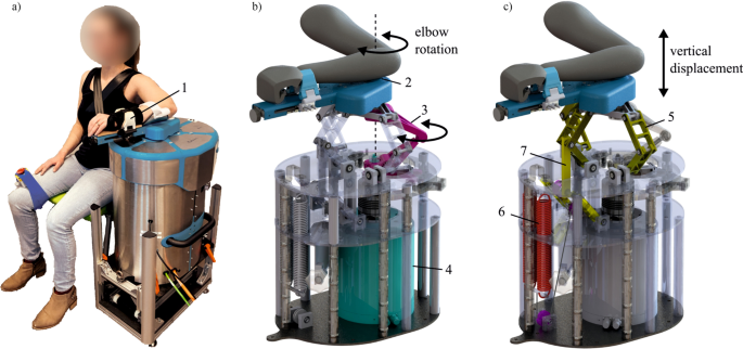
The Shoulder-Elbow-Perturbator (SEP—Hankamp Rehab, Enschede, The Netherlands) was used to assess participants' elbow dynamics. The design of the SEP enables independent manipulation of elbow angle and weight support of the human arm [12]. A direct-drive servo motor (HIWIN TMS3C, Offenburg, Germany) attached to a lever supporting the lower arm controlled the elbow angle by aligning its axis to the medial epicondyle of the humerus (Fig. 1). A computer with Etherlab and MATLAB Simulink was used to control the SEP and capture elbow torque and angle with a sample rate of 1 kHz.

Fig. 1
figure 1

The Shoulder Elbow Perturbator (SEP), a robotic device to quantify multiple upper limb impairments. a Participant positioned in the SEP with the shoulder abducted in 80°, the forearm strapped (1) to the lever arm of the SEP, and the medial epicondyle of the humerus aligned with the motor rotation axis. b Internals of the SEP showing that the motor (4) transmits a torque through the torque link (3), allowing elbow rotation (2). c Internals of the SEP showing the shoulder abductor manipulation mechanism. The sarrus linkages (5) allow vertical displacement of the arm, and the arm is supported by two springs (6) with an upward force. With the cable routing and pulley configuration, the upward force can be manipulated independently of the linkage position (7). (Modified – with permission from [20])

Wednesday, April 19, 2023

Quantifying Nonuse in Chronic Stroke Patients: A Study Into Paretic, Nonparetic, and Bimanual Upper-Limb Use in Daily Life

Quantifying DOES NOTHING to get survivors recovered! Useless. I can tell you exactly why I don't use my left arm/hand. SPASTICITY! Cure that and I'll recover in no time. Solve the fucking correct problem, not this useless quantification!  Leaders solve problems, not like the mouse behavior you see here.

Quantifying Nonuse in Chronic Stroke Patients: A Study Into Paretic, Nonparetic, and Bimanual Upper-Limb Use in Daily Life

2012, Archives of Physical Medicine and Rehabilitation
Marian E. Michielsen, MSc
†
 , Ruud W. Selles, PhD, Henk J. Stam, MD, PhD Gerard M. Ribbers, MD, PhD, Johannes B. Bussmann, PhD
ABSTRACT. Michielsen ME, Selles RW, Stam HJ, RibbersGM, Bussmann JB. Quantifying nonuse in chronic stroke pa-tients: a study into paretic, nonparetic, and bimanual upper-limb use in daily life. Arch Phys Med Rehabil 2012;xx:xxx.
Objective:
To quantify uni- and bimanual upper-limb use inpatients with chronic stroke in daily life compared with healthycontrols.
Design:
Cross-sectional observational study.
Setting:
 Outpatient rehabilitation center.
Participants:
 Patients with chronic stroke (n=38) and healthy controls (n=18).
Intervention:
 Not applicable.
Main Outcome Measures:
 Upper-limb use in daily life was measured with an accelerometry-based upper-limb activity monitor, an accelerometer based measurement device. Uni-manual use of the paretic and the nonparetic side and bimanual upper-limb use were measured for a period of 24 hours. Out-comes were expressed in terms of both duration and intensity.
Results:
 Patients used their unaffected limb much more than their affected limb (5.3h vs 2.4h), while controls used bothlimbs a more equal amount of time (5.4h vs 5.1h). Patients used their paretic side less than controls used their nondominant sideand their nonparetic side more than controls their dominant side. The intensity with which patients used their paretic side was lower than that with which controls used their nondominant side, while that of the nonparetic side was higher than that of the dominant side of controls. Finally, patients used their paretic side almost exclusively in bimanual activities. During bimanual activities, the intensity with which they used their affected side was much lower than that of the nonaffected side.
Conclusion:
 Our data show considerable nonuse of the paretic side, both in duration and in intensity, and both during unimanual and bimanual activities in patients with chronic stroke. Patients do compensate for this with increased use of the nonparetic side.

Sunday, April 17, 2022

Reliability and diagnostic accuracy of corrected slack angle derived from 2D-SWE in quantitating muscle spasticity of stroke patients

Survivors don't give a flying fuck about quantifying their spasticity, they want it cured. ARE YOU THAT FUCKING CLUELESS?

Reliability and diagnostic accuracy of corrected slack angle derived from 2D-SWE in quantitating muscle spasticity of stroke patients

Abstract

Background

To explore the feasibility of corrected slack angle acquired from two-dimensional shear wave elastography (2D-SWE) for quantitating the spasticity of medial gastrocnemius (MG) in stroke patients.

Methods

Consecutive stroke patients with spastic MG and matched healthy controls were recruited. Intra- and interobserver reliability of 2D-SWE measurement were evaluated, and the correlation between corrected slack angle and modified Ashworth scale (MAS) score was examined. The corrected slack angle before and after botulinum toxin A (BoNT-A) injection was compared and its diagnostic performance in classifying the severity of spasticity were assessed with receiver operating characteristic (ROC) curve analysis.

Results

The intra- (0.791 95% CI 0.432–0.932) and interobserver (0.751 95% CI 0.382–0.916) reliability of slack angle acquired with 2D-SWE were good. Significant correlation was found between corrected slack angle and MAS score (R = − 0.849, p < 0.001). The corrected slack angle increased after BoNT-A injection. The cutoff value of MAS ≥ 3 had the highest sensitivity (100%) and specificity (93.33%). The positive predictive value (PPV) for classification of MAS ≥ 1+ and the negative predictive value (NPV) for classification of MAS ≥ 3 were greater than 90%.

Conclusion

2D-SWE was a reliable method to quantitate the post-stroke spasticity. The corrected slack angle had advantage in classifying the severity of spasticity, especially in early identification of mild spasticity and confirmation of severe spasticity.

Background

Spasticity is a common complication of central nervous system injury including cerebrovascular accident, brain trauma and spinal cord injury, etc. [1]. It has been estimated that spasticity affects approximately 19% to 45% of patients suffering from stroke [2, 3]. Quantitation of spasticity is necessary, in order to assess the impact of therapies and to determine appropriate medicine dosage. The modified Ashworth scale (MAS) is the most feasible measurement of spasticity in clinical practice [4]. However, its dependency on subjective assessment restricts its application. Magnetic resonance elastography is capable of objectively evaluating the spasticity of individual muscle [5], but it is limited by a variety of contraindications (e.g., cardiac pacemaker, metal implants, claustrophobia).

Ultrasound elastography had been widely used to examine tissue elasticity in various organs, including breast [6], prostate [7] and liver [8]. Recently, it has been developed as a quantitative method for the evaluation of skeletal muscle. Several studies have attempted to investigate the mechanical properties of the spastic muscles in post-stroke patients [9,10,11]. However, various measurements (strain ratio [9], elasticity index [10], shear elastic modulus [11]) were reported because of different elastography techniques they used. As the newest modality of ultrasound elastography that uses acoustic radiation force and generate quantitative elastograms [12], although the technical assumption may not perfectly be met in skeletal muscle because skeletal muscle is not a homogeneous or isotropic material [13], two-dimensional shear wave elastography (2D-SWE) has also been used in evaluation of skeletal muscle cautiously. But its standard protocol is still lacking, which makes it difficult to achieve consensus on the cut-off elastic value of spastic muscles. Besides, the correlation between shear modulus and the clinical assessment such as MAS for stroke patients remains equivocal [14, 15].

Slack angle, defined as the angle of joint from where the muscle becomes tensioned and the shear modulus begins to rise as the joint was passively moved, has been a crucial parameter to characterize the mechanical property of skeletal muscles [16, 17]. Because skeletal muscle is a kind of active and deformable tissue, continuous 2D-SWE recording would make sense. It has been revealed that the slack angle of gastrocnemius occurred at a more plantarflexed angle in stroke patients than control subjects [18]. However, the slack angle was visually determined in previous studies [17,18,19]. Its inter- and intraobserver reliability and its correlation with clinical assessment have not been reported yet. Moreover, the inter-individual variability of muscle elasticity, caused by gender, age, physical activity and biological structure [20], may be a confounding factor when using the slack angle to evaluate the spasciticy of muscle. Instead of comparing with the spastic slack angle directly, we suggest that it would be reasonable to propose the corrected slack angle, which was performed a self-correction by subtracting the shear modulus of the unaffected muscle from that of the spastic muscle. It could be beneficial to rule out the confounding factors related to the passive extensibility of skeletal muscle.

Therefore, the present study aims to explore the feasibility of quantitating the spasticity of stroke patients with corrected slack angle acquired from 2D-SWE. For this purpose, whether corrected slack angle correlated with MAS and its change after botulinum toxin A (BoNT-A) injection are examined, as well as its diagnostic performance for classifying the severity of spasticity.


Sunday, February 6, 2022

Motor and Premotor Cortices in Subcortical Stroke: Proton Magnetic Resonance Spectroscopy Measures and Arm Motor Impairm

Survivors don't care about quantifying their impairment, they want it cured. This was almost totally fucking useless. After you measured this what protocols are needed to cure the impairment? THAT IS WHAT SURVIVORS NEED. GET THERE!


Motor and Premotor Cortices in Subcortical Stroke: Proton Magnetic Resonance Spectroscopy Measures and Arm Motor Impairment

First Published January 8, 2013 Research Article Find in PubMed

Background. 

Although functional imaging and neurophysiological approaches reveal alterations in motor and premotor areas after stroke, insights into neurobiological events underlying these alterations are limited in human studies.  

Objective. 

We tested whether cerebral metabolites related to neuronal and glial compartments are altered in the hand representation in bilateral motor and premotor areas and correlated with distal and proximal arm motor impairment in hemiparetic persons.  

Methods. 

 In 20 participants at >6 months postonset of a subcortical ischemic stroke and 16 age- and sex-matched healthy controls, the concentrations of N-acetylaspartate and myo-inositol were quantified by proton magnetic resonance spectroscopy. Regions of interest identified by functional magnetic resonance imaging included primary (M1), dorsal premotor (PMd), and supplementary (SMA) motor areas. Relationships between metabolite concentrations and distal (hand) and proximal (shoulder/elbow) motor impairment using Fugl-Meyer Upper Extremity (FMUE) subscores were explored.  

Results. 

N-Acetylaspartate was lower in M1 (P = .04) and SMA (P = .004) and myo-inositol was higher in M1 (P = .003) and PMd (P = .03) in the injured (ipsilesional) hemisphere after stroke compared with the left hemisphere in controls. N-Acetylaspartate in ipsilesional M1 was positively correlated with hand FMUE subscores (P = .04). Significant positive correlations were also found between N-acetylaspartate in ipsilesional M1, PMd, and SMA and in contralesional M1 and shoulder/elbow FMUE subscores (P = .02, .01, .02, and .02, respectively). 

Conclusions. 

Our preliminary results demonstrated that proton magnetic resonance spectroscopy is a sensitive method to quantify relevant neuronal changes in spared motor cortex after stroke and consequently increase our knowledge of the factors leading from these changes to arm motor impairment.

Human imaging studies have revealed that early after subcortical stroke, restoration of paretic arm function is associated with a greater involvement of radiologically normal-appearing (or spared) motor (primary motor cortex or M1) and premotor (dorsal premotor cortex or PMd, supplementary motor area or SMA) areas in both injured (ipsilesional) and uninjured (contralesional) hemispheres.1-3 Later, successful recovery occurs in stroke survivors who exhibit relatively normal patterns of ipsilesional activation and less contralesional motor activation, whereas patients, who often show bilateral cortical activation, typically have less complete recovery.4-6 These results should be viewed in the context of the anatomic structures and pathways of these areas. Although M1 motor pathways are critical, the premotor areas also contribute to motor control and might be recruited during motor recovery after stroke. The parallel nature of the direct (corticospinal) pathways from premotor areas and M1 emphasizes that PMd and SMA are, in some respects, at a similar level of hierarchical organization as M1,7 although these projections to spinal cord motor neurons are less numerous and less efficient than those from M1.8-10 Another possibility is the indirect (corticoreticulospinal) projections to cervical propriospinal premotoneurons, which have divergent projections to muscle groups operating at multiple joints.11,12 Finally, corticocortical connections between these areas might also play an important role in poststroke recovery.7,13-15 Thus, understanding the neural events associated with the functional changes in these areas could provide critical insight into successful treatments of patient’s impairment.

Proton magnetic resonance spectroscopy (1H-MRS) provides a noninvasive means to measure concentrations of certain metabolites associated with a specific cell type16 after stroke.17 Most clinical stroke studies report lower levels of N-acetylaspartate (NAA, putative marker of neuronal integrity) in spared ipsilesional M1 and PMd.18-21 In some instances, the NAA levels were related to clinical severity. In a series of studies of stroke survivors, we also found higher myo-inositol (mI, putative marker of glial cells) in ipsilesional and contralesional M1.21 However, none of these studies addressed the changes in key metabolites related to neuronal and glial compartments, that is, NAA and mI, in motor and premotor areas in stroke.

The first aim of the current study was to quantify NAA and mI concentrations in ipsilesional and contralesional motor and premotor areas in chronic subcortical stroke. Since neuronal integrity might be compromised in these remote areas,21,22 we expected NAA to be lower, especially in the ipsilesional areas. Given the role of glia in plastic brain changes,23-25 we also expected mI to be higher. The second aim was to explore correlations between metabolite concentrations and arm motor impairment. Since the premotor projections are significantly stronger on the proximal muscles than distal muscles compared with M18,9, we predicted that metabolite measures in ipsilesional PMd and SMA would be correlated with proximal (shoulder/elbow) motor impairment whereas those in M1 would be correlated with both proximal and distal (hand) impairments. Since both direct and indirect pathways from the contralesional M1 project to axial and proximal muscles rather than hand muscles,26,27 relationships between contralesional M1 metabolites and proximal impairment were also expected.

More at link.