Changing stroke rehab and research worldwide now.Time is Brain! trillions and trillions of neurons that DIE each day because there are NO effective hyperacute therapies besides tPA(only 12% effective). I have 523 posts on hyperacute therapy, enough for researchers to spend decades proving them out. These are my personal ideas and blog on stroke rehabilitation and stroke research. Do not attempt any of these without checking with your medical provider. Unless you join me in agitating, when you need these therapies they won't be there.

What this blog is for:

My blog is not to help survivors recover, it is to have the 10 million yearly stroke survivors light fires underneath their doctors, stroke hospitals and stroke researchers to get stroke solved. 100% recovery. The stroke medical world is completely failing at that goal, they don't even have it as a goal. Shortly after getting out of the hospital and getting NO information on the process or protocols of stroke rehabilitation and recovery I started searching on the internet and found that no other survivor received useful information. This is an attempt to cover all stroke rehabilitation information that should be readily available to survivors so they can talk with informed knowledge to their medical staff. It lays out what needs to be done to get stroke survivors closer to 100% recovery. It's quite disgusting that this information is not available from every stroke association and doctors group.

Showing posts with label beta-amyloid. Show all posts

Tuesday, September 9, 2025

FDA OKs first blood test that can help diagnose Alzheimer’s disease


With your extra risk of dementia post stroke; has this been rolled out in your hospital so EXACT DEMENTIA PREVENTION PROTOCOLS CAN BE INITIATED?

1. A documented 33% dementia chance post-stroke from an Australian study?   May 2012.

2. Then this study came out and seems to have a range from 17-66%. December 2013.`    

3. A 20% chance in this research.   July 2013.

4. Dementia Risk Doubled in Patients Following Stroke September 2018 

FDA FDA OKs first blood test that can help diagnose Alzheimer’s diseaseOKs first blood test that can help diagnose Alzheimer’s disease

WASHINGTON (AP) — U.S. health officials on Friday endorsed the first blood test that can help diagnose Alzheimer’s and identify patients who may benefit from drugs that can modestly slow the memory-destroying disease.

The test can aid doctors in determining whether a patient’s memory problems are due to Alzheimer’s or a number of other medical conditions that can cause cognitive difficulties. The Food and Drug Administration cleared it for patients 55 and older who are showing early signs of the disease.

More than 6 million people in the United States and millions more around the world have Alzheimer’s, the most common form of dementia.

The new test, from Fujirebio Diagnostics, Inc., identifies a sticky brain plaque, known as beta-amyloid, that is a key marker for Alzheimer’s. Previously, the only FDA-approved methods for detecting amyloid were invasive tests of spinal fluid or expensive PET scans.

The lower costs and convenience of a blood test could also help expand use of two new drugs, Leqembi and Kisunla, which have been shown to slightly slow the progression of Alzheimer’s by clearing amyloid from the brain. Doctors are required to test patients for the plaque before prescribing the drugs, which require regular IV infusions.

Saturday, September 6, 2025

FDA OKs first blood test that can help diagnose Alzheimer’s disease


With your extra risk of dementia post stroke; has this been rolled out in your hospital so EXACT DEMENTIA PREVENTION PROTOCOLS CAN BE INITIATED?

1. A documented 33% dementia chance post-stroke from an Australian study?   May 2012.

2. Then this study came out and seems to have a range from 17-66%. December 2013.`    

3. A 20% chance in this research.   July 2013.

4. Dementia Risk Doubled in Patients Following Stroke September 2018 

The latest here:

FDA OKs first blood test that can help diagnose Alzheimer’s disease

WASHINGTON (AP) — U.S. health officials on Friday endorsed the first blood test that can help diagnose Alzheimer’s and identify patients who may benefit from drugs that can modestly slow the memory-destroying disease.

The test can aid doctors in determining whether a patient’s memory problems are due to Alzheimer’s or a number of other medical conditions that can cause cognitive difficulties. The Food and Drug Administration cleared it for patients 55 and older who are showing early signs of the disease.

More than 6 million people in the United States and millions more around the world have Alzheimer’s, the most common form of dementia.

The new test, from Fujirebio Diagnostics, Inc., identifies a sticky brain plaque, known as beta-amyloid, that is a key marker for Alzheimer’s. Previously, the only FDA-approved methods for detecting amyloid were invasive tests of spinal fluid or expensive PET scans.

The lower costs and convenience of a blood test could also help expand use of two new drugs, Leqembi and Kisunla, which have been shown to slightly slow the progression of Alzheimer’s by clearing amyloid from the brain. Doctors are required to test patients for the plaque before prescribing the drugs, which require regular IV infusions.

“Today’s clearance is an important step for Alzheimer’s disease diagnosis, making it easier and potentially more accessible for U.S. patients earlier in the disease,” said Dr. Michelle Tarver, of FDA’s center for devices.

A number of specialty hospitals and laboratories have already developed their own in-house tests for amyloid in recent years. But those tests aren’t reviewed by the FDA and generally aren’t covered by insurance. Doctors have also had little data to judge which tests are reliable and accurate, leading to an unregulated marketplace that some have called a “wild west.”

Several larger diagnostic and drug companies are also developing their own tests for FDA approval, including Roche, Eli Lilly and C2N Diagnostics.

The tests can only be ordered by a doctor and aren’t intended for people who don’t yet have any symptoms.

Saturday, June 6, 2020

Experts identify new protein that leads to Alzheimer’s disease

You just might want your doctor to be closely following this so when interventions become available you are the first to know.  What procedures are in place at your hospital to keep staff up-to-date on research? 

Your chances of getting dementia.

1. A documented 33% dementia chance post-stroke from an Australian study?   May 2012.

2. Then this study came out and seems to have a range from 17-66%. December 2013.

3. A 20% chance in this research.   July 2013.

4. Dementia Risk Doubled in Patients Following Stroke September 2018 

5. Parkinson’s Disease May Have Link to Stroke March 2017

The latest here:

Experts identify new protein that leads to Alzheimer’s disease


Experts at the University of Tokyo have identified a new protein in the pathway that leads to Alzheimer's disease.
Researchers used the "molecular scissors" of CRISPR/Cas9 to search for new genes related to the neurodegenerative disease.
The exact causes of Alzheimer's disease remain unknown, but one of the most well-supported theories focuses on a protein called amyloid beta.
Aggregation, or clumping together, and the depositing of two proteins, amyloid beta and tau, throughout a patient's brain are a signature of Alzheimer's disease.
CRISPR/Cas9 allows scientists to make specific changes to the DNA inside cells. Researchers used the CRISPR/Cas9 system to delete individual genes in mouse cells growing in a dish and then measured the amount of amyloid beta that the cells produced.
We believe this is the first time anyone has used this CRISPR/Cas9 genetic screening technique to look for changes in amyloid beta production."
Yukiko Hori, Study Co-First Author and Lecturer, University of Tokyo
The research paper was published in FASEB Journal.
Researchers tested a total of 19,150 individual genes for their effect on amyloid beta levels and ruled out all but one: calcium and integrin-binding protein 1 (CIB1).
Cells without functional CIB1 genes produced abnormally high levels of amyloid beta protein.
"Nobody knows why the deposition of amyloid beta occurs in Alzheimer's disease patients' brains, but we think a starting point of the process could be CIB1," said Professor Taisuke Tomita, an expert in pathological biochemistry at the University of Tokyo and leader of the research lab that performed the study.
In healthy cells, CIB1 is not directly involved with processing amyloid beta, but CIB1 stays attached to a protein called gamma secretase both inside cells and at the cell membrane. In cells without CIB1, gamma secretase spends more time inside the cell and does not move to the membrane.
Amyloid beta protein goes through a multistep process of trimming before it reaches its final form. Under healthy conditions, gamma secretase processes amyloid beta precursors to produce the final amyloid beta protein.
That processing activity occurs in an internal compartment within the cell, then gamma secretase moves to the cell's surface membrane.
Additional experiments in mouse cells allowed researchers to track how CIB1 regulates gamma secretase. In healthy cells, CIB1 is not directly involved in gamma secretase's activity to process amyloid beta, but CIB1 is attached to gamma secretase both in the internal compartment and at the cell surface membrane.
In cells without CIB1, gamma secretase remains in the internal compartment inside the cell and does not move to the surface membrane. More time in that internal compartment allows gamma secretase to produce more amyloid beta proteins.
"Our results show that regulating the location of CIB1 and gamma secretase could be a new target for Alzhemier's disease therapy," said Hori.
Convinced by their cellular experiments, Tomita's research team then decided to search directly for changes in the amount of CIB1 in the brains of Alzheimer's disease patients.
The patient data they examined comes from a long-term project based in the U.S., the Religious Orders Study and Memory and Aging Project (ROSMAP).
The project tracks the health of volunteers who are all professional religious leaders (nuns, priests, brothers) and agree to donate their organs for research after their death.
People diagnosed with early-stage Alzheimer's disease had lower levels of CIB1 in their brains than healthy people. Paradoxically, people diagnosed with late-stage Alzheimer's disease had higher-than-healthy levels of CIB1.
"We cannot say for certain why CIB1 is increased in late-stage Alzheimer's disease. What is important is that in both the early and late stages of Alzheimer's disease, something is abnormal about the regulation of CIB1," said Tomita.
Future research projects will uncover more details about the role of CIB1 in the cellular processes that lead to unhealthy levels of amyloid beta and Alzheimer's disease.
Researchers also plan to use their CRISPR/Cas9 screening technique to search for new genes that affect the other major Alzheimer's disease protein, tau.
Source:
Journal reference:
Chiu, Y. W., et al. (2020) Identification of calcium and integrin‐binding protein 1 as a novel regulator of production of amyloid β peptide using CRISPR/Cas9‐based screening system. FASEB Journal. doi.org/10.1096/fj.201902966RR.

Blood Clotting and Alzheimer’s


You'll have to ask your doctors how they will use this to prevent your likely chance of Alzheimers/dementia.  

Your chances of getting dementia.

1. A documented 33% dementia chance post-stroke from an Australian study?   May 2012.

2. Then this study came out and seems to have a range from 17-66%. December 2013.

3. A 20% chance in this research.   July 2013.

4. Dementia Risk Doubled in Patients Following Stroke September 2018 

5. Parkinson’s Disease May Have Link to Stroke March 2017

 The latest here:

Blood Clotting and Alzheimer’s


Alzheimer’s disease (AD) is the most common form of dementia, a condition in which the individual suffers the loss of cognitive faculties such as thinking, logic, judgment, language, problem-solving, and memory.
Image Credit: Lightspring/Shutterstock.com
It is a progressive condition and is thought to be linked to a reduced number of interneuronal connections in the brain.

Fibrinogen and AD

The brain in a patient with AD is the presence of abnormal proteins within and around the neurons, called beta-amyloid plaques and tau neurofibrillary plaques. The accumulation of these proteins is correlated with synaptic degeneration and cognitive decline.
Earlier studies have demonstrated abnormalities in the complex vascular network of the brain, but scientists have not been able to clarify if the vascular deficit caused the synaptic breakdown or if it only led to inflammation in the nervous system. Both of these conditions end in cognitive deterioration.
A study from the Gladstone Institutes demonstrated that fibrinogen, a key blood coagulation component, is involved in a cascade of events at both molecular and cellular events which lead to the loss of memory storage neurons within the brain. This is the pathophysiological basis of memory loss, according to the researchers.
The fibrinogen comes from the blood within the brain’s blood vessels. It leaks out of the blood, leading to activation of the microglia, which are the brain’s immune cells. This leads to their attack on the neurons at the synapses, causing their destruction.
The study employed cutting-edge technology to generate the first 3D volume images of AD, from humans and mouse experiments. The researchers then manipulated the mice, preventing the leakage of fibrinogen, which in turn kept the brain’s immune cells quiescent. This protected the brain against memory loss.
In a healthy brain, too, the presence of fibrinogen induced the same type of changes as in AD, but without any evidence of amyloid plaques. This was surprising, as amyloid has been the target of many research efforts to find a therapy that works for AD.
These findings lend support to the hypothesis that dementia is caused by two separate mechanisms. Or in other words, the symptoms of AD and vascular dementia could be due to independently operating underlying disease processes.

Vascular and Amyloid Hypotheses

On the one hand, vascular insufficiency causes dementia, as is seen in elderly people following a stroke. On the other hand, AD occurs in people whose brains show the appearance of amyloid plaques.
Alternatively, both could be part of a linked process. Both sets of people show the deterioration of cognitive abilities and when both pathologies are present, the decline in cognition is more profound than for either alone.

How are Amyloid-Beta and Fibrinogen Linked?

The additive effect of fibrinogen in AD could be partly explained by the fact that when amyloid interacts with fibrinogen or its successor, fibrin, increased amounts of fibrin are deposited within the blood vessels of the brain.
The amyloid-fibrinogen interaction leads to the formation of clots that are resistant to the thrombolytic action of tissue plasminogen activator (tPA) when they are formed directly from thrombin.
This resistance to breakdown could be due to the formation of a tighter fibrin network when associated with amyloid, or because amyloid interferes with the binding of plasminogen to fibrin.
In short, the interaction of fibrinogen or fibrin with amyloid could initiate or aggravate the hypoperfusion and inflammation in the brain.
Fibrin build-up interferes with the normal cerebral perfusion and induces microinfarcts, which lead to neuroinflammation, disruption of the blood-brain barrier, and dysfunction of the neurons.
Simultaneously, the amyloid activates fibrinogen, causing more fibrin to be generated via the intrinsic coagulation pathway. Amyloid also activates factor XII which triggers thrombosis, besides increasing the level of inflammation and vascular permeability through other soluble inflammatory mediators like kallikrein and bradykinin.

Fibrinogen-Linked Prothrombotic State in AD

AD patients may have a higher risk of thrombosis, as shown by higher levels of activated factors VII and V, factor V Leiden (a mutant and more degradation-resistant form of factor V), von Willebrand factor, and prothrombin fragments.
Patients with AD were tested for the risk of spontaneous emboli within the cerebral circulation, compared to healthy age-matched controls. A full 40% of them showed such events, vs only 15% in the controls, and this subset showed a faster cognitive decline over the next 6 months.
Again, microbleeds from cerebral blood vessels occur more frequently in AD than with non-AD controls, perhaps due to ischemia-induced hemorrhage.
Research in mice shows that by reducing the fibrinogen levels, the incidence of cerebral amyloid angiopathy (CAA) declines. With CAA, amyloid is deposited around cerebral blood vessels, causing narrowing and ischemia, as well as blood-brain barrier damage. CAA is present in up to 90% of AD patients.
Reduction in the level of fibrinogen also reduces microglial activation and improves cognitive performance in mouse models. On the other hand, AD is associated with a prothrombotic state: there is a higher tendency to form clots, reduced fibrin degradation, increased platelet activation, and higher levels of clotting factors.
Platelet activation might deliver fibrin directly to forming clots, contributing to the formation of resistant clots.

Directions for Future Therapy in AD

The presence of fibrin in cerebrospinal fluid helps distinguish a patient with AD from mild dementia, or non-dementia patients, and is linked with a more severe disease course. These findings could change the way the condition is perceived and treated.
The use of coagulation data in AD patients could be a novel biomarker, and research suggests that the inhibition of fibrinogen effects in AD could be a new therapeutic target.
Since anticoagulant therapy is a challenge in elderly patients, and AD increases the risk of major cerebral bleeds due to the presence of CAA, novel inhibitors that act on the amyloid-fibrinogen interaction could be useful in the therapy of this devastating condition.
Image Credit: Juan Gaertner/Shutterstock.com

Source

  • Strickland, S. (2017). Impact of the Coagulation System on the Pathogenesis of Alzheimer's Disease. Blood (2017) 130 (Supplement 1): SCI-3. https://doi.org/10.1182/blood.V130.Suppl_1.SCI-3.SCI-3. ashpublications.org/.../Impact-of-the-Coagulation-System-on-the
  • Suidan, G. L., et al. (2018). Abnormal Clotting of the Intrinsic/Contact Pathway in Alzheimer Disease Patients Is Related to Cognitive Ability. Blood Advances; 2(9): 954–963. doi: 10.1182/bloodadvances.2018017798. https://www.ncbi.nlm.nih.gov/pmc/articles/PMC5942003/
  • Cortes-Cantelli, et al. (2012). Fibrinogen and Altered Hemostasis in Alzheimer's Disease. Journal of Alzheimer's disease: JAD 32(3). DOI: 10.3233/JAD-2012-120820.
  • Merlini, M. et al. (2019). Fibrinogen Induces Microglia-Mediated Spine Elimination and Cognitive Impairment in an Alzheimer’s Disease Model. Neuron. DOI: https://doi.org/10.1016/j.neuron.2019.01.014

Friday, April 24, 2020

Study: Moderate lifetime drinking may lead to lower Alzheimer-related beta amyloid deposits in the brain

You do know your doctor will never approve so don't try this.

Your doctor will never tell you about this benefit, you'll get this instead. Of course you'll have to figure out the dose needed yourself. 

Safest level of alcohol consumption is none, worldwide study shows

  I however will be doing this because social engagement is also proven to prevent Alzheimers.

With this, however I have to substitute women friends.

Men must drink with male friends twice a week to stay healthy, study finds

December 2017

Study: Moderate lifetime drinking may lead to lower Alzheimer-related beta amyloid deposits in the brain


“Korean researchers studied 414 men and women, average age 71, who were free of dementia or alcohol-related disorders. All underwent physical exams, tests of mental acuity, and positron emission tomography (PET) and magnetic resonance imaging (MRI) scans. They were carefully interviewed about their drinking habits.
The study, in PLOS Medicine, measured drinking in “standard drinks” — 12 ounces of beer, five ounces of wine, or one-and-a-half ounces of hard liquor. Compared with abstainers, those who drank one to 13 standard drinks a week had a 66 per cent lower rate of beta amyloid deposits in their brains.
The results applied only to those who drank moderately for decades, and not to those who recently began drinking moderately or drank more than 13 drinks a week.”

The Study:

Association of moderate alcohol intake with in vivo amyloid-beta deposition in human brain: A cross-sectional study (PLOS Medicine). From the Abstract:

  • Background: An emerging body of literature has indicated that moderate alcohol intake may be protective against Alzheimer disease (AD) dementia. However, little information is available regarding whether moderate alcohol intake is related to reductions in amyloid-beta (AB) deposition, or is protective via amyloid-independent mechanisms in the living human brain. Here we examined the associations of moderate alcohol intake with in vivo AD pathologies, including cerebral A? deposition, neurodegeneration of AD-signature regions, and cerebral white matter hyperintensities (WMHs) in the living human brain.
  • Conclusions: In this study, we observed in middle- and old-aged individuals with neither dementia nor alcohol-related disorders that moderate lifetime alcohol intake was associated with lower cerebral AB deposition compared to a lifetime history of not drinking. Moderate lifetime alcohol intake may have a beneficial influence on AD by reducing pathological amyloid deposition rather than amyloid-independent neurodegeneration or cerebrovascular injury.

The Study in Context:

Saturday, April 18, 2020

Sleep deprivation increases Alzheimer’s protein

Maybe this to counteract sleep deprivation? Good thing I'm retired now, I was vastly sleep deprived while working.

Flavanol-rich chocolate acutely improves arterial function and working memory performance counteracting the effects of sleep deprivation in healthy individuals

Notice this says healthy individuals, none of us stroke survivors are healthy.

Sleep deprivation increases Alzheimer’s protein

At a Glance

  • In a small study, losing just one night of sleep led to an increase in beta-amyloid, a protein in the brain associated with impaired brain function and Alzheimer’s disease. 
  • The results suggest that sleep deprivation may increase the risk for beta-amyloid build-up.
Tired man drinking coffee The study adds to growing evidence that sleep deprivation can affect buildup of beta-amyloid in the brain. lolostock/iStock/Thinkstock
Beta-amyloid is a metabolic waste product that’s found in the fluid between brain cells (neurons). A build-up of beta amyloid is linked to impaired brain function and Alzheimer’s disease. In Alzheimer’s disease, beta-amyloid clumps together to form amyloid plaques, which hinder communication between neurons.
Impaired sleep has been associated with Alzheimer’s disease. Studies suggest that sleep plays a role in clearing beta-amyloid out of the brain. Moreover, lack of sleep has been shown to elevate brain beta-amyloid levels in mice. Less is known about the impact of sleep deprivation on beta-amyloid levels in people.
To investigate the possible link between beta-amyloid and sleep in people, lead author Dr. Ehsan Shokri-Kojori, in a team led by Drs. Nora D. Volkow and Gene-Jack Wang of NIH’s National Institute on Alcohol Abuse and Alcoholism (NIAAA), used positron emission tomography (PET) to scan the brains of 20 healthy participants, aged 22 to 72. To measure beta-amyloid they used a radiotracer called 18F-florbetaben that has been shown to bind beta-amyloid. Results were published online on April 9, 2018, in the Proceedings of the National Academy of Sciences.
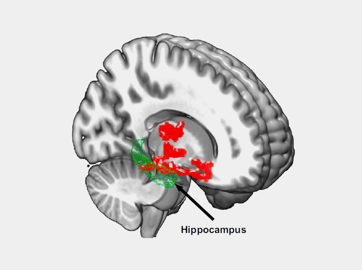
Illustration of beta-amyloid accumulationBrain imaging after one night of sleep deprivation revealed beta-amyloid accumulation (red) in the hippocampus and nearby regions. Proceedings of the National Academy of Sciences
The researchers scanned participants’ brains after getting a full night’s rest and after a night of sleep deprivation (about 31 hours without sleep). Beta-amyloid increased about 5% in the participants’ brains after losing a night of sleep. These changes occurred in brain regions that included the thalamus and hippocampus, which are especially vulnerable to damage in the early stages of Alzheimer’s disease.
The scientists also found that study participants with larger increases in beta-amyloid reported worse mood after sleep deprivation. These findings support other studies that have found that the hippocampus and thalamus play a role in mood disorders.
“Even though our sample was small, this study demonstrated the negative effect of sleep deprivation on beta-amyloid burden in the human brain,” Shokri-Kojori says. “Future studies are needed to assess the generalizability to a larger and more diverse population.”
“This research provides new insight about the potentially harmful effects of a lack of sleep on the brain and has implications for better characterizing the pathology of Alzheimer's disease,” says Dr. George F. Koob, director of NIAAA.
More studies are needed to identify the precise biological mechanism underlying the observed increase in beta-amyloid. It’s also important to note that the link between sleep disorders and Alzheimer’s risk is thought to go both ways. Elevated beta-amyloid may also lead to trouble sleeping.

Tuesday, March 3, 2020

Associations of Widowhood and β-Amyloid With Cognitive Decline in Cognitively Unimpaired Older Adults

Does this mean I shouldn't get married again? Even though this is widows not widowers?

Associations of Widowhood and β-Amyloid With Cognitive Decline in Cognitively Unimpaired Older Adults

JAMA Netw Open. 2020;3(2):e200121. doi:10.1001/jamanetworkopen.2020.0121
Key Points español 中文 (chinese) Question  Is widowhood a specific risk factor associated with more rapid cognitive decline among cognitively unimpaired older adults with higher levels of brain β-amyloid, the Alzheimer disease biomarker?
Findings  In this cohort study of 257 community-dwelling cognitively unimpaired older adults, widowhood and β-amyloid were additively and interactively associated with cognitive decline. These results were independent of demographic factors, cardiovascular disease risk, depression, health-related behaviors, and social support factors.
Meaning  These findings suggest that widowhood may be an understudied risk factor for cognitive decline associated with Alzheimer disease and highlight the need for increased research and clinical attention to this high-risk group.
Abstract
Importance  To reduce the rising incidence of clinical impairment due to Alzheimer disease, it is essential to define older adults at highest risk. Widowhood may be an unrecognized factor contributing to accelerated clinical progression along the Alzheimer disease pathway among cognitively unimpaired older adults.
Objective  To determine whether widowhood status and level of brain β-amyloid (ie, the Alzheimer disease pathologic protein) are additively or interactively associated with cognitive decline among cognitively unimpaired older adults.
Design, Setting, and Participants  In this cohort study, 257 married, widowed, and unmarried (ie, never married, divorced, or separated) participants from the Harvard Aging Brain Study longitudinal cohort underwent baseline evaluation of neocortical β-amyloid levels using Pittsburgh compound B positron emission tomography and 4 annual cognitive assessments. Data were collected from September 2010 to February 2017 and analyzed from July 2018 to July 2019.
Main Outcomes and Measures  Cognitive performance was measured using the Preclinical Alzheimer Cognitive Composite.
Results  Of the 257 participants, 153 (59.5%) were women, and the mean (SD) age was 73.5 (6.1) years; 145 participants (56.4%) were married (66 [45.5%] women), 77 (30.0%) were unmarried (56 [72.7%] women), and 35 (13.6%) were widowed (31 [88.6%] women). Compared with married participants, widowed participants demonstrated worsening cognitive performance after adjusting for age, sex, socioeconomic status, depression, and β-amyloid levels (β = −0.11; 95% CI, −0.19 to −0.04; P = .002) with no difference observed between married and unmarried participants. Furthermore, widowed participants with higher baseline β-amyloid levels exhibited steeper cognitive decline (β = −0.22; 95% CI, −0.42 to −0.03; P = .02), indicating both independent and interactive associations of β-amyloid levels and widowhood with cognition. In a secondary model using dichotomous β-amyloid–marital status groupings, the rate of cognitive decline among widowed participants with high β-amyloid was nearly 3 times faster than among married participants with high β-amyloid (widowed, high β-amyloid: β, −0.33; 95% CI, −0.46 to −0.19; P < .001; married, high β-amyloid: β, −0.12; 95% CI, −0.18 to −0.01; P < .001).
Conclusions and Relevance  In a sample of cognitively unimpaired older adults, being widowed was associated with accelerated β-amyloid–related cognitive decline during 3 years. Cognitively unimpaired, widowed older adults were particularly susceptible to Alzheimer disease clinical progression, emphasizing the need for increased research attention and evidenced-based interventions for this high-risk group.

Wednesday, April 3, 2019

Blocking protein that impairs brain’s clean-up crew improves old mice’s smarts

We need this. Or is young blood a better way to improve cognition?

Young Blood Revitalizes the Aging Brain June 2014 

I bet this from Feb. 2016 was never followed up on.

 Improving Brain’s Garbage Disposal May Slow Alzheimer’s Disease

Or this from Sept. 2013?

When brain’s trash collectors fall down on the job, neurodegeneration risk picks up


Blocking protein that impairs brain’s clean-up crew improves old mice’s smarts

Stanford neuroscientist Tony Wyss-Coray, PhD, has been working for several years on the question of what causes the brain to lose its acuity with advancing age. One focus of his research has been a class of brain cells called microglia, which serve as the brain's immune cells.
A less-vaunted microglial function is more akin to that of a garbage crew. Among the many different things microglia do to keep the brain healthy is scarfing up bits of cellular debris and protein deposits that build up like so much dandruff in the course of normal metabolic activity.
Bits of myelin debris -- remnants of decaying nerve-cell coatings that, by providing electrical insulation, speed the transmission of impulses along nerve tracts -- commonly accumulate in aging brains. Aging-associated aggregation of a protein fragment called beta-amyloid into gummy deposits in the brain are considered a hallmark of Alzheimer's disease. Likewise with clumps of yet another substance, alpha-synuclein, associated with Parkinson's disease.
These accumulations occur over time largely because, as I wrote in my news release about a study published in Nature by Wyss-Coray and his colleagues:
[T]he garbage-collecting performance of microglia diminishes in aging brains. Why this happens, and the extent to which the faulty garbage service is actually responsible for age-related cognitive losses, are unclear. But it's a decent bet that one way or another, microglial malperformance plays a role in neurodegeneration.
In a series of sophisticated experiments, Wyss-Coray's team identified a single protein called CD22 that, in the brain, is found only on the surfaces of microglial cells; becomes increasingly abundant with increasing age (in mice, at least); and, when activated, profoundly impairs these cells from performing their debris-cleanup role.
Then, the researchers conducted another set of experiments. Into one side of mice's brains, they injected specialized proteins (called antibodies) capable of blocking CD22's activity without otherwise disrupting brain function. Into the mirror-image spot on the other side of the mice's brains, they injected otherwise identical antibodies that, however, lacked the ability to block CD22.
Along with the antibodies, the scientists injected bits of myelin -- like sprinkling some confetti around to test a new vacuum cleaner. On inspection two days later, the myelin debris was much less prevalent on the side of mice's brains where "working" antibodies rather than "dummy" antibodies to CD22 had been injected.
The same thing happened when instead of myelin debris, the researchers injected beta-amyloid or alpha-synuclein, the Alzheimer's- and Parkinson's-associated substances.
In all three cases, microglia exposed to CD22-blocking antibodies outperformed their fellow microglia on the opposite side of the brain in ingesting the neurodegeneration-linked substances.
Then, the researchers lengthened the period of exposure from 48 hours to a full month. From my news release:
They reconfigured their injection technique to provide continuous CD22-blocking antibody infusion on both sides of the brain over this period. ... [O]ld mice receiving these infusions outperformed control mice of the same age on two different tests of learning and memory that are commonly used to assess mice's cognitive ability.
"The mice became smarter," Wyss-Coray told me. "Blocking CD22 on their microglia restored their cognitive function to the level of younger mice."
He thinks CD22 could prove to be a brand-new target for treatment of neurodegenerative diseases.

Monday, April 1, 2019

Association Between CSF Beta-Amyloid and Apathy in Early-Stage Alzheimer Disease

You likely need to know about this and your doctors' protocol to counteract apathy.  I don't give a shit that no treatments exist for apathy. It is your doctors responsibility to initiate research that finds a treatment. Or don't you care that you have an incompetent doctor? My dad in the midst of parkinsons dementia had apathy, I couldn't help him with any of my suggestions because of that. 

You will need this.

Your chances of getting dementia.
1. 33% dementia chance post-stroke from an Australian study?   May 2012.
2. Then this study came out and seems to have a range from 17-66%. December 2013.
3. A 20% chance in this research.   July 2013.

Association Between CSF Beta-Amyloid and Apathy in Early-Stage Alzheimer Disease 

Show all authors
First Published March 26, 2019 Research Article


Apathetic syndrome is a common clinical feature in patients with Alzheimer diseases (AD), from preclinical phases to late dementia stages, and it is strongly related to major disease outcomes. Unfortunately, no specific pharmacological treatments for apathy have been accomplished so far. Translational evidences have previously shown that a link between apathy and hallmarks of AD-related pathophysiological, that is, β-amyloid (Aβ) plaques, and neurofibrillary pathology exists. However, only few studies investigated the association between cerebrospinal fluid (CSF) core biomarkers of AD and apathy scores, finding conflicting results.

Thirty-seven patients were identified as having AD dementia according to National Institute on Aging–Alzheimer Association 2011 criteria. All participants underwent an extensive diagnostic workup including CSF assessment to assess the concentrations of the core biomarkers of AD pathophysiology, that is, Aβ42, t-tau, and pTau181. To follow, they were stratified as apathy absence, apathy mild, and apathy severe according the score of the Neuro Psychiatric Inventory-apathy item. We investigated for potential associations between apathy scores and CSF core biomarkers concentrations as well as for differences in terms of clinical and CSF biomarkers data across the 3 apathy groups.

The CSF Aβ42 values were negatively correlated with apathy scores. In addition, patients with severe apathy had significantly lower Aβ42 levels compared to nonapathetic ones.

Based on our results, we encourage further studies to untangle the potential association between the complex pathophysiological dynamics of AD and apathy. Apathy may represent an innovative reliable clinical outcome measure to use in clinical trials, investigating treatments with either a symptomatic or a disease-modifying effect.

Monday, February 13, 2017

New Alzheimer’s Treatment Fully Restores Memory Function

What is YOUR doctor and stroke hospital doing to ensure that this is tested in humans? You will likely need this so you don't want incompetent personnel passing on this because SOMEONE ELSE WILL SOLVE THE PROBLEM.

1. A documented 33% dementia chance post-stroke from an Australian study?   May 2012.

2. Then this study came out and seems to have a range from 17-66%. December 2013.

3. A 20% chance in this research.   July 2013.

4. A 2-fold increase in dementia risk in this study    Jan. 2017 


New Alzheimer’s Treatment Fully Restores Memory Function

Australian researchers have come up with a non-invasive ultrasound technology that clears the brain of neurotoxic amyloid plaques - structures that are responsible for memory loss and a decline in cognitive function in Alzheimer’s patients.
If a person has Alzheimer’s disease, it’s usually the result of a build-up of two types of lesions - amyloid plaques, and neurofibrillary tangles. Amyloid plaques sit between the neurons and end up as dense clusters of beta-amyloid molecules, a sticky type of protein that clumps together and forms plaques.
Neurofibrillary tangles are found inside the neurons of the brain, and they’re caused by defective tau proteins that clump up into a thick, insoluble mass. This causes tiny filaments called microtubules to get all twisted, which disrupts the transportation of essential materials such as nutrients and organelles along them, just like when you twist up the vacuum cleaner tube.
As we don’t have any kind of vaccine or preventative measure for Alzheimer’s - a disease that affects 343,000 people in Australia, and 50 million worldwide - it’s been a race to figure out how best to treat it, starting with how to clear the build-up of defective beta-amyloid and tau proteins from a patient’s brain. Now a team from the Queensland Brain Institute (QBI) at the University of Queensland have come up with a pretty promising solution for removing the former.
Publishing in Science Translational Medicine, the team describes the technique as using a particular type of ultrasound called a focused therapeutic ultrasound, which non-invasively beams sound waves into the brain tissue. By oscillating super-fast, these sound waves are able to gently open up the blood-brain barrier, which is a layer that protects the brain against bacteria, and stimulate the brain’s microglial cells to activate. Microglial cells are basically waste-removal cells, so they’re able to clear out the toxic beta-amyloid clumps that are responsible for the worst symptoms of Alzheimer’s.
The team reports fully restoring the memory function of 75 percent of the mice they tested it on, with zero damage to the surrounding brain tissue. They found that the treated mice displayed improved performance in three memory tasks - a maze, a test to get them to recognise new objects, and one to get them to remember the places they should avoid.
"We’re extremely excited by this innovation of treating Alzheimer’s without using drug therapeutics," one of the team, Jürgen Götz, said in a press release. "The word ‘breakthrough’ is often misused, but in this case I think this really does fundamentally change our understanding of how to treat this disease, and I foresee a great future for this approach."
The team says they’re planning on starting trials with higher animal models, such as sheep, and hope to get their human trials underway in 2017. 
You can hear an ABC radio interview with the team here.

Wednesday, August 17, 2016

Diet and exercise can reduce protein build-ups linked to Alzheimer’s, UCLA study shows

I'm positive your doctor and hospital will not follow this up by creating diet stroke protocols for you. You are completely on your own. Guess wrong and you might get Alzheimers and your doctor will never acknowledge their complicity in you getting Alzheimers. 

Diet and exercise can reduce protein build-ups linked to Alzheimer’s, UCLA study shows


UCLA Health System
A study by researchers at UCLA’s Semel Institute for Neuroscience and Human Behavior has found that a healthy diet, regular physical activity and a normal body mass index can reduce the incidence of protein build–ups that are associated with the onset of Alzheimer’s disease. In the study, 44 adults ranging in age from 40 to 85 (mean age: 62.6) with mild memory changes but no dementia underwent an experimental type of PET scan to measure the level of plaque and tangles in the brain. Researchers also collected information on participants’ body mass index, levels of physical activity, diet and other lifestyle factors. Plaque, deposits of a toxic protein called beta–amyloid in the spaces between nerve cells in the brain; and tangles, knotted threads of the tau protein found within brain cells, are considered the key indicators of Alzheimer’s. The study found that each one of several lifestyle factors — a healthy body mass index, physical activity and a Mediterranean diet — were linked to lower levels of plaques and tangles on the brain scans. “The fact that we could detect this influence of lifestyle at a molecular level before the beginning of serious memory problems surprised us,” said Dr. David Merrill, the lead author of the study, which appeared in the September issue of the American Journal of Geriatric Psychiatry.