Changing stroke rehab and research worldwide now.Time is Brain! trillions and trillions of neurons that DIE each day because there are NO effective hyperacute therapies besides tPA(only 12% effective). I have 523 posts on hyperacute therapy, enough for researchers to spend decades proving them out. These are my personal ideas and blog on stroke rehabilitation and stroke research. Do not attempt any of these without checking with your medical provider. Unless you join me in agitating, when you need these therapies they won't be there.

What this blog is for:

My blog is not to help survivors recover, it is to have the 10 million yearly stroke survivors light fires underneath their doctors, stroke hospitals and stroke researchers to get stroke solved. 100% recovery. The stroke medical world is completely failing at that goal, they don't even have it as a goal. Shortly after getting out of the hospital and getting NO information on the process or protocols of stroke rehabilitation and recovery I started searching on the internet and found that no other survivor received useful information. This is an attempt to cover all stroke rehabilitation information that should be readily available to survivors so they can talk with informed knowledge to their medical staff. It lays out what needs to be done to get stroke survivors closer to 100% recovery. It's quite disgusting that this information is not available from every stroke association and doctors group.

Showing posts with label train your doctor. Show all posts

Tuesday, May 19, 2026

Towards precision medicine for brain arteriovenous malformations

 If you have one it will be good to train your doctor on this. I wouldn't take any chances that your doctor actually keeps up with stroke research.

Towards precision medicine for brain arteriovenous malformations


Published May 15, 2026 -

View PDF 
Abstract

Recent advances in cerebrovascular genomics, single-cell biology, pharmacology, and gene editing technology are transforming our understanding of brain arteriovenous malformations (bAVMs) — a leading cause of pediatric hemorrhagic stroke. Once considered static anatomical defects, bAVMs are now recognized as dynamic, genetically driven lesions resulting from somatic mutations in KRAS, BRAF, and pathways involved in arteriovenous specification, angiogenesis, and vascular remodeling. By integrating human genetics, animal models, and endovascular innovations, researchers have uncovered convergent mechanisms that link endothelial Ras/MAPK hyperactivation to abnormal vessel growth and higher rupture risk. These insights provide a foundation for precision medicine approaches that combine molecular diagnostics — such as liquid or endoluminal biopsies — with mutation-specific pharmacotherapies and emerging CRISPR-based gene editing strategies. We suggest that genotype-guided interventions, tailored by spatial and developmental cerebrovascular context, could ultimately reclassify bAVMs from surgically incurable malformations to treatable molecular conditions.


More at link.

Sunday, April 6, 2025

Scientists Just Discovered This Surprising Side Effect Of Cold Plunges

 I'm sure your doctor told you all about the benefits of cold and heat shock proteins and how to get them.

Hasn't your competent? doctor implemented cold showers for you in the hospital? Or even a sauna?
Your doctor knows all about autophagy and how to have you experience it? RIGHT? Or maybe you should read up on it and train your doctor.
  • autophagy (18 posts to October 2011)
  • The latest here:

    Scientists Just Discovered This Surprising Side Effect Of Cold Plunges

    Korin Miller
    4 min read

    Cold Plunges Can Impact You On A Cellular Level Stocksy

    Everyone and their mom seems to be talking about cold plunging right now. Celebs like Kate Middleton and Brooke Shields have even openly shared their experiences with the practice. But despite all the positive testimonials surrounding this chilly health hack, scientific research into its actual health perks are still ongoing.

    Now, there’s a new study that suggests that cold plunging can literally impact you on a cellular level, triggering them to start a process called autophagy that can have total-body benefits. In fact, doctors swear this cellular impact could help support your overall health and longevity. Women's Health asked experts for their insights on the new study, and the latest health findings to better understand the phenomenon.

    Meet the experts: Joseph J. Ciotola, MD, is an orthopedic surgeon at Baltimore’s Mercy Medical Center who has studied with cold plunge expert Wim Hof; Bert Mandelbaum, MD, is a sports medicine specialist and co-director of the Regenerative Orthobiologic Center at Cedars-Sinai Orthopaedics in Los Angeles; Phillip Williams, MD, is an assistant professor of orthopedic surgery at Baylor College of Medicine; lead study author Kelli King, PhD, is a postdoctoral fellow at University of Ottawa’s School of Human Kinetics.

    What did the study find?

    The study, which was published in the journal Advanced Biology, had 10 healthy(So, unhealthy older persons like us need to have this research done on us!) young men do a cold plunge for an hour every day for a week straight.

    The researchers collected blood samples before and after the plunges to look at how the participants’ cells responded to the cold plunges.

    “We found that in as little as four days, cold water plunges can reduce cellular stress and increase a cellular protective mechanism called autophagy,” says lead study author Kelli King, PhD, a postdoctoral fellow at University of Ottawa’s School of Human Kinetics. “This mechanism helps remove damaged proteins and organelles in the body and is critical during stressful conditions—such as cold exposure—to facilitate cell survival.”

    How do cold plunges change your cells?

    Exposure to cold is what’s known to doctors as a hormetic stressor, explains Joseph J. Ciotola, MD, an orthopedic surgeon at Baltimore’s Mercy Medical Center who has studied with cold plunge expert Wim Hof. And this "stressor" is actually good for you in moderation, as it helps your body build resilience and greater tolerance to distress.

    “This uses your body’s natural adaptive abilities and stimulates the cells with the cold,” he says. “These hormetic stressors, which also include fasting and heat exposure from saunas, use your body’s own potential to heal, ultimately making it more resistant to disease.”

    In this instance, by repeatedly exposing yourself to cold temperatures like you’d experience with a cold plunge, your body learns to more effectively deal with extreme environmental conditions, King says.

    Doing things like cold plunges “trains the body to adapt and survive,” says Bert Mandelbaum, MD, sports medicine specialist and co-director of the Regenerative Orthobiologic Center at Cedars-Sinai Orthopaedics in Los Angeles. “Hormetic stressors like cold plunges are really getting us to perform at the highest level and to adapt to different stresses,” he adds.

    What are the health benefits?

    The health benefits from this particular study are all thanks to a process called autophagy. Autophagy specifically helps clear out damaged proteins, which are called protein aggregates and are a precursor to several chronic conditions, she explains. This allows healthier cells to thrive.

    “There is growing literature that autophagy is a key component in disease prevention, including metabolic conditions such as type 2 diabetes, neurodegenerative diseases such as Alzheimer’s and Parkinson’s, as well as certain cancers,” King says.

    High rates of autophagy can even help to regulate uncontrolled cell growth, i.e. cancer, King says.

    What else can cold plunges help with?

    A lot of the perks around cold plunges so far have been linked to inflammation and mental health. “Cold plunges can reduce inflammation, accelerate muscle recovery, and ease joint pain—key for athletic performance and post-surgical rehab,” says Phillip Williams, MD, assistant professor of orthopedic surgery at Baylor College of Medicine. “They also trigger endorphin release, improving mood and stress resilience.”

    Again, research into the potential perks of cold plunges is ongoing. So, it's entirely possible that there are more health benefits still to be discovered in the future.

    What counts as a “cold plunge”?

    This particular study had participants do cold plunges in water that was 57.2 degrees Fahrenheit. Most research into cold plunges focuses on temperatures between 50 and 60 degrees Fahrenheit.

    For beginners, it can be good to start with 30 seconds to a minute of exposure to the water. Once you've done it more often, you can work up to five to 10 minutes at a time, per the Mayo Clinic. In this study, the participants did 60-minute cold plunges, but you don't need to stay in that long to reap some of the other established brain and anti-inflammation benefits!

    You Might Also Like

    Cold Plunges Can Impact You On A Cellular Level Stocksy

    Everyone and their mom seems to be talking about cold plunging right now. Celebs like Kate Middleton and Brooke Shields have even openly shared their experiences with the practice. But despite all the positive testimonials surrounding this chilly health hack, scientific research into its actual health perks are still ongoing.

    Now, there’s a new study that suggests that cold plunging can literally impact you on a cellular level, triggering them to start a process called autophagy that can have total-body benefits. In fact, doctors swear this cellular impact could help support your overall health and longevity. Women's Health asked experts for their insights on the new study, and the latest health findings to better understand the phenomenon.

    Meet the experts: Joseph J. Ciotola, MD, is an orthopedic surgeon at Baltimore’s Mercy Medical Center who has studied with cold plunge expert Wim Hof; Bert Mandelbaum, MD, is a sports medicine specialist and co-director of the Regenerative Orthobiologic Center at Cedars-Sinai Orthopaedics in Los Angeles; Phillip Williams, MD, is an assistant professor of orthopedic surgery at Baylor College of Medicine; lead study author Kelli King, PhD, is a postdoctoral fellow at University of Ottawa’s School of Human Kinetics.

    What did the study find?

    The study, which was published in the journal Advanced Biology, had 10 healthy young men do a cold plunge for an hour every day for a week straight.

    The researchers collected blood samples before and after the plunges to look at how the participants’ cells responded to the cold plunges.

    “We found that in as little as four days, cold water plunges can reduce cellular stress and increase a cellular protective mechanism called autophagy,” says lead study author Kelli King, PhD, a postdoctoral fellow at University of Ottawa’s School of Human Kinetics. “This mechanism helps remove damaged proteins and organelles in the body and is critical during stressful conditions—such as cold exposure—to facilitate cell survival.”

    How do cold plunges change your cells?

    Exposure to cold is what’s known to doctors as a hormetic stressor, explains Joseph J. Ciotola, MD, an orthopedic surgeon at Baltimore’s Mercy Medical Center who has studied with cold plunge expert Wim Hof. And this "stressor" is actually good for you in moderation, as it helps your body build resilience and greater tolerance to distress.

    “This uses your body’s natural adaptive abilities and stimulates the cells with the cold,” he says. “These hormetic stressors, which also include fasting and heat exposure from saunas, use your body’s own potential to heal, ultimately making it more resistant to disease.”

    In this instance, by repeatedly exposing yourself to cold temperatures like you’d experience with a cold plunge, your body learns to more effectively deal with extreme environmental conditions, King says.

    Doing things like cold plunges “trains the body to adapt and survive,” says Bert Mandelbaum, MD, sports medicine specialist and co-director of the Regenerative Orthobiologic Center at Cedars-Sinai Orthopaedics in Los Angeles. “Hormetic stressors like cold plunges are really getting us to perform at the highest level and to adapt to different stresses,” he adds.

    What are the health benefits?

    The health benefits from this particular study are all thanks to a process called autophagy. Autophagy specifically helps clear out damaged proteins, which are called protein aggregates and are a precursor to several chronic conditions, she explains. This allows healthier cells to thrive.

    “There is growing literature that autophagy is a key component in disease prevention, including metabolic conditions such as type 2 diabetes, neurodegenerative diseases such as Alzheimer’s and Parkinson’s, as well as certain cancers,” King says.

    High rates of autophagy can even help to regulate uncontrolled cell growth, i.e. cancer, King says.

    What else can cold plunges help with?

    A lot of the perks around cold plunges so far have been linked to inflammation and mental health. “Cold plunges can reduce inflammation, accelerate muscle recovery, and ease joint pain—key for athletic performance and post-surgical rehab,” says Phillip Williams, MD, assistant professor of orthopedic surgery at Baylor College of Medicine. “They also trigger endorphin release, improving mood and stress resilience.”

    Again, research into the potential perks of cold plunges is ongoing. So, it's entirely possible that there are more health benefits still to be discovered in the future.

    What counts as a “cold plunge”?

    This particular study had participants do cold plunges in water that was 57.2 degrees Fahrenheit. Most research into cold plunges focuses on temperatures between 50 and 60 degrees Fahrenheit.

    For beginners, it can be good to start with 30 seconds to a minute of exposure to the water. Once you've done it more often, you can work up to five to 10 minutes at a time, per the Mayo Clinic. In this study, the participants did 60-minute cold plunges, but you don't need to stay in that long to reap some of the other established brain and anti-inflammation benefits!

    Korin Miller
    4 min read

    Cold Plunges Can Impact You On A Cellular Level Stocksy

    Everyone and their mom seems to be talking about cold plunging right now. Celebs like Kate Middleton and Brooke Shields have even openly shared their experiences with the practice. But despite all the positive testimonials surrounding this chilly health hack, scientific research into its actual health perks are still ongoing.

    Now, there’s a new study that suggests that cold plunging can literally impact you on a cellular level, triggering them to start a process called autophagy that can have total-body benefits. In fact, doctors swear this cellular impact could help support your overall health and longevity. Women's Health asked experts for their insights on the new study, and the latest health findings to better understand the phenomenon.

    Meet the experts: Joseph J. Ciotola, MD, is an orthopedic surgeon at Baltimore’s Mercy Medical Center who has studied with cold plunge expert Wim Hof; Bert Mandelbaum, MD, is a sports medicine specialist and co-director of the Regenerative Orthobiologic Center at Cedars-Sinai Orthopaedics in Los Angeles; Phillip Williams, MD, is an assistant professor of orthopedic surgery at Baylor College of Medicine; lead study author Kelli King, PhD, is a postdoctoral fellow at University of Ottawa’s School of Human Kinetics.

    What did the study find?

    The study, which was published in the journal Advanced Biology, had 10 healthy young men do a cold plunge for an hour every day for a week straight.

    The researchers collected blood samples before and after the plunges to look at how the participants’ cells responded to the cold plunges.

    “We found that in as little as four days, cold water plunges can reduce cellular stress and increase a cellular protective mechanism called autophagy,” says lead study author Kelli King, PhD, a postdoctoral fellow at University of Ottawa’s School of Human Kinetics. “This mechanism helps remove damaged proteins and organelles in the body and is critical during stressful conditions—such as cold exposure—to facilitate cell survival.”

    How do cold plunges change your cells?

    Exposure to cold is what’s known to doctors as a hormetic stressor, explains Joseph J. Ciotola, MD, an orthopedic surgeon at Baltimore’s Mercy Medical Center who has studied with cold plunge expert Wim Hof. And this "stressor" is actually good for you in moderation, as it helps your body build resilience and greater tolerance to distress.

    “This uses your body’s natural adaptive abilities and stimulates the cells with the cold,” he says. “These hormetic stressors, which also include fasting and heat exposure from saunas, use your body’s own potential to heal, ultimately making it more resistant to disease.”

    In this instance, by repeatedly exposing yourself to cold temperatures like you’d experience with a cold plunge, your body learns to more effectively deal with extreme environmental conditions, King says.

    Doing things like cold plunges “trains the body to adapt and survive,” says Bert Mandelbaum, MD, sports medicine specialist and co-director of the Regenerative Orthobiologic Center at Cedars-Sinai Orthopaedics in Los Angeles. “Hormetic stressors like cold plunges are really getting us to perform at the highest level and to adapt to different stresses,” he adds.

    What are the health benefits?

    The health benefits from this particular study are all thanks to a process called autophagy. Autophagy specifically helps clear out damaged proteins, which are called protein aggregates and are a precursor to several chronic conditions, she explains. This allows healthier cells to thrive.

    “There is growing literature that autophagy is a key component in disease prevention, including metabolic conditions such as type 2 diabetes, neurodegenerative diseases such as Alzheimer’s and Parkinson’s, as well as certain cancers,” King says.

    High rates of autophagy can even help to regulate uncontrolled cell growth, i.e. cancer, King says.

    What else can cold plunges help with?

    A lot of the perks around cold plunges so far have been linked to inflammation and mental health. “Cold plunges can reduce inflammation, accelerate muscle recovery, and ease joint pain—key for athletic performance and post-surgical rehab,” says Phillip Williams, MD, assistant professor of orthopedic surgery at Baylor College of Medicine. “They also trigger endorphin release, improving mood and stress resilience.”

    Again, research into the potential perks of cold plunges is ongoing. So, it's entirely possible that there are more health benefits still to be discovered in the future.

    What counts as a “cold plunge”?

    This particular study had participants do cold plunges in water that was 57.2 degrees Fahrenheit. Most research into cold plunges focuses on temperatures between 50 and 60 degrees Fahrenheit.

    For beginners, it can be good to start with 30 seconds to a minute of exposure to the water. Once you've done it more often, you can work up to five to 10 minutes at a time, per the Mayo Clinic. In this study, the participants did 60-minute cold plunges, but you don't need to stay in that long to reap some of the other established brain and anti-inflammation benefits!


    Wednesday, March 19, 2025

    The Evolution of Poststroke Recovery: Advances and Challenges

     WOW! Completely missing the possibility of vastly increasing the chances of recovery by just stopping the 5 causes of the neuronal cascade of death in the first week!  to save hundreds of millions to billions of neurons! Do you people ever think at all?

    I lost 5.4 billion neurons that first week. If I'd only lost 177 million neurons in the 90 minutes it took to get tPA I'd easily be recovered by now.

    Stroke rehab is a complete fucking failure as proven by this!

    The Evolution of Poststroke Recovery: Advances and Challenges

    A seismic shift is underway in how experts view functional recovery after a stroke.

    For years, the prevailing belief was that regaining limb mobility beyond 6 months was nearly impossible, as damaged brain circuits were thought to be irreparable. But today, a wave of renewed optimism — fueled by groundbreaking research — is challenging that notion. From brain stimulation to stem cell therapy, scientists are exploring new ways to push the boundaries of recovery.

    It’s still early days, but the potential impact of these emerging interventions could be profound. Currently, approximately seven million Americans live with chronic stroke, with an estimated 75% experiencing some level of impairment.

    As the population ages, stroke rehabilitation is poised to take center stage. By 2050, the number of stroke survivors in the United States is expected to nearly triple to 20 million, underscoring the urgent need for more effective recovery strategies.

    Now, a growing body of research is challenging long-held beliefs about the limits of stroke recovery. Breakthroughs in neuroplasticity, brain stimulation, and regenerative medicine are opening new doors — offering fresh hope to patients and transforming stroke rehabilitation.

    Brain Stimulation

    Noninvasive brain stimulation — such as transcranial direct current stimulation (tDCS) or transcranial magnetic stimulation (TMS) — is being explored as a way to enhance stroke recovery by pairing physical rehabilitation with targeted brain stimulation to modulate disrupted neural networks.

    However, key questions remain about the optimal dose, duration, and patient population. For example, findings from the TRANSPORT 2 trial, presented at the International Stroke Conference (ISC) 2025, revealed that tDCS, delivered at two different doses, did not improve upper extremity function beyond the gains achieved through physical therapy alone.

    Despite the disappointing results from this relatively small phase 2 trial, the research is far from over. “This doesn’t close the door on tDCS,” said Lauren H. Sansing, MD, professor of neurology at Yale School of Medicine, New Haven, Connecticut, and chair of ISC. “It didn’t show a benefit, but it provided an enormous amount of data to help us rethink how to test this therapy.”

    Joseph Broderick, MD, professor of neurology and rehabilitation medicine, University of Cincinnati in Cincinnati and director of the UC Gardner Neuroscience Institute, noted that determining the correct dose is essential as too much stimulation “can cause side effects that you don’t want.”

    In addition, Wayne Feng, MD, professor of neurology and biomedical engineering, Duke University School of Medicine, Durham, North Carolina, who presented the TRANSPORT 2 results, acknowledged the lack of solid evidence for tDCS but insisted “it has an edge over TMS. [tDCS] is portable, easy to use, low cost, and can be paired with rehab therapy” which can’t be said for TMS, he said.

    Another noninvasive brain stimulation technique, low-intensity transcranial focused ultrasound stimulation, targets cortical, subcortical, and deep brain regions.

    While being explored for stroke recovery, it is also under investigation for a range of other neurological and psychiatric conditions.

    Researchers are also exploring more direct methods of neuromodulation.

    Deep brain stimulation (DBS) delivers constant electrical stimulation to a particular brain region through a surgically implanted thin wire or electrode.

    One small trial targeting the dentate nucleus in the cerebellum — an area involved in coordinating muscle movements — showed that when combined with physical therapy, the approach improved upper limb function in stroke patients with persistent moderate to severe upper-extremity impairment. Participants showed a median seven-point improvement on the upper-extremity Fugl-Meyer scale.

    Gary K. Steinberg, MD, PhD, Lacroute-Hearst professor of neurosurgery and neurosciences at Stanford University in California, believes electrical brain stimulation will play a key role in poststroke rehabilitation. Among the various approaches, he sees DBS as particularly promising.

    However, some experts caution that DBS is relatively invasive, requires hospitalization, and carries potential neurosurgical risks.

    Joel Stein, MD, professor and chair, Department of Rehabilitation and Regenerative Medicine, Columbia University, New York City, said he’s somewhat skeptical about DBS. “I have found it’s hard to convince patients to undergo a surgical intervention when the benefits may be modest,” he told Medscape Medical News. 

    This hesitation around invasive procedures has led researchers to explore less invasive alternatives for stroke rehabilitation.

    Vagus nerve stimulation (VNS) has been shown to be safe and effective in several neurological conditions, including epilepsy and stroke. Following a positive phase 3 sham-controlled trial in 2021, the US Food and Drug Administration approved a VNS device for patients in the chronic stage of ischemic stroke (> 9 months poststroke) with persistent moderate to severe upper extremity impairment.

    Steinberg emphasized the benefit is modest and noted VNS is only for upper limb weakness, requires repeated stimulation, and has to be paired with intensive rehabilitation.“Nomatter what form of energy you use to stimulate the brain, whether it’s electrical or magnetic, the brain is going to be a better target than the vagal nerve,” he said.

    Some experts argue that the precise mechanisms behind VNS and the consistency of stimulation parameters in stroke treatment require further investigation.

    Exploring Stem Cell Therapy

    This ongoing need for refinement in neuromodulation therapies has also driven interest in regenerative approaches, such as stem cell therapy.

    Stem cell therapy involves injecting human neural stem cells through a small hole in the brain, a process designed to support neurorestoration. These stem cells secrete powerful growth factors, along with other proteins and molecules, that promote brain repair.(Use the correct word: exosomes!)

    These factors help neurons grow new axons and dendrites, stimulate the formation of blood vessels in stroke-damaged areas, and reduce inflammation, all of which contribute to the recovery process.

    The first in-human phase 1/2a study, conducted by Steinberg and colleagues, enrolled 18 patients at least 6 months poststroke but some years out from their stroke.

    All patients showed improvement in their total Fugl-Meyer motor score at 12 months, with 10 out of 16 achieving clinically meaningful recovery — defined as an increase of 10 or more points on the scale.

    Gains were observed in both upper and lower limb functions. Upper limb improvement at 12 months (+6.9; P < .0002) exceeded that seen with VNS which showed an improvement of only 5.3 points, according to Steinberg. He emphasized that this was despite “our patients being much more severe” than those in the VNS trial.

    The intervention also improved gait, quality of life, and even benefited patients with aphasia. Additionally, factors such as age, gender, and stroke volume “didn’t matter” in terms of the extent of improvement, Steinberg noted.

    All adverse events, which included incisional pain, nausea, and fatigue, resolved spontaneously and none were related to the stem cells.

    Steinberg and his colleagues are now planning a randomized multi-center, blinded study of stem cell therapy.

    Broderick cautioned that the study is too small to determine whether patients would have improved without the intervention. He noted that many treatments have generated excitement early on, only to show diminished impact when tested in larger patient populations.

    Stem cell therapy has also been explored in Parkinson’s disease, a condition that may be more suited to such an approach because it targets a specific population of cells, Broderick noted. However, even in that context, “it hasn’t worked out that well” and has been associated with side effects, he added.

    This uncertainty also extends to stroke treatment, where many fundamental questions remain unanswered.

    Feng emphasized the need to determine which cells should be injected or infused, in what quantities, and which stroke patients are most likely to benefit.

    Robotics, Virtual Reality

    As researchers continue to refine biological interventions like stem cell therapy, technological advancements are also playing a growing role in stroke rehabilitation.

    This fast-growing field involves using robots or programmable devices designed to deliver high intensity task-specific training. Growing evidence suggests robotic therapy can boost limb function and motivate patients to stick to rehab exercises.

    This is important as sustained repetitive exercises are crucial for poststroke recovery, but many patients drop out due to cognitive and other stroke-related issues.

    However, Feng noted that research on robotic therapy for severely impaired patients has been less than encouraging, potentially highlighting the limits of brain plasticity. “As much as we have fantasies about brain plasticity, it’s quite limited in stroke patients once the brain has been ‘broken’ or injured by stroke,” he said.

    Despite these challenges, researchers continue to explore alternative rehabilitation strategies. One emerging approach is innovative virtual reality (VR) therapy, along with interactive video gaming, which is gaining popularity as a neurorehabilitation modality.

    A recent meta-review of 57 systematic reviews, encompassing 1033 randomized controlled trials, concluded that VR can improve upper and lower limb function, balance, gait, and possibly cognition.

    VR offers several advantages over other rehabilitation approaches. It is relatively affordable, enhances patient satisfaction and engagement, and has no significant side effects.

    “Exercise is a really critical piece of recovery, but it’s very hard to get people to comply, so if you can make it more engaging, that’s really important,” said Stein.

    In addition, VR is relatively accessible, using, for example, video games offered by Nintendo Wii and Xbox Kinect. Some companies are attempting to make games more stroke-specific, said Stein.

    Broderick pointed out that VR and other poststroke interventions do not address deficits in language and vision caused by stroke. “There are limits to what we can recover from,” he said.(Why are you giving up on 100% recovery?)

    A variety of compounds — including levodopa, fluoxetine, D-amphetamine, citicoline, niacin, and inosine — are being tested in animal models and clinical trials or are already in use to aid motor recovery after stroke. However, study results have been mixed.

    Lots more details in these for those looking to train their doctors! Look how long your doctor and hospital have been INCOMPETENT!

  • fluoxetine (22 posts to May 2014)

  • simvastatin and fluoxetine drug combination (1 post to May 2016)

  • Looking ahead, Broderick believes gene therapy could eventually help “spark motor recovery” in some stroke patients. He cited a study suggesting that certain genetic variants may influence stroke recovery outcomes.

    Feng reported he is a consultant for NAMSA, and an advisor for Burke rehabilitation Institute.Stein reported he is on the advisory board of Dessintey, which develops and markets intensive rehabilitation technologies. He is also involved in a clinical research trial with Brain Q involving low intensity, noninvasive brain stimulation and his center is a participating site for a registry study of the vagal nerve stimulation device.

    Steinberg’s stem cell work has received support from the California Institute for Regenerative Medicine. All other sources reported no relevant disclosures.


    Thursday, February 20, 2025

    Life-changing ways to restore movement after stroke

     You tell us NOTHING ABOUT THE PROTOCOLS AND WHERE THEY ARE LOCATED! So, completely fucking useless! Survivors need that information so they can train their doctors and therapists into how to get them recovered! Top down dissemination of this information doesn't work, otherwise you would hear numerous stories of survivor recovery! 

    Life-changing ways to restore movement after stroke

              Leading rehabilitation specialists share breakthrough techniques that help stroke survivors regain independence, with remarkable success stories of recovery
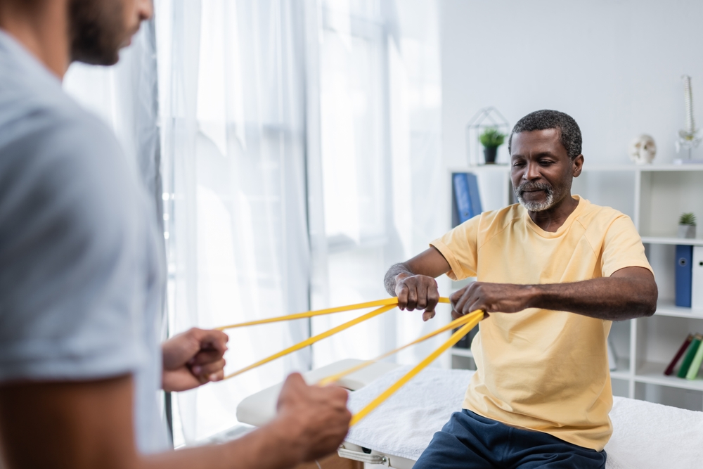
    resistance band, TO
    Photo credit: Shutterstock.com / LightField-Studios-2

    Leading rehabilitation specialists share breakthrough techniques that help stroke survivors regain independence, with remarkable success stories of recovery

    When Sarah Thompson woke up one morning unable to move her left side, her life changed forever. Now, two years later, she walks confidently through her local park, sharing her recovery journey with other stroke survivors. Her story represents hope for the nearly 800,000 Americans who experience a stroke each year, demonstrating that recovery isn’t just possible—it’s happening every day through innovative rehabilitation approaches.


    Understanding the impact of stroke on movement

    Recent research from the National Stroke Association reveals that stroke remains the leading cause of long-term disability in America. However, neuroplasticity—the brain’s remarkable ability to reorganize and form new neural connections—offers hope for recovery. Medical experts now understand that the brain can adapt and rewire itself, even years after a stroke occurs.

    The science behind stroke recovery

    The human brain possesses an extraordinary capacity for healing and adaptation. When stroke damages one area, other regions can sometimes take over those functions through dedicated rehabilitation. This process, known as neurological reorganization, forms the foundation of modern stroke recovery programs.


    Revolutionary approaches in physical therapy

    Modern physical therapy for stroke recovery has evolved significantly beyond traditional exercise routines. Today’s approaches incorporate cutting-edge technology and evidence-based techniques that maximize recovery potential.

    Constraint-induced movement therapy has emerged as a groundbreaking technique. This approach involves restricting the unaffected limb while intensively training the affected one, forcing the brain to relearn movement patterns. Studies show that this method can significantly improve upper limb function, even years after a stroke.

    Virtual reality systems now play an increasingly important role in rehabilitation. These interactive programs create engaging environments that motivate patients while providing real-time feedback on their progress. Research indicates that VR-enhanced therapy can improve balance and walking ability more effectively than traditional methods alone.

    Innovative occupational therapy techniques

    Occupational therapy has undergone a revolution in recent years, incorporating new technologies and approaches that help stroke survivors regain independence in daily activities. Advanced robotics now assist with fine motor training, while smart home technology provides new opportunities for independent living.

    Therapists now utilize task-specific training, focusing on real-world activities that matter most to each individual. This personalized approach helps patients rebuild practical skills while maintaining motivation throughout their recovery journey.

    The role of speech and swallowing therapy

    Communication and swallowing difficulties often accompany mobility challenges after a stroke. Modern speech therapy programs utilize biofeedback devices and electrical stimulation to strengthen oral muscles and improve swallowing function. These techniques have shown remarkable success in helping patients regain their ability to eat and drink safely.

    Breakthrough assistive technologies

    The field of assistive technology has experienced unprecedented advancement. New devices ranging from lightweight carbon fiber braces to brain-computer interfaces are transforming possibilities for stroke survivors.

    Exoskeleton technology, once confined to research laboratories, now helps patients relearn walking patterns. These wearable robots provide precise support and feedback, allowing for more intensive and effective gait training.

    Smart home systems, controlled through voice commands or simple gestures, enable greater independence. These technologies adapt to each user’s abilities, providing customized support for daily activities.

    The crucial role of mental health support

    Recovery extends beyond physical rehabilitation. Mental health professionals specializing in stroke recovery now recognize the profound impact of emotional well-being on physical progress. Support groups incorporating both in-person and virtual meetings provide essential connection and encouragement.

    Nutrition and lifestyle factors in recovery

    Recent research highlights the critical role of nutrition in stroke recovery. Anti-inflammatory diets rich in omega-3 fatty acids, antioxidants, and lean proteins support brain healing and muscle recovery. Exercise physiologists work alongside nutritionists to develop comprehensive wellness plans that optimize recovery potential.

    Home modification strategies

    Creating an enabling environment proves crucial for successful rehabilitation. Home modification experts now use 3D modeling technology to design spaces that support independence while maintaining safety. These modifications range from simple grab bar installations to complete room reorganizations that facilitate movement and daily activities.

    The power of community support

    Community-based rehabilitation programs have shown remarkable success in supporting long-term recovery. These programs combine exercise classes, skill-building workshops, and social activities to create comprehensive support networks for stroke survivors.

    Alternative and complementary therapies

    Many stroke survivors find additional benefit from complementary therapies such as acupuncture, aquatic therapy, and mindfulness practices. While these approaches shouldn’t replace traditional rehabilitation, they often provide valuable support for overall recovery.

    The future of stroke rehabilitation

    Emerging technologies promise even more advanced recovery options. Brain stimulation techniques, stem cell therapies, and artificial intelligence-driven rehabilitation programs are currently under development, offering hope for enhanced recovery outcomes in the future.

    Understanding the recovery timeline

    Recovery progresses differently for each individual, but research shows that intensive rehabilitation in the first six months after stroke yields significant results. However, improvement can continue for years with dedicated effort and appropriate support.

    Financial considerations and resources

    Navigating the financial aspects of stroke recovery challenges many families. Insurance specialists and social workers now play crucial roles in rehabilitation teams, helping families access necessary resources and support services.

    The importance of caregiver support

    Caregivers require support and education to effectively assist in the recovery process. Modern rehabilitation programs include comprehensive caregiver training and support services, recognizing the vital role family members play in successful recovery.

    Hope for continued progress

    While stroke recovery presents significant challenges, modern rehabilitation approaches offer more opportunities for recovery than ever before. Through combination of traditional therapies, innovative technologies, and comprehensive support systems, stroke survivors can work toward reclaiming their independence and quality of life.

    The journey of stroke recovery continues to evolve as new research and technologies emerge. For survivors like Sarah Thompson, these advances offer not just hope, but tangible paths toward recovery and renewed independence.

    Monday, December 2, 2024

    Visceral Fat May Indicate Alzheimer’s Risk Decades Before Symptoms

     Your competent? doctor needs to first prevent this visceral fat and then if it occurs, remove it! You need excellent brain blood flow for recovery, so don't let your doctor weasel out of this requirement!

    In case your doctor is incompetent in this matter, use this to train them:


    With your chances of getting dementia post stroke, you need prevention solutions. YOUR DOCTOR IS RESPONSIBLE FOR PREVENTING THIS!

    1. A documented 33% dementia chance post-stroke from an Australian study?   May 2012.

    2. Then this study came out and seems to have a range from 17-66%. December 2013.`    

    3. A 20% chance in this research.   July 2013.

    4. Dementia Risk Doubled in Patients Following Stroke September 2018 

    The latest here:

    Visceral Fat May Indicate Alzheimer’s Risk Decades Before Symptoms

    Summary: A new study links visceral fat, a deep layer of abdominal fat, to increased brain amyloid and tau proteins—key markers of Alzheimer’s disease—decades before dementia symptoms arise. Researchers found that visceral fat accounted for 77% of the relationship between high BMI and amyloid accumulation, while other fat types showed no similar impact.

    The study highlights how managing visceral fat through lifestyle changes or medication could reduce Alzheimer’s risk. Midlife interventions are especially critical, as participants in their 40s and 50s exhibited these pathologies early on. The findings further reveal that metabolic factors like insulin resistance and cholesterol levels amplify obesity-related brain damage. This research emphasizes the urgent need for targeted prevention strategies against obesity-related Alzheimer’s risk.

    Key Facts:

    • Visceral fat is strongly associated with amyloid and tau protein buildup in the brain.
    • Insulin resistance and low HDL cholesterol worsen obesity-related Alzheimer’s pathology.
    • Managing visceral fat through weight loss or metabolic treatments may reduce dementia risk.

    Source: RSNA

    Researchers have linked a specific type of body fat to the abnormal proteins in the brain that are hallmarks of Alzheimer’s disease up to 20 years before the earliest symptoms of dementia  appear, according to a study being presented today at the annual meeting of the Radiological Society of North America (RSNA).

    The researchers emphasized that lifestyle modifications targeted at reducing this fat could influence the development of Alzheimer’s disease.

    This shows a brain.
    The effects of visceral fat on amyloid pathology were partially reduced in people with higher HDL. Credit: Neuroscience News

    “This crucial result was discovered because we investigated Alzheimer’s disease pathology as early as midlife—in the 40s and 50s—when the disease pathology is at its earliest stages, and potential modifications like weight loss and reducing visceral fat are more effective as a means of preventing or delaying the onset of the disease,” said lead study author Mahsa Dolatshahi, M.D., M.P.H., post-doctoral research associate at Mallinckrodt Institute of Radiology (MIR) at Washington University School of Medicine in St. Louis, Missouri.

    An estimated 6.9 million Americans, aged 65 and older, are living with Alzheimer’s disease, according to the Alzheimer’s Association. The association estimates this number could grow to 13 million by 2050, barring the development of medical breakthroughs to prevent or cure the disease.

    For the study, the researchers focused on the link between modifiable lifestyle-related factors, such as obesity, body fat distribution and metabolic aspects, and Alzheimer’s disease pathology.

    A total of 80 cognitively normal midlife individuals (average age: 49.4 years, female: 62.5%,) were included in the study. Approximately 57.5% of participants were obese, and the average body mass index (BMI) of the participants was 32.31.

    The participants underwent brain positron emission tomography (PET), body MRI and metabolic assessment (glucose and insulin measurements), as well as a lipid (cholesterol) panel.

    MRI scans of the abdomen were performed to measure the volume of the subcutaneous fat (the fat under skin) and visceral fat (deep hidden fat surrounding the organs).

    “We investigated the association of BMI, visceral fat, subcutaneous fat, liver fat fraction, thigh fat and muscle, as well as insulin resistance and HDL (good cholesterol), with amyloid and tau deposition in Alzheimer’s disease,” said Dr. Dolatshahi, a member of the Raji Lab at MIR’s Neuroimaging Labs Research Center.

    Thigh muscle scans were used to measure volume of muscle and fat. Alzheimer’s disease pathology was measured using PET scans with tracers that bind to amyloid plaques and tau tangles that accumulate in the brains of people with Alzheimer’s disease.

    The findings revealed that higher levels of visceral fat were related to increased amyloid, accounting for 77% of the effect of high BMI on amyloid accumulation. Other types of fat did not explain obesity-related increased Alzheimer’s pathology.

    “Our study showed that higher visceral fat was associated with higher PET levels of the two hallmark pathologic proteins of Alzheimer’s disease—amyloid and tau,” Dr. Dolatshahi said.

    “To our knowledge, our study is the only one to demonstrate these findings at midlife where our participants are decades out from developing the earliest symptoms of the dementia that results from Alzheimer’s disease.”

    The study also showed that higher insulin resistance and lower HDL were associated with high amyloid in the brain. The effects of visceral fat on amyloid pathology were partially reduced in people with higher HDL.

    “A key implication of our work is that managing Alzheimer’s risk in obesity will need to involve targeting the related metabolic and lipid issues that often arise with higher body fat,” said senior study author Cyrus A. Raji, M.D., Ph.D., associate professor of radiology at MIR.

    Although previous studies have shown the role of high BMI in damaging the cells of the brain, no similar study has investigated the differential role of visceral and subcutaneous fat or metabolic profile, especially in terms of Alzheimer’s amyloid pathology as early as midlife, Dr. Dolatshahi pointed out.

    “This study goes beyond using BMI to characterize body fat more accurately with MRI and, in so doing, reveals key insights about why obesity can increase risk for Alzheimer’s disease,” Dr. Dolatshahi said.

    Drs. Raji, Dolatshahi and colleagues are also presenting a study at RSNA 2024 that shows how obesity and visceral fat reduce blood flow in the brain.

    In that study, the researchers performed brain and abdominal MRI on cognitively normal midlife individuals with a wide range of BMI and compared whole-brain and regional cerebral blood flow on brain MRI in individuals with high vs. low visceral and subcutaneous fat. 

    The high visceral fat group showed lower whole-brain blood flow. No significant difference was observed in cerebral blood flow in the groups with high vs. low subcutaneous fat.

    “This work will have a considerable impact on public health because nearly three out of four Americans are overweight or obese,” Dr. Raji said.

    “Knowing that visceral obesity negatively affects the brain opens up the possibility that treatment with lifestyle modifications or appropriate weight-loss drugs could improve cerebral blood flow and potentially lower the burden of and reduce the risk for Alzheimer’s disease.”

    Other co-authors are Paul K. Commean, B.E.E., Mahshid Naghashzadeh, M.S., Sara Hosseinzadeh Kassani, Ph.D., Jake Weeks, B.S., Caitlyn Nguyen, B.S., Abby McBee-Kemper, B.S., Nancy Hantler, B.S., LaKisha Lloyd, M.Sc., Shaney Flores, M.S., Yifei Xu, M.S., Jingxia Liu, Ph.D., Claude B. Sirlin, M.D., Bettina Mittendorfer, Ph.D., Joseph E. Ippolito, M.D., Ph.D., John C. Morris, M.D., and Tammie L.S. Benzinger, M.D., Ph.D.

    This study was awarded the RSNA Trainee Research Prize.

    About this Alzheimer’s disease research news

    Author: Linda Brooks
    Source: RSNA
    Contact: Linda Brooks – RSNA
    Image: The image is credited to Neuroscience News

    Original Research: The findings will be presented at the 110th Scientific Assembly and Annual Meeting of the Radiological Society of North America