http://scopeblog.stanford.edu/2017/11/16/nanoparticles-help-track-human-heart-muscle-cells-in-mice-in-stanford-study/
 This
beautiful image shows human heart muscle cells called cardiomyocytes
that have been derived from embryonic stem cells and then reintroduced
into the beating heart of a living mouse. Understanding where these
reintroduced cells go in the heart, and what they do when they get
there, is a critically important step toward using the cells to repair
heart disease.
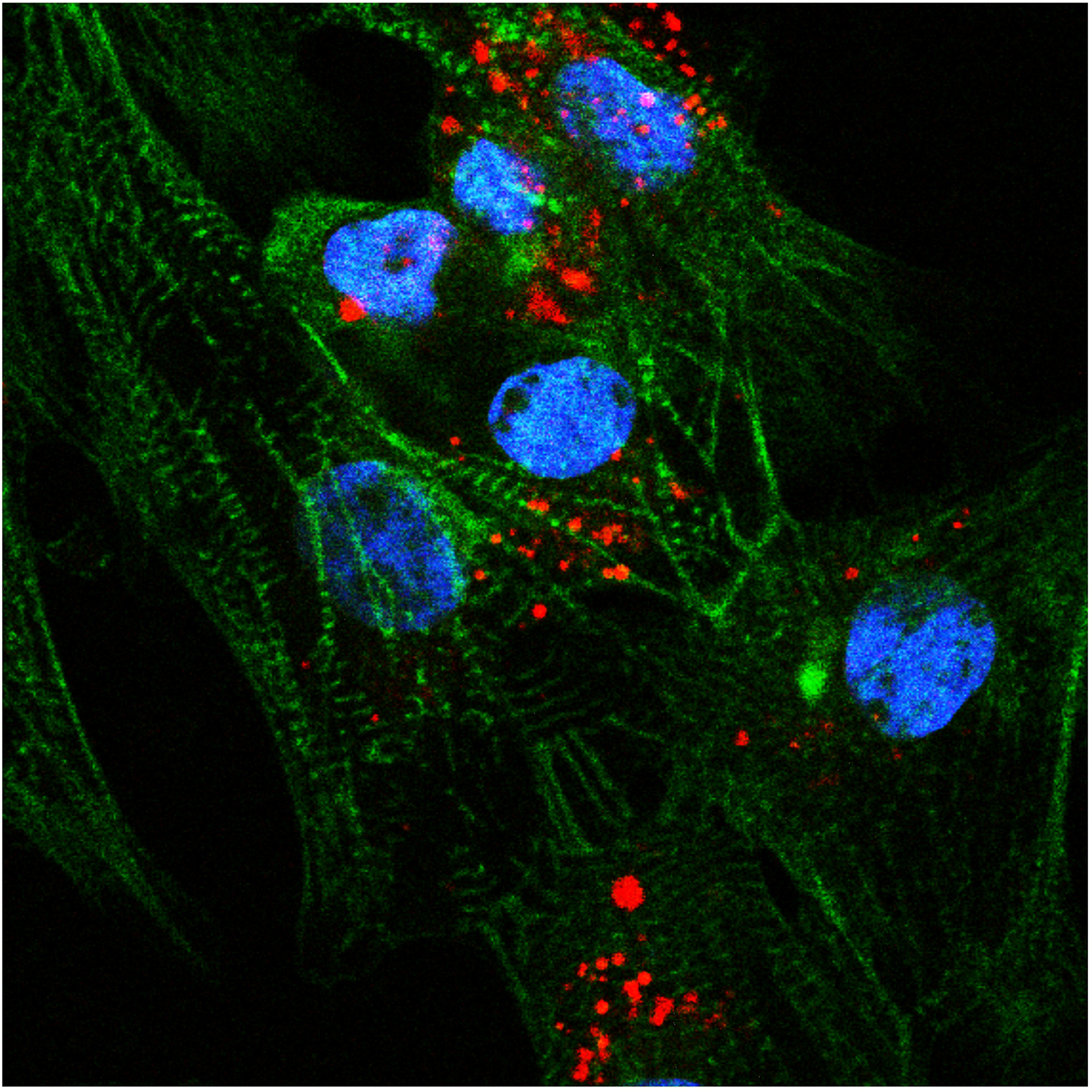
This
beautiful image shows human heart muscle cells called cardiomyocytes
that have been derived from embryonic stem cells and then reintroduced
into the beating heart of a living mouse. Understanding where these
reintroduced cells go in the heart, and what they do when they get
there, is a critically important step toward using the cells to repair
heart disease.Here Stanford Cardiovascular Institute instructor Xulei Qin, PhD, and cardiologist Joseph C. Wu, MD, PhD, along with radiologist Heike Daldrup-Link used a new technique called photoacoustic imaging to visualize semiconducting polymer nanoparticles they’ve latched onto the cardiomyocytes (whose nuclei are shown in blue) like microscopic ankle bracelets. The nanoparticles, indicated in the photo in red, absorb laser light and emit acoustic signals that be used to quickly and accurately track the cells’ location with unprecedented sensitivity and resolution — even when they are buried by several millimeters of tissue. They published their results earlier this month in Advanced Functional Materials.
As Wu explained to me in an email:
This technique provides a much better way to follow how the cells integrate in small animal models because these nanoparticles have strong photoacoustic signals and specific spectral features to sensitively detect and distinguish a small number of labeled cardiomyocytes from native heart tissues.Previously: A new label will allow physicians to pinpoint locations of bacterial infections, Nano-hitchhikers ride stem cells into heart, let researchers watch in real time and weeks later and Stem cells create faithful replicas of native tissue, according to Stanford study
Photo by Xulei Qin
No comments:
Post a Comment