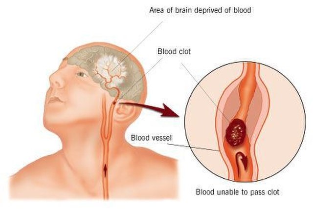
Stroke is the leading cause of serious long-term disability,
significantly reducing mobility in almost half of the affected patients
aged 65 years and older. There are currently no proven neurorestorative
treatments for chronic stroke. To address the complex problem of
restoring function in ischemic brain tissue, stem cell
transplantation-based therapies have emerged as potential restorative
therapies. Aligning with the major cell types found within the ischemic
brain, stem-cell-based clinical trials for ischemic stroke have fallen
under three broad cell lineages: hematopoietic, mesenchymal, and neural.
In this review article, we will discuss the scientific rationale for
transplanting cells from each of these lineages and provide an overview
of published and ongoing trials using this framework.
Introduction
Stroke is the leading cause of serious long-term
disability, significantly reducing mobility in almost half of affected
patients aged 65 years and older (Benjamin et al., 2017).
Each year, 795,000 strokes occur in the US alone, and the annual
economic impact of stroke is estimated at $33.9 billion. The current
standard of care for ischemic stroke is acutely time-sensitive:
administration of intravenous tPA is recommended within 4.5 h of stroke
onset, and endovascular therapy in select patients within 24 h of stroke
onset (Hacke et al., 2008; Berkhemer et al., 2015; Campbell et al., 2015; Goyal et al., 2015; Jovin et al., 2015; Saver et al., 2015, 2016; Albers et al., 2018; Nogueira et al., 2018).
Beyond the acute period, there are currently no proven neurorestorative
treatments for stroke. Despite numerous clinical trials, drug-based
therapies, including selective serotonin reuptake inhibitors,
amphetamines, and ion channel modulators, have not yielded significant
benefits, perhaps due to the complex cellular disruption that occurs
within damaged ischemic tissue (Chollet et al., 2011; Mead et al., 2015; Simpson et al., 2015; Yeo et al., 2017).
Unlike other organs, the brain responds to ischemia by undergoing
liquefactive necrosis, a process in which dead tissue liquefies and is
cleared by brain resident phagocytes over months. This long-lasting
inflammatory process results in substantial neurotoxicity, myelin
degradation, and glial scarring, as well as releasing a host of
neuroinflammatory mediators, including cytokines (TNF-a, IL-1b, IL-6,
IL-20),chemokines (MCP-1, MIP1a), cellular adhesion molecules
(immunoglobulins, cadherins, integrins), reactive oxygen species, and
matrix metalloproteases (Lakhan et al., 2009; Ceulemans et al., 2010; Stonesifer et al., 2017; Chung et al., 2018; Zbesko et al., 2018).
At the liquefactive core of the infarct, hematopoietic lineage (myeloid
and lymphoid), mesenchymal lineage (endothelial and other stromal), and
neural lineage (neurons, astrocytes, and oligodendrocytes) cells
undergo extreme stress, interacting and dying within this inflammatory,
acidic, and hypoxic milieu (Chung et al., 2018).To
address the complex problem of restoring function in ischemic tissue,
stem cell transplantation-based therapies have been investigated as
potential restorative treatments for chronic stroke. Aligning with the
major cell types found within the ischemic brain, stem-cell-based
clinical trials for ischemic stroke have fallen under three broad cell
lineages: hematopoietic, mesenchymal, and neural (Table 1).
In this review, we will discuss the scientific rationale for
transplanting cells from each of these lineages and provide an overview
of published and ongoing trials using this framework.
Hematopoietic Lineage
Hematopoietic stem cells (HSCs) at rest reside within the
bone marrow niche but can be mobilized to the general blood circulation
in response to stimulant factors such as granulocyte colony-stimulating
factor (G-CSF). A majority of trials conducted using HSC lineage cells
have relied on autologous bone marrow transplantation, favoring the lack
of immunogenicity and ethical challenges to obtaining a reliable source
of hematopoietic cells. In these studies, bone marrow was obtained from
the patient and purified, either by density gradient alone or combined
with immunosorting to obtain the cell population for transplantation.
The cell surface marker CD34 characterizes a population enriched for
HSCs, and the proportion of CD34+ cells found in the peripheral blood
immediately after stroke has been found to directly correlate with
functional recovery (Dunac et al., 2007).
Bone Marrow Derived Cells
Bone Marrow Mononuclear Cell (BM-MNC) Transplantation
Early Phase I Trials (2009–2012)
The first Phase I trials of bone-marrow-derived cells demonstrated
unequivocally that BM-MNCs could safely be transplanted in stroke
patients at varying time points after stroke onset, and via various routes of administration. Suarez-Monteagudo et al. (2009)
implanted 14–55 million autologous BM-MNCs by stereotactic
intralesional injection in five chronic stroke patients at least 1 year
and no more than 10 years post-stroke onset. This trial found that
intracranially-injected autologous BM-MNCs were well-tolerated and safe,
with several patients reporting long-term neuropsychiatric
improvements. Targeting the subacute phase of ischemic stroke, Barbosa da Fonseca et al. (2010) infused 125–500 million autologous BM-MNCs in six patients via intra-arterial injection 8–12 weeks after stroke onset and reported no cases of neurologic worsening. In the acute setting, Savitz et al. (2011)
determined that intravenous infusion of 70–100 million cells per
kilogram weight of autologous BM-MNCs in 10 patients with acute stroke
(24–72 h after onset) was both safe and feasible. Additionally, Prasad et al.’s (2012) research group intravenously infused 11 patients with 80 million autologous BM-MNCs (mean 0.92 × 10⁶ CD34+ cells) within 7–30 days of stroke onset and also confirmed the safety and feasibility of this treatment protocol. Friedrich et al. (2012) administered 50–600 million autologous BM-MNCs via
intra-arterial (MCA) infusion to 20 patients within 3–10 days of stroke
onset and found this method to be safe. Of note, while not known at the
time, the brain biodistribution of intra-arterially and intravenously
transplanted BM-MNCs was eventually determined to be comparable in a
separate clinical trial completed a year later (Rosado-de-Castro et al., 2013).
In both cases, brain biodistribution was low compared to that of lung
or spleen, or liver. A summary of different routes of administration and
their relative advantages and disadvantages concerning reaching
ischemic brain tissue have been included in Table 2.
In the context of intracranial
cell administration, transplantation into peri-ischemic vs. directly
lesioned areas was extensively investigated in preclinical stroke
models. The lesioned area was determined to be a poor injection target
site due to unstable vascular supply and a highly inflammatory
microenvironment. Moreover, the lesioned area was shown to eventually
become a non-functional, fluid-filled cyst, suggesting that post-stroke
recovery would be due to changes at the periphery of the lesion, not the
cystic core (Veizovic et al., 2001; Modo et al., 2002; Smith et al., 2012).
Of the clinical trials that utilized intracranial administration, only
the porcine neural cell transplantation study conducted by Savitz et al. (2005)
injected directly into the infarct. The study, discussed in a later
section of this review, was terminated by the FDA after two of the five
patients developed adverse events including cortical vein occlusion,
complex partial seizures, and ring-enhancing lesions.
Phase I/II, Phase II Trials (2014–2015)
As Phase I/II trials progressed, the safety of these treatment
protocols was consistently demonstrated, but BM-MNC administration was
not found to significantly improve neurologic outcomes in transplant
cohorts. In a follow-up to their initial Phase I trial, Prasad et al. (2014)
performed a Phase II randomized study in which 120 patients received
either 280 million autologous BM-MNCs or placebo intravenously within
7–30 days of stroke onset; this study yielded no clinical benefit for
BM-MNC transplantation over placebo. Sharma et al.’s (2014)
research group pursued a different route of administration, identifying
24 patients with chronic stroke (onset between 4 months to 10 years) to
receive 1 million BM-MNCs per kilogram body weight intrathecally.
Intrathecal administration proved safe, and functional improvement was
noted in treated patients, however, there was no control group for
comparison. In a phase I/IIa trial, Taguchi et al. (2015)
intravenously administered either 250 million or 340 million cells of
autologous BM-MNCs and found a trend toward improved neurologic outcomes
and cerebral perfusion in the high dose group. Studies by Bhasin et al. (2016)
additionally found that positive neurologic outcomes in patients given
autologous BM-MNCs could be due to paracrine effects of secreted
vascular endothelial growth factor (VEGF) and brain-derived neurotrophic
factor (BDNF).
Sorted CD34+ From Bone Marrow
Hypothesizing that the hematopoietic stem cell-enriched
CD34+ fraction of BM-MNCs contained the functional subset of cells
responsible for repair during ischemic CNS injury, Moniche et al. (2012)
conducted a Phase I/II study in which 160 million autologous CD34+
BM-MNCs were infused intra-arterially (MCA) into 10 patients within 5–9
days of stroke onset. The comparison group consisted of 10 subacute
stroke patients who did not receive the intervention. In this trial,
there was no clinical benefit at 180 days, but there was a statistically
significant increase in b-NGF among the treated cohort (p < 0.02). Similarly, Banerjee et al.’s (2014) research group intra-arterially infused (via
the MCA) 10 million autologous sorted CD34+ cells in five patients
within 1–7 days of stroke onset, and found the intervention to be safe,
and associated with a decrease in infarct size over time.
Sorted Aldehyde Dehydrogenase-Bright (ALDH-BR) From Bone Marrow
Aldehyde dehydrogenase (ALDH) was among the first markers
used by immunologists to identify populations of human hematopoietic
stem and progenitor cells in the 1990s. HSCs were found to express the
highest levels of ALDH, while lymphocytes expressed the lowest (Kastan et al., 1990).
Transplant of BM-derived ALDH-br cells was reported to improve
functional recovery in limb ischemia and ischemic heart failure (Keller, 2009; Perin et al., 2011, 2012). Based on these findings, Savitz et al. (2019) transplanted up to eight million autologous ALDH-br BM-MNCs (dubbed autologous ALD-401 cells) via
intracarotid infusion in 29 patients and compared this cohort to 19
control patients who received a sham procedure. Disappointingly, the
study found no significant difference in primary or secondary efficacy
measures between treatment and placebo groups.
Mobilized Peripheral Blood Stem Cells
Shyu’s research group previously demonstrated peripheral
blood-derived stem-cell (PBSC) transplant efficacy in treating chronic
ischemia in rats (Shyu et al., 2006). To further evaluate the efficacy of this cell type in human subjects, Chen et al. (2014)
conducted a Phase II trial in which autologous PBSCs were mobilized
with G-CSF, and 3–8 million sorted CD34+ cells were stereotactically
transplanted into 15 chronic stroke patients (stroke onset ranging from 6
months to 5 years prior). The authors noted improvement in multiple
neurologic and functional outcome scores in the treated cohort.
Umbilical Cord Blood Derived Cells
Allogeneic umbilical cord blood is an immunologically
tolerant source of readily available cells. Unlike other sources of
HSCs, HLA-matching is not required, and its safety and efficacy as a
blood donor graft have been well established (Zhou et al., 2012). In this context, Laskowitz et al. (2018)
conducted a Phase I study to establish the safety and feasibility of
administering a single intravenous infusion of allogeneic (non-HLA
matched) umbilical cord blood in stroke patients. The study included 10
patients treated between 3 and 9 days post-stroke onset, and noted an
improvement in neurological and functional outcome, although there was
no control group for comparison. A list of pending and ongoing clinical
trials using cells of hematopoietic origin have been compiled in Table 3.
Mesenchymal Lineage
Mesenchymal stem cells (MSCs) are stromal cell
precursors to cells of osteogenic, chondrogenic, and/or adipogenic
lineages, and can be harvested and expanded from a variety of tissue
types, including bone marrow (BM-MSCs, a cell population distinct from
hematopoietic origin BM-MNCs), adipose tissue (adipose-derived MSCs, or
AD-MSCs), and umbilical cord (UB-MSCs). This versatility, combined with
low immunogenicity due to low expression of human leukocyte antigens,
makes MSCs excellent candidates for allogeneic stem cell transplants (Le Blanc et al., 2003; Klyushnenkova et al., 2005).
Unlike bone marrow-derived hematopoietic cells, MSCs from a single
source can be expanded for transplant into many individuals, providing
both standardization and scalability for large clinical studies.
BM-MSCs have been shown to cross the blood-brain barrier
and improve functional recovery after acute ischemic stroke in animal
models (Chen et al., 2001; Lee et al., 2016),
likely due to a paracrine effect by secreting neurotrophic, mitogenic,
and angiogenic factors, including VEGF, BDNF, nerve growth factor, basic
fibroblast growth factor, and insulin-like growth factor 1 (Eckert et al., 2013; Shichinohe et al., 2015; Stonesifer et al., 2017). One hypothesized mode of delivery for these factors is via the secretion of membrane fragments (extracellular vesicles, EVs) from transplanted cells (Bang and Kim, 2019; Surugiu et al., 2019).
Recent preclinical data in organoids suggest that EVs alone may be
sufficient to significantly decrease injury in a hypoxia-starvation
model of injury, opening the possibility for future early phase clinical
trials of EV delivery for ischemic stroke (Zheng et al., 2020).
Additionally, AD-MSC transplants have also demonstrated
success in experimental ischemic stroke models, in which animals treated
with AD-MSCs demonstrated increased expression of BDNF and enhanced
nerve regeneration, and simultaneously reduced expression of
pro-apoptotic proteins such as BCL-2 and BAX within the ischemic lesion (Li et al., 2016).
Multiple preclinical studies of UB-MSCs showed that transplanted cells
quickly homed to the site of injury in rat ischemia models and resulted
in improved long-term neurologic outcomes (Zhang et al., 2017; Wu et al., 2018). These results were also most likely due to paracrine effects, as transplanted cells did not persist long term.
Bone Marrow Derived MSCs
Autologous Transplant
Bang et al. (2005)
were the first to conduct autologous MSC transplants in stroke
patients, to introduce cells with the potential to provide trophic
support for neurogenesis and/or neuromodulatory effects—functions that
are limited with hematopoietic cells. This group conducted a Phase I/II
trial in which 100 million culture-expanded autologous BM-MSCs (grown in
fetal bovine serum-containing media) were intravenously infused in five
patients at 5–7 weeks post-stroke onset. Compared to a control cohort
of 25 patients, treated patients demonstrated consistent neurologic
improvement at 3, 6, and 12 months post-transplantation. Based on the
success of this first study, the same group subsequently conducted a
larger observer-blinded trial consisting of 52 patients (16
transplanted, 36 control; Lee et al., 2010).
Due to the significant time required to expand MSCs in culture, the
investigators opted to decrease the time-to-transplant by administering
an initial dose of 50 million MSCs, and subsequently administering an
additional 50 million MSCs 2 weeks later (rather than a one-time
treatment with 100 million MSCs as in the Phase I/II trial). Notably,
clinical improvement was found to be correlated with intactness of the
subventricular zone—a known neurogenic site—as demonstrated on
diffusion-weighted MR imaging. The MSC-transplanted cohort exhibited a
higher rate of functional recovery and lower mortality compared to the
control cohort. The authors reported that the use of bovine serum
proteins to expand MSCs in culture did not appear to result in zoonoses
or other adverse effects.
Due to ongoing concern regarding the use of bovine
serum, particularly in the context of infectious diseases such as
Creutzfield-Jakob, a Phase I study using MSCs expanded with autologous
human serum was also conducted and published by a different group (Honmou et al., 2011). Honmou et al.’s (2011)
research group demonstrated the safety and feasibility of BM-MSCs
expanded in culture with human autologous serum, injected intravenously
in 12 patients. Human serum resulted in more rapid MSC expansion in vitro
compared to bovine serum, and at 1-week post-transplant, the mean
infarct volume, as measured on MRI, was reduced by approximately 20% in
patients treated according to this protocol. Circumventing the issue of
serum altogether, Bhasin et al.’s (2011)
research group conducted a Phase I/II trial in which autologous MSCs
expanded in culture under serum-free conditions were intravenously
administered to 20 chronic stroke patients between 3 months and 2 years
following stroke onset. While both the control (n = 20) and
transplanted groups experienced statistically significant improvement,
there was no statistically significant difference between the groups in
terms of functional outcome. Bhasin et al. (2013)
presented a follow-up study in 2013 in which mesenchymal cells were
compared with hematopoietic/mononuclear cells. Again, stem cell
transplantation was found to be safe and feasible, with no conclusive
evidence for efficacy but a trend toward functional improvement. Their
2016 study, again transplanting BM-MNCs, was discussed previously.
In a separate Phase I/IIa study, Fang et al. (2019)
compared BM-MSCs to autologous endothelial progenitor cells (EPCs), as
well as to placebo in patients within 5 weeks of stroke onset. This
study included 18 total patients: six transplanted with BM-MSC, six with
EPCs, and six with saline (placebo). The BM-MSCs were expanded in fetal
bovine serum culture, and EPCs were derived by seeding bone marrow
mononuclear cells on fibronectin plates to select for adherent cells,
and subsequently maintained in bovine serum culture with endothelial
cell media. Two injections of 2.5 million cells per kilogram body weight
were given approximately 1 week apart, and patients were followed for 4
years. While the trial was deemed safe, no functional or neurological
difference was observed between the BM-MSC, EPC, and placebo groups.
Allogeneic Transplant
The fact that MSCs express low levels of human leukocyte
antigen and are easily expanded in culture is a significant advantage
over other cell-based therapies about stroke therapy (Le Blanc et al., 2003).
These features allow for the development of large quantities of
standardized single-source cells for allogeneic transplant and also
mitigate many of the challenges of autologous cell therapy in terms of
timing of treatment. This is of particular benefit in patients who are
unable to provide autologous cells. Moreover, because MSCs may be
cultured and expanded in vitro without sacrificing stemness potential (Reyes et al., 2001; Jiang et al., 2002),
genetic modification of these cells prior to transplantation in order
to enhance supportive properties has also opened new possibilities.
Unmodified Cell Transplants
In the largest MSC trial to date, Hess et al. (2017;
MASTERS, Athersys) conducted a phase II randomized, double-blind,
placebo-controlled dose-escalation trial of intravenous adult BM-MSCs
for acute ischemic stroke at 33 centers across the US and UK. In this
trial, patients were randomized to receive either 400 million or 1.2
billion allogeneic BM-MSCs (n = 65) or placebo (n = 61)
within 24–48 h after stroke onset. The allogeneic BM-MSC product used in
this study—MultiStem—was derived from two independent donors (Boozer et al., 2009).
No dose-dependent toxicity was observed and the treatment was deemed
safe, but the transplanted group and placebo groups exhibited no
significant difference in terms of functional outcome at 90 days
post-stroke. Of note, a post hoc analysis of those patients
achieving an “Excellent Outcome” defined as mRS ≤1 and NIHSS ≤1 and
Barthel ≥95, demonstrated a statistically significant benefit for all
patients treated at 1 year (23.1% transplant vs. 8.2% placebo; p =
0.02) and for all patients treated within the originally planned time
window of ≤36 h post-stroke the benefit at 1 year was even greater
(29.0% transplant vs. 8.2% placebo; p < 0.01). This finding
was encouraging enough for Athersys to initiate another Phase III
prospective, randomized, placebo-controlled, double-blind trial treating
patients between 18–36 h of stroke (MASTERS-2).
In a separate Phase I/II trial using allogeneic single-donor adult mesenchymal BM-MSCs (Levy et al., 2019;
Stemedica) transplanted up to 1.5 million BM-MSCs per kg body weight
intravenously in 38 patients with chronic stroke (>6 months
post-stroke). The highest dose (1.5 million/kg) was found to be safe,
and significant behavioral gains were observed. Excellent functional
outcome (Barthel score >95) was reported in 35.5% of patients at 12
months post-transplant, compared to in 11.4% at baseline; however, there
was no control group included in this study.
Modified MSC Transplants
SB623 is a BM-MSC line that has been transiently transfected with a
plasmid containing the human Notch1 intracellular domain, which results
in constitutive Notch1 expression. Importantly, the Notch1 plasmid is
not replicated during mitosis and is therefore rapidly lost during cell
division. In in vitro preclinical models, Notch1-modified MSCs
promoted neural cell growth and rescued neural cell survival after
ischemia by providing trophic support via the secreted extracellular matrix, promoting angiogenesis, and playing a protective, anti-inflammatory role (Aizman et al., 2009; Tate et al., 2010; Dao et al., 2011, 2013).
Experimental stroke models transplanted with Notch-1-modified MSCs have
demonstrated functional recovery and peri-infarct neuroprotection as
measured by a reduction in cell loss. Despite these benefits, however,
the SB623 cells themselves are short-lived in vivo (Yasuhara et al., 2009; Tajiri et al., 2013).
This finding suggests that observed improvements are a result of
supportive trophic activity rather than engraftment, and alleviates
concerns over the challenges of achieving long-term allogeneic cell
engraftment.
Based on this encouraging preclinical data, Steinberg et al. (2016)
completed a Phase I/IIa trial in which 18 chronic stroke patients (6
months to 3 years post-stroke onset) received a stereotactic
intracranial injection of either 2.5, 5, or 10 million allogeneic
modified SB623 BM-MSCs (six patients per cohort). Each patient received
five stereotactic image-guided injections of 20 μl each surrounding the
infarct. Of note, the intracranial administration of SB623 was chosen
because of the desire to prioritize trophic factor delivery by
transplanted cells over cell engraftment (Bliss et al., 2010).
In comparison to intra-arterial and intravenous delivery, the
intracranial injection was shown to result in greater delivery of
transplanted cells to the lesion, although cells are unequally
distributed throughout (Rosado-de-Castro et al., 2013).
Patients who received intracranial injections reported several
treatment-emergent adverse events (TEAEs), including headache, nausea,
and vomiting. It was determined that most, if not all, of the TEAEs,
were due to the surgical procedure rather than the cell transplantation.
No link between TEAE and cell dosage was identified, and all TEAEs
recovered without sequelae. No antibody response to SB623 cells was
observed, and a significant improvement in neurological function was
noted after 3, 6 (the pre-determined efficacy endpoint), and 12 months.
In a follow-up article detailing 2-year outcomes, the initial
improvements were found to be stable at 2 years post-transplantation (Steinberg et al., 2018).
Interestingly, the authors noted that the size of a transient T2-FLAIR
signal (DWI negative) on MRI at an early time point (1–2 weeks
post-transplant) was correlated with the degree of long-term functional
improvements, and could be a possible indicator of functional transplant
activity. A larger Phase IIb randomized, double-blind,
placebo-controlled study, ACTIsSIMA, has been completed but the results
are as yet unpublished. Additional pending clinical trials using cells
of mesenchymal origin have been detailed in Table 4.
Neural Lineage
Neural stem cells (NSCs) are multipotent cells that can
differentiate into neurons, astrocytes, and oligodendrocytes. This
native capacity to repopulate and support endogenous cell types within
the brain has generated much interest in NSC transplantation for stroke.
The subventricular zone of the lateral ventricle and the dentate gyrus
of the hippocampus have been identified as neurogenic sites, and in
murine models, NSC’s have been shown to migrate from these niches to
promote neurogenesis and vascular remodeling in response to ischemic
stroke (Zhang et al., 2014; Hao et al., 2015).
One of the greatest barriers to developing an NSC
transplantation model is the challenge of harvesting cells for
transplantation. Clinical trials utilizing NSCs have attempted to
overcome this barrier by: (1) modifying a human teratoma cell line
[teratomas and cancer lines were the only non-embryonic source of
pluripotent human cells before the development of induced pluripotent
stem cells (iPSCs)] to induce neuronal differentiation by introducing
the morphogen retinoic acid; (2) using NSCs harvested from human fetal
tissue and/or clonal lines derived from these cells; or (3) using cells
of non-human origin (i.e., porcine fetal NSCs).
Retinoic Acid-Induced Differentiated Tumor Cells
Kondziolka et al. (2000)
were the first to conduct NSC transplants for chronic stroke. In a
Phase I trial published in 2000, 12 patients received either two or six
million human “LBS-Neurons” via intracranial injection between 6
months and 6 years post-stroke. These cells were derived by
differentiating the NT2/D1 human cell line, originally derived from a
lung metastasis of testicular embryonal carcinoma, into neurons using a
10 μM dose of retinoic acid. Before the discovery of iPSCs in 2007,
NT2/D1 was among the lines widely used to represent human pluripotent
cells and was shown to be capable of terminal differentiation into
multiple cell types, including neurons and astrocytes (Bani-Yaghoub et al., 1999).
Preclinical studies demonstrated that NT2/D1-derived neurons resulted
in improved functional outcomes after ischemia when injected
intracranially (Borlongan et al., 1998).
In humans, treatment with NT2/D1-derived neurons was found to be safe
and associated with significantly improved functional outcome at 6
months.
Subsequently, the same University of Pittsburg group,
with the addition of Stanford University investigators, conducted a
Phase II randomized observer-blinded trial in which 14 chronic stroke
patients received an intracranial injection of either five or 10 million
LBS-Neurons; four patients acted as nonsurgical controls. While the
primary efficacy endpoint—improvement of European Stroke Scale score at 6
months — was not achieved, one of the prespecified secondary outcome
measures (the Action Research Arm Test designed to measure gross
hand-movement) improved significantly compared with controls and with
baseline scores on the same test.
Porcine
Given the concerns surrounding implanting cells derived
from malignant human tumors, compounded with the ethical implications
and difficulty of obtaining fetal tissue, Savitz et al. (2005)
attempted a Phase I trial to xenotransplant up to 50 million fetal
porcine cells in five patients with chronic stroke, 18 months to 10
years post-stroke onset. Before intracranial transplantation, the cells
were treated with anti-MHC to prevent rejection. While two patients
reported clinical improvement, one patient developed seizures, and one
experienced a temporary worsening of motor symptoms. As a result, this
trial was halted due to safety concerns.
Human Primary Fetal Brain Derived
The human fetal brain represents a source of actively
dividing NSCs that have demonstrated proven engraftment and functional
capacity in both preclinical and clinical studies for disorders such as
leukodystrophies (Uchida et al., 2000, 2012; Tamaki et al., 2002; Kelly et al., 2004; Gupta et al., 2012, 2019);
however, fetal tissue is difficult to obtain in the US due to
governmental policy. CTX0E03 is one such fetal tissue line, derived by a
research group in the UK and is currently being studied in ischemic
stroke in the PISCES clinical trials (ReNeuron). CTX0E03 cells are a
clonally derived human fetal cortical cell line that was transfected
with a single copy of c-mycERTAM, an immortalizing gene dependent on
tamoxifen administration for function (Pollock et al., 2006).
Despite c-myc’s role as a known oncogene, preclinical stroke models
indicated that CTX0E03 cells were safe to transplant, and promoted
behavioral recovery via enhanced neurogenesis and angiogenesis in a dose-dependent fashion after ischemia (Stroemer et al., 2009). It is unclear if CTX0E03 cells exhibit long-term engraftment in the brain (Hicks et al., 2013; Baker et al., 2019).
The PISCES 1 clinical trial for ischemic stroke transplanted 2.5, 5,
10, or 20 million CTX0E03 cells intracranially in 11 patients who had
experienced a stroke 6–24 months prior (Kalladka et al., 2016).
The treatment was found to be safe, and treated patients exhibited
improved neurologic outcomes on several scales. However, the initial
study was limited to cisgender male patients due to concerns about the
potential for estrogen to activate c-myc. The PISCES 2 open-label Phase 2
trial transplanted 20 million CTX0E03 cells into the putamen of 23
patients, 13 males, and 10 females, 2–13 months after subcortical
ischemic stroke (individuals actively taking tamoxifen were excluded).
While the primary endpoint [two patients improving two points in the
Action Research Arm Test (ARAT) subtest 2 at 3 months] was not met,
there were substantial improvements in this metric as well as the mRS
and Barthel Index (Muir et al., 2020).
A Phase III prospective, randomized, controlled, double-blinded study
(PISCES 3) treating 130 patients 6–12 months post-stroke is currently
ongoing in the US.
Cotransplant Studies of Primary Fetal NSC and UB-MSCs
Given the significant trophic support provided by
mesenchymal cells and the regenerative capacity of NSCs, combined
engraftment of fetal-derived neural and cord-blood-derived mesenchymal
lineage cells together has been investigated as a potential therapeutic
strategy to improve and support stem cell engraftment. Chen et al. (2013)
sought to establish the safety and feasibility, as well as the optimal
route of cell administration, of a multiple cell type co-transplantation
in a group of 10 patients. The investigators isolated three distinct
neural cell types from a single fetal donor: (1) olfactory ensheathing
cells (OECs) from the fetal olfactory bulb; (2) neural progenitor cells
(NPCs) from the subependymal zone; and (3) Schwann cells (SCs) from the
sciatic nerve. Cord blood from a separate donor was used to derive
UB-MSCs, and all transplanted UB-MSCs were derived from a single cord.
Chronic stroke patients who were 6 months to 20 years post-stroke onset
were included, and were divided amongst five distinct treatment
protocols: (1) OECs alone (intracranial, n = 2); (2) OECs + NPCs (intracranial, n = 2); (3) OECs + NPCs (intracranial) with a second dose of NPCs at a later time point (intrathecal, n = 4); (4) OECs + NPCs (intracranial) + later doses of NPCs (intrathecal) and UB-MSCs (intravenous, n = 1); and (5) OECs and NPCs (intracranial) + later doses of SC and NPCs (intrathecal) and UB-MSCs (intravenous, n
= 1). Treatment was found to be safe and feasible in all cases,
however, due to the variety of cell types and routes of administration
evaluated, the authors concluded that the study was not sufficiently
powered to conclude that co-transplantation is safe under all studied
conditions. The authors also noted a trend towards functional benefits
with intracranial injections, but not with intrathecal and intravenous
cell administration.
Another Phase I study conducted by Qiao et al. (2014)
involved co-transplantation of UB-MSCs with human fetal cells. Enrolled
patients received either four intravenous doses of 0.5 × 10⁶ UB-MSC cells per kg body weight or one intravenous dose of 0.5 × 10⁶ UB-MSC cells per kg body weight followed by three intrathecal doses of 0.5 × 10⁶ UB-MSC cells per kg body weight and 6 × 10⁶ human fetal derived NPCs (Qiao et al., 2014).
In total, the investigators treated six subacute to chronic stroke
patients between 1 week and 2 years post-stroke onset. Two of these
patients received UB-MSCs only. The trial demonstrated that
co-transplantation was safe and feasible; no malignancies were observed
from the use of multipotent fetal cells. Further, each treated patient
experienced clinical improvement that was stable at 2 years
post-transplant. A list of pending clinical trials using cells of neural
origin has been compiled in Table 5.
Conclusions
The regenerative properties of stem cells have brought
cell-based transplantation studies into the spotlight as appealing
therapies for otherwise recalcitrant disorders such as subacute and
chronic ischemic stroke. In this review article, we have detailed the
clinical trials to date, which have featured transplantation of various
cell types, administered via a variety of routes and in a variety
of doses, to treat ischemic stroke of varying chronicity. We have
discussed the history of and scientific rationale for the different cell
types transplanted, their routes of administration, and associated
trial outcomes, and have provided snapshots of current ongoing trials.
Additional studies are necessary to strengthen our understanding of the
relationship between neural cells and their surrounding stromal,
endothelial, and immune landscape in both the healthy state and in
pathologic conditions. As discussed in this review, specific attention
should be given to the paracrine mechanisms by which transplanted cells
exert their therapeutic effect, especially in light of data that
suggests that these benefits persist even after the clearance of the
originally transplanted cell type.
Furthermore, promising preclinical studies require
adequate support and prudent design to overcome the “translational
roadblock,” a notable decrease in efficacy between preclinical studies
and their clinical trial counterparts. The difference is thought to be
due to a series of factors. First, a combination of publication bias and
overstated efficacy in preclinical studies has led to overly optimistic
preclinical data that fail to result in statistically meaningful
clinical interventions (Dirnagl et al., 2009; Macleod et al., 2009).
Poor translation to the clinic has also resulted from the differences
in primary endpoints between animal models and trials, lengthening of
time-to-treatment in the clinic compared to in animal models, nuances in
translating dosage, and heterogeneity of patient characteristics such
as age (Dirnagl et al., 2009; Hermann et al., 2019).
Recent studies have shown patient age to be a significant prognostic
factor, and have suggested that, for clinical improvement, the timing of
interventions must be increased to account for increased age (Sandu et al., 2017).
Additionally, the underpowered clinical trial design has also
contributed significantly to the difficulty of translating otherwise
promising preclinical interventions to the bedside (Dirnagl and Macleod, 2009; Schmidt-Pogoda et al., 2020).
Judiciously guiding the development of future stem-cell-based clinical
interventions, including those harnessing recent advances in cellular
regeneration, trophic support, immunomodulation, and perhaps as-of-yet
undiscovered mechanisms of repair, will be essential in achieving
successful clinical trials of promising neurorestorative therapies.





 Joy Q. He
 Eric S. Sussman2 and
  Gary K. Steinberg