This is where stroke leaders would look at this
and mutter to themselves; 'We could use this to detect brain signals
occurring during stroke recovery; neurogenesis and neuroplasticity, and
thus figure out how to make those signals repeatable'. But we have to do
human testing first.
But the leaders would already have started listening to brain signals using one of these already.
1. Use nanowires to listen in on single neurons
2. Or lay a grid across the cortex
to listen in.
But we have NO stroke leaders, nothing will get done until we get survivors in charge.
Leaders solve problems, they don't run away from them.
The latest here:
New Tool Maps Brain Signals with Unprecedented Clarity
Summary: Researchers developed an innovative chemical tool to explore how signals like dopamine and epinephrine interact with neurons via G protein-coupled receptors (GPCRs).
This new tool allows for precise detection of neuromodulators across various brain regions with high spatial resolution. It marks cells with a permanent fluorescent signal, facilitating the study of signal pathways and interactions at a cellular level across the entire brain.
This advancement could significantly enhance our understanding of neuronal signaling and improve targeting GPCRs in drug development.
Key Facts:
- The tool enables detailed visualization of GPCR-related signals across the entire brain with high spatial resolution, a balance previously unachievable in neuroscience.
- It has been tested on opioids and epinephrine, utilizing both green and red fluorescence to track multiple molecules simultaneously.
- While the fluorescence takes several hours to manifest and is not suitable for real-time tracking, the tool provides valuable postmortem insights into neuronal pathways and drug targeting.
Source: University of Michigan
University of Michigan researchers have developed a new tool to better understand how chemicals like dopamine and epinephrine interact with neurons.
These chemicals are among a wide variety of signals that get processed in the brain through G protein-coupled receptors (GPCRs), proteins that sit on the surface of neurons to receive messages—in the forms of proteins, sugars, fats, even light—that inform cellular behavior.
GPCRs are involved in an enormous number of biological functions, making them a prime target for treating diseases; more than one-third of FDA-approved drugs target GPCRs. But to fully understand how various molecules interact with GPCRs, researchers need to be able to detect those molecules across the whole brain with high spatial resolution.
“The challenge in our field has been achieving the right balance between a detailed view and the whole picture across the brain,” said Wenjing Wang, a neuroscientist at the U-M Life Sciences Institute.
LSI faculty member Peng Li said most existing tools can detect a neural modulator either in a small part of the brain with high spatial resolution or in the whole brain with very low resolution.
“But we need to identify the cells that respond to the neuromodulators across various brain regions, in high resolution,” he said.
In a study published in the Proceedings of the National Academy of Sciences, Wang, Li and colleagues unveiled a new chemical tool that achieves both goals for three chemicals that all target GPCRs.
Wang’s lab at LSI uses protein engineering to develop technologies that can detect how signaling molecules travel within the brain to reach and interact with specific neurons. They previously created a tool to reveal the presence of opioids, another GPCR binding partner, at a cellular level.
When the molecule is detected, the tool creates a permanent fluorescent mark in the cells. Thus, researchers can see the specific cells that are highlighted, as well as the whole picture of cells across the brain.
This latest work broadens the utility of that sensor to detect multiple types of GPCR activators, beyond just opioids. So far, the team has tested the tool with opioids and epinephrine in cultured neurons and in mouse models. The team also expanded the tool to use both green and red fluorescence, enabling the tracking of multiple molecules at once.
“Coming from detecting just opioids, we now have a tool that we can begin to easily modulate for various signals that interact with GPCRs,” said Wang, who also is an assistant professor of chemistry at the U-M College of Literature, Science, and the Arts.
“The goal is eventually to even study the interplays of different signaling pathways simultaneously.”
The team cautions that while the tool provides important visualizations of how signals travel across neurons for analysis postmortem, it cannot be used to track chemicals in real time, as it takes several hours for the fluorescence to appear. But it does offer a new path forward for improving understanding of neuronal signaling and the role of GPCRs as drug targets.
“Ideally, we aim to be able to create a brain map for multiple neuromodulators concurrently, offering a comprehensive understanding of the sites of neuromodulation,” said Li, who also is an assistant professor at the U-M School of Dentistry.
About this brain mapping and neurotech research news
Author: Morgan Sherburne
Source: University of Michigan
Contact: Morgan Sherburne – University of Michigan
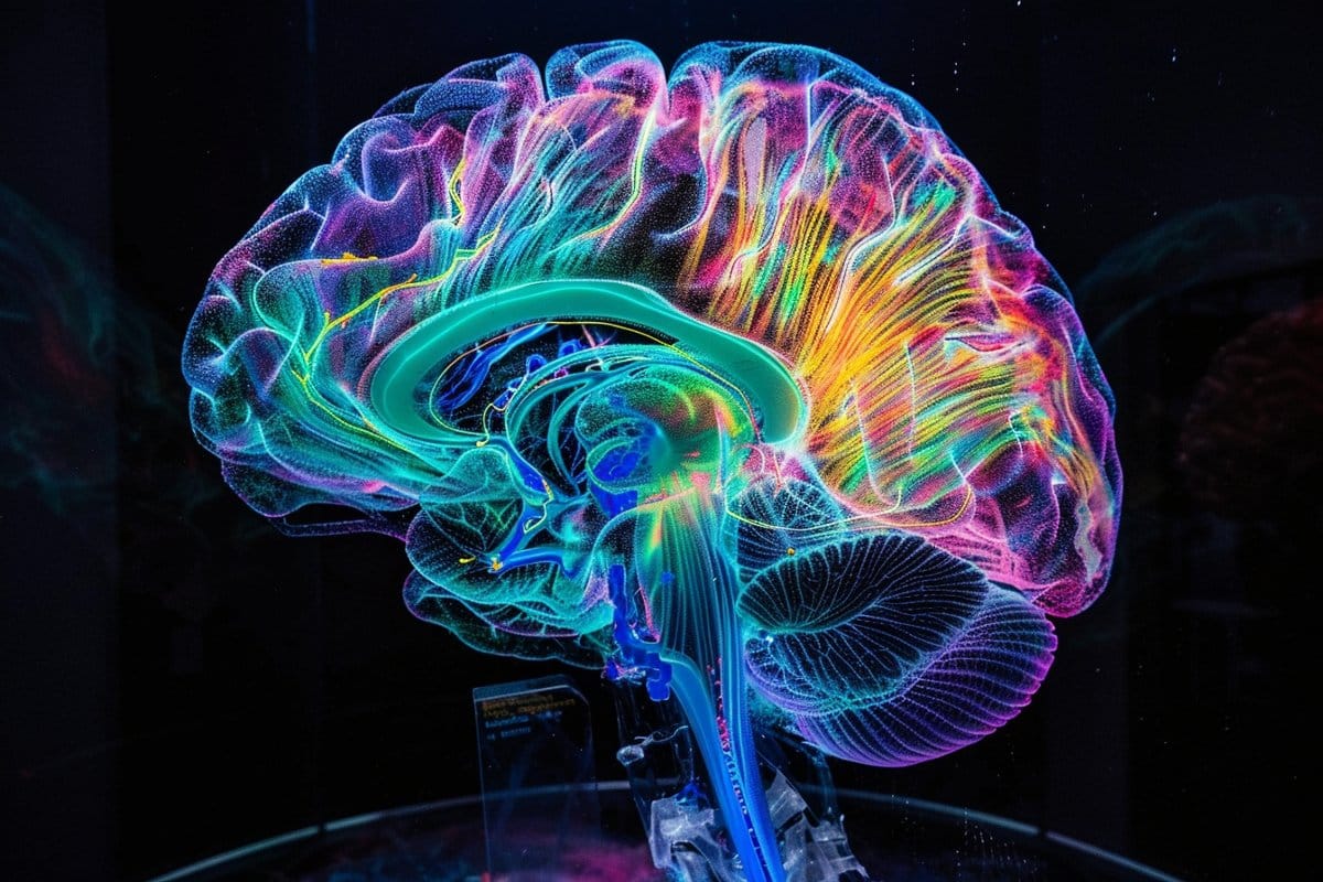
Image: The image is credited to Neuroscience News
Original Research: Closed access.
“Single-chain fluorescent integrators for mapping G-protein-coupled receptor agonists” by Wenjing Wang et al. PNAS
No comments:
Post a Comment