Use the labels in the right column to find what you want. Or you can go thru them one by one, there are only 34,134 posts. Searching is done in the search box in upper left corner. I blog on anything to do with stroke. DO NOT DO ANYTHING SUGGESTED HERE AS I AM NOT MEDICALLY TRAINED, YOUR DOCTOR IS, LISTEN TO THEM. BUT I BET THEY DON'T KNOW HOW TO GET YOU 100% RECOVERED. I DON'T EITHER BUT HAVE PLENTY OF QUESTIONS FOR YOUR DOCTOR TO ANSWER.
Changing stroke rehab and research worldwide now.Time is Brain!trillions and trillions of neuronsthatDIEeach day because there areNOeffective hyperacute therapies besides tPA(only 12% effective). I have 523 posts on hyperacute therapy, enough for researchers to spend decades proving them out. These are my personal ideas and blog on stroke rehabilitation and stroke research. Do not attempt any of these without checking with your medical provider. Unless you join me in agitating, when you need these therapies they won't be there.
What this blog is for:
My blog is not to help survivors recover, it is to have the 10 million yearly stroke survivors light fires underneath their doctors, stroke hospitals and stroke researchers to get stroke solved. 100% recovery. The stroke medical world is completely failing at that goal, they don't even have it as a goal. Shortly after getting out of the hospital and getting NO information on the process or protocols of stroke rehabilitation and recovery I started searching on the internet and found that no other survivor received useful information. This is an attempt to cover all stroke rehabilitation information that should be readily available to survivors so they can talk with informed knowledge to their medical staff. It lays out what needs to be done to get stroke survivors closer to 100% recovery. It's quite disgusting that this information is not available from every stroke association and doctors group.
Each year, approximately 265,000 Americans have a stroke that causes
visual impairment. New research, which appears in the journal Proceedings of the Royal Society B,
sheds light on how the damage in the brain caused by a stroke can lead
to permanent vision impairment. The findings could provide researchers
with a blueprint to better identify which areas of vision are
recoverable, facilitating the development of more effective
interventions to encourage vision recovery.
"This study breaks new ground by describing the cascade of processes
that occur after a stroke in the visual center of the brain and how this
ultimately leads to changes in the retina," said senior study author
Brad Mahon, an associate professor at Carnegie Mellon University and the
University of Rochester. "By more precisely understanding which
connections between the eye and brain remain intact after a stroke, we
can begin to explore therapies that encourage neuroplasticity with the
ultimate goal of restoring more vision in more patients."
When a stroke occurs in the primary visual cortex, the neurons
responsible for processing vision can be damaged. Depending upon the
extent of the damage, this can result in blind areas in the field of
vision. While some patients spontaneously recover vision over time, for
most the loss is permanent. A long-known consequence of damage to
neurons in this area of the brain is the progressive atrophy of cells in
the eyes, called retinal ganglion cells.
"While the eye is not injured in the stroke, cells in the retina that
send projections to parts of the brain that are damaged will degenerate
over time," Mahon said. "Once this occurs, it becomes more and more
unlikely for vision to recover at that location."
The new research sought to understand the mechanisms of vision loss
after stroke and whether it was possible to identify areas in the field
of vision that could be recovered. The study involved 15 patients
treated at Strong Memorial and Rochester General hospitals for a stroke
that affected the primary visual processing area of the brain. The
participants took vision tests, underwent scans in an MRI to identify
areas of brain activity and were administered a test that evaluated the
integrity of cells in their retina.
The team found that the survival of the retinal ganglion cells
depended upon whether or not the primary visual area of the brain to
which they are connected remained active. Eye cells that were connected
to areas of visual cortex that were no longer active would atrophy and
degenerate, leading to permanent visual impairment.
However, the researchers observed that some cells in the eye remained
healthy, even though the patient could not see at the corresponding
field of vision. This finding suggests that these eye cells remain
connected to unscathed neurons in the visual cortex and that visual
information was making its way from the eyes to the visual cortex, even
though this information was not being interpreted by the brain in a
manner that allowed sight.
"The integration of a number of cortical regions of the brain is
necessary in order for visual information to be translated into a
coherent visual representation of the world," said study co-author Dr.
Bogachan Sahin, an assistant professor in the University of Rochester
Medical Center (URMC) Department of Neurology. "And while the stroke may
have disrupted the transmission of information from the visual center
of the brain to higher order areas, these findings suggest that when the
primary visual processing center of the brain remains intact and
active, clinical approaches that harness the brain's plasticity could
lead to vision recovery."
The
research has formed the basis of a new clinical trial for stroke
patients with vision loss that is now under way at URMC and lead by
Sahin. The study involves a class of drugs called selective serotonin
reuptake inhibitors, the most common of which is the antidepressant
Prozac, which are known to enhance neuroplasticity - the brain's ability
to rewire itself and form new connections to restore function after
damage. The hypothesis is that the drug will help restore vision by
fostering the development of new connections between areas of the brain
necessary for interpreting signals from the healthy eye cells.
The study also suggests new clinical approaches to maximize the
potential for recovery by more effectively targeting blind regions in
the field of vision. URMC researchers Krystel Huxlin and Dr. James V.
Aquavella have developed a visual training regime that has been shown to
help with vision recovery after stroke and the new study could help
refine how this technology is employed.
"These findings suggest a treatment protocol that involves a visual
field test and an eye exam to identify discordance between the visual
deficit and retinal ganglion cell degeneration," said Colleen Schneider,
an M.D./Ph.D. student at the University of Rochester School of Medicine
and Dentistry and the first author of the study. "This could identify
areas of vision with intact connections between the eyes and the brain
and this information could be used to target visual retraining therapies
to regions of the blind field of vision that are most likely to
recover."
Data from this study is openly available in KiltHub, CMU's
comprehensive institutional repository hosted within figshare. In the
future, it will be incorporated into The Open Brain Project, a new,
digital platform for exploration of the human brain. Ana Van Gulick,
research liaison for psychology and brain sciences and program director
for Open Science at Carnegie Mellon University Libraries, is a key
contributor to this joint effort of CMU and the University of Rochester.
"The field of neuroscience is currently undergoing a dramatic shift
toward open science that will encourage new collaborations and methods
of research inspired by data science," Van Gulick said. "A cornerstone
of this is providing open access to datasets in a standard format so
that they can be aggregated and reused to extend scientific discovery.
The data currently available in KiltHub and the larger collection that
will later be discoverable through The Open Brain Project will provide a
rich open access resource for education and research in neuroscience."
This study also is part of a larger research program being carried
out by the Translational Brain Mapping Program at the University of
Rochester Medical Center. Mahon and Sahin were recently awarded a $1.7
million grant from National Eye Institute to continue their
investigations into vision loss after stroke. The funding will support a
multi-institution research effort that includes CMU, URMC, Rochester
Regional Health and the University of Pittsburgh Medical Center.
And the next study should be how to cure these. But that won't occur since we have NO stroke leadership and NO stroke strategy. You're screwed if you have a stroke. NO one in the world knows how to get you to 100% recovery. I had minor left neglect which spontaneously recovered.
https://news.liverpool.ac.uk/2017/10/12/new-study-examines-full-range-of-post-stroke-visual-impairments/
A new University of Liverpool study, published today in Wiley Brain and Behaviour, examines the wide range of visual impairments developed by stroke survivors.
Approximately 65% of acute stroke survivors have visual impairment
which typically relates to impaired central or peripheral vision, eye
movement abnormalities, or visual perceptual defects.
Symptoms can include blurred or altered vision, double or jumbled
vision, loss of visual field, reading difficulty, inability to recognize
familiar objects or people and glare.
Post stroke visual impairment (PSVI) is currently an under researched
area. However the full range of impairments is currently unknown. 915 post-stroke patients
In order to profile the full range of visual disorders researchers
from the University’s Department of Health Services Research, led by Dr Fiona Rowe, examined the visual impairment screening/referral forms from 915 post-stroke patients from 20 NHS hospital trusts.
The researchers found that the average number of days post-stroke onset before a visual assessment was conducted was 22.
Once assessed 92% were confirmed to have a visual impairment, of these:
• 24% had reduced clarity of vision (central visual acuity)
• 16% percent of those with a visual impairment had developed a squint (strabismus)
• 68% had impairments to the way their eye or eyes moved (ocular motility disorders)
• Peripheral visual field loss was present in 52%
• 15% had developed a condition causing them to ignore
everything on one side of their visual world. The condition, known as
visual inattention, usually affects people who have had a right sided
stroke and they ignore things on their left side
Overall 84% were visually symptomatic with visual field loss the most
common complaint followed by blurred vision, reading difficulty, and
diplopia. Wide range of disorders
Treatment options were provided to all with confirmed visual
impairment. Targeted advice was most commonly provided along with
refraction, prisms, and occlusion.
Of the research Dr Rowe,
said: “There are a wide range of visual disorders that occur following
stroke and, frequently, with visual symptoms. There are equally a wide
variety of treatment options available for these individuals.
“Our research highlights the fact that ALL stroke survivors require
early screening for visual impairment and warrant referral for
specialist assessment and targeted treatment specific to the type of
visual impairment.”
The full paper, entitled ‘Vision In Stroke cohort: Profile overview of visual impairment’, can be found here. DOI: 10.1002/brb3.771
She discusses a problem but offers no solutions. That great stroke association president should be contacting all these researchers that never propose solutions to the problems they describe. http://www.paneuropeannetworks.com/health/stroke-and-visual-disturbances/
Kristin Modalsli Sand is a member of the stroke research group at the
Center for Neurovascular Disease at Haukeland University Hospital in
Bergen, Norway, and project manager of the multicentre prospective study
NOR-OCCIP (Norwegian Occipital Ischemic Stroke Study), which focuses on
the management and outcome of visual field defects in occipital
cerebral infarction.
Speaking at the 3rd European Stroke Organisation Conference (ESOC)
2017, which Pan European Networks attended in Prague, Czech Republic, in
May, Sand took as her topic visual disturbances in ischaemic stroke
patients. Her presentation centred on three questions, namely ‘why
should we care about visual disturbances?’, ‘when should we suspect a
visual disturbance is actually an ischaemic lesion?’, and ‘should we
thrombolyse the patient?’ Why should we care about visual disturbances?
The first answer to this question, Sand explained, is simple: because
they happen frequently. Given the way that the brain is organised – the
eyes being at the very front, the occipital lobe being at the very
back, and the two of them very intricately communicating – “it’s not
difficult to understand that a lesion in … say, any part of the brain
could give some sort of problem with vision”.
This is also reflected in the literature, she continued, noting that
61% of the 1,200 patients included in a large study on VISTA (Virtual
International Stroke Trials Archive) had a vision problem and 50% a
visual field defect. “This is something that affects a lot of patients,
and we have to deal with it,” Sand said.
Poor functional patient outcomes are the second reason that we should
care about visual disturbances, she added, explaining that patients who
experience vision problems after a stroke have higher scores on the
National Institutes of Health Stroke Scale (NIHSS), higher modified
Rankin Scale scores, and lower Barthel Index scores compared to patients
who experience other problems or deficits after a stroke (and no vision
problems – something which has been confirmed by numerous studies).
Multiple pieces of evidence have also shown that patients who
experience vision problems after a stroke have a poor quality of life,
Sand added, pointing to one of her own studies as an example and
highlighting the “dose-response relationship” between increasing vision
problems and an increasingly poor quality of life.
“We were quite surprised when we did a study on mortality and visual
field defects, and we saw at first, in the acute phase with the severe
stroke patients, that there was a clear tendency for visual field defect
with hemianopia to have a higher mortality rate,” she continued.
“We then wanted to look at the mild strokes, asking: what about those
who have an NIHSS score of four or below and have a visual field
defect? We looked at them and in the acute phase we found what we
expected: there was not really any difference. Then we looked at the
long-term outcomes for these patients. Something happens after about
four years. Those who have a hemianopia after four years as their only
deficit after a small ischaemic stroke have higher mortality rates, and
this was also still significant after trending for confounding factors.
When you think about it, having a hemianopia, you’re prone to accidents.
When you cross the street, you might be hit by a car or you might fall,
so it’s not so difficult to imagine that this could actually be the
case.”
The third reason that we should care about visual disturbances is
because we can “fix” them, Sand said. Highly significant results from
VISTA show that patients who experience visual problems and are treated
with thrombolytic agents improve compared to patients who don’t receive
thrombolytic agents. This makes treatment “really important”.
Visual disturbances can also be fixed in the sense that patients can
receive training and visual rehabilitation. Sand explained: “We know
that when you have a motor problem in the tongue, or in the arm or the
leg, this can be trained. But somehow there’s a conception that a motor
problem in an eye muscle is not available for training, and this is a
really grave misconception.
“We have a short period of a ten-day programme to try to work with
[patients’] eye muscles and strengthen them, and they have a really
miraculous recovery, and we know that compensation techniques improve
reading speeds for patients, improve their search strategies, and
improve their activities in daily lifestyle function.”
She added that vision restitution therapy (VRT) is more
controversial, as some studies have demonstrated it has an effect (but
not necessarily as positive an effect as some had hoped), while others
have not. VRT is nonetheless important to consider, Sand said, because
“we know that VRT also improves reading speeds and significantly
improves the quality of life for the patient”. When should we suspect that a visual disturbance is actually an ischaemic lesion?
Sand then turned her attention to how to tell whether a patient
presenting with an isolated vision problem is actually presenting with a
stroke.
“The hallmark of any acute stroke is the acute onset, but in vision
problems we have to be very critical … because the patient might just
present with nausea or a headache, and not really recognise at all that
they have a vision problem. So, you have to remember, in the acute
setting, to examine the visual field.”
Sand explained that most of the information designed to help people
recognise a stroke – for example FAST (Facial drooping, Arm weakness,
Speech difficulties and Time to call emergency services) – don’t say
anything about vision, so most people “don’t realise that an acute onset
of a vision problem is or could be a stroke”.
However, many of the symptoms which persist do provide an indication
of whether a stroke is more likely – for instance “if you have a
hemifield where [the patient] just can’t see as opposed to a hemifield
with flickering lights, which could of course also be a stroke”. Other
things to consider include whether the lesion is localisable and whether
you can you pinpoint a lesion from the patient’s symptoms. “Many times –
for example, with a migraine – the patient has more global symptoms,
and it’s more difficult to pinpoint the precise lesion,” Sand explained,
“so that’s important to consider.”
Of course, there are cases where you might not be able to tell if a
stroke has occurred, in which case you have to consider the patient’s
“comorbidity and risk factors for stroke” when deciding whether or not
to thrombolyse, and any of the numerous “differential diagnoses” which
might be more likely, among them migraine, epilepsy and other ocular
conditions. Should we thrombolyse the patient?
Sand then returned to her final question: should we thrombolyse
patients with visual disturbances? Such patients often score zero or
else very low on the NIHSS, which might result in thrombolytic treatment
being withheld, but “of course we know better,” she said. “We know that
there’s actually increased mortality, poor post-stroke outcome, and
poor quality of life. So, no, it’s not too much to treat and we really
should do a lot of work to do better by these patients, because they
often don’t get the treatment that they deserve.”
Drawing her presentation to a close, Sand summed up her “take-home
message” to the ESOC audience: “When you have a visual disturbance in
the ER, you need to assess whether it’s an acute onset and be very
critical. Don’t forget to examine the patient when [they] present with
acute headache or acute vertigo or acute nausea … Where is the lesion?
Try to be critical. Can I explain all the patient’s symptoms with one
lesion?” she asked, urging her listeners to consider the whole picture –
that is, other risk factors for ischaemic stroke and whether a
differential diagnosis is more likely.
“Of course, my main message here today is that, no, visual disturbances are not too much to thrombolyse,” she concluded.
This article will appear in issue two of Pan European Networks: Health, which will be published at the end of August.
You will notice that none of the treatment options have the doctor doing one damn thing. Adaptation, not cure. You are on your own. http://journal.www.cehjournal.org/article/visual-effects-and-rehabilitation-after-stroke-2/
trokes, or cerebrovascular accidents (CVA) are common, particularly
in older people. The problems of motor function and speech are well
known. This article explains the common visual problems which can occur
with a stroke and gives information about diagnosis and management.
What is a stroke?
A stroke occurs when there is an interruption to blood flow to the
brain either because of a blood clot blocking the blood vessel or a
haemorrhage in the brain.1
Strokes can cause signs which are obvious, such as loss of speech,
drooping of one side of their face, or weakness or paralysis of the arm
and/or leg on one side of the body.1
The vision is affected in about two thirds of people who have had a
stroke, but this is often not obvious to the patient or their carers.
For example, someone who has weakness down one side may bump into things
or not eat all the food on their plate, not realising that this may
also be because they have visual field loss.2
What causes a stroke?
A stroke or cerebrovascular accident, (CVA) is the result of a
blocked blood vessel in the brain (thrombosis or embolus), or
haemorrhage into the brain.1 Strokes are more likely in the elderly, and those who have high blood pressure, diabetes or cardiovascular disease.
Types of visual loss in people who have had a stroke
There are four ways in which vision can be affected following a stroke:
Loss of central vision
Visual field loss
Visual perceptual abnormalities
Eye movement abnormalities
These may occur in isolation but more frequently occur in combination.3 Problems with central vision
are quite common after a stroke. The symptoms include blurred or
altered vision. In many the vision improves, but the visual loss can be
permanent. Visual field loss occurs in up to half of people
with a stroke, with the commonest defect being homonymous hemianopia in
which vision is lost in the right or the left visual fields (Figure 1).4
Patients may not be aware of this, and bump into door frames or trip
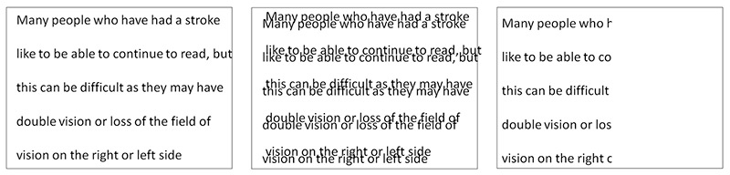
over things on the affected side. Reading can also be difficult (Figure
2).
Figure 1. Right homonymous hemianopia: the right-hand field of view is lost in both eyesFigure 2. Impact of vertical double vision (central image) and right hemianopia (right image) on readingVisual perceptual deficits are many and varied
affecting about a third of people with a stroke. Problems that may
develop include neglect one side of their body; difficulty recognising
faces or objects, or difficulties with colour vision, depth perception
and motion.5
Eye movement abnormalities can also be varied, including strabismus
(misaligned eyes), difficulty in converging the eyes to look at near
objects, or double vision due to the cranial nerves which control eye
movement being affected.6 Typical symptoms include double vision, or jumbled, blurred and/or juddery vision (Figure 2).
Impact
Blurred vision, double vision and lossand loss of visual field are
significant symptoms that impair daily functioning.7
The patient or their close relatives may report that they frequently
bump into objects such as door frames; have difficulty finding things on
surfaces; are unsure of their footing while walking and stumble; may
leave food uneaten on one side of the plate and have difficulty with
reading. Other impacts on the quality of life include loss of
confidence, fear of falling, fear of going out alone, social isolation
and loss of independence.8
How to assess visual function in someone who has had a stroke
Examination for visual loss is essential for stroke survivors.9 There are various assessment tools which can be used to examine visual function after a stroke:
Treatment options aim to restore visual function to as normal as possible.10 For eye movement abnormalities,prisms and patching one eye can be effective in reducing double vision.6
For visual field loss a Cochrane systematic review reports favourable
evidence of visual scanning training which aims to compensate for the
visual field loss.11 It is available as a paper training option (www.strokevision.org.uk) or through
computer training (www.eyesearch.ucl.ac.uk; www.readright.ucl.ac.uk.
Stroke survivors with persistent impairment of central vision may be
helped by low vision services which can include magnifiers, reading
aids, computerised adaptations and improved lighting.12
Furthermore, simple adaptations can be made by stroke survivors such as
using large print, ensuring good lighting at home, putting labels or
coloured stickers on cooking equipment, decluttering areas and having a
companion when going out, particularly in busy, crowded places.10
Conclusion
Post-stroke difficulties in visual function are an under-recognised
problem that cause significant impact to the quality of life of stroke
survivors. Carers and health workers need to be aware that problems with
vision are a common consequence of stroke that is not outwardly
obvious.
Assessment including visual functioning is best provided as part of a
multi-disciplinary team on acute stroke units, or in
neuro-rehabilitation units. A careful history about visual problems from
the patient and carers followed by examination of visual acuity, eye
movements and visual field are important in understanding the
difficulties in visual functioning.
Management should be tailored to each individual, their visual
difficulties and visual needs. With about one quarter of stroke
survivors being of working age, rehabilitation in the conext of
adaptation of the work place environment is vital if younger people are
to return to work after stroke. Rehabilitation requires patience and
perseverance on the side of the client, relatives and the health
provider.
Despite improvement in stroke prevention and acute stroke management,
the increasing ageing population will result in more stroke survivors
requiring rehabilitation. Policy makers need to understand the
importance of providing post-stroke rehabilitation services including
visual functioning.
Well there is NO standardized way to identify ANY stroke damage diagnosis because everyone uses impairment to try to guess where the damage was. Totally fucking useless. You have no way of knowing which of these 9 causes of deficits is responsible. Without knowing what caused the deficit your therapist is just whistling in the dark. We will never get stroke protocols properly correlated with damage until we get of objective damage diagnosis. Your doctor is totally incompetent for not coming up with a damage diagnosis and properly instructing your therapists in the stroke protocols that will get you back to 100%. Your doctor will whine and complain and give the fucking excuse: 'All strokes are different, all stroke recoveries are different'. If s/he gives you that load of bullshit, call up the hospital president and ask for someone competent. That probably means firing the complete stroke medical team, but we have to start cleaning out the deadwood sometime. http://www.news-medical.net/news/20160927/Review-highlights-urgent-demand-for-tool-to-identify-stroke-survivors-with-visual-impairments.aspx
A University of Liverpool led review of the methods available to
screen for post-stroke visual impairments has found there is an urgent
demand for the development of a tool.
Currently, over 65% of stroke survivors will suffer from a visual impairment while 45% of stroke units do not assess vision.
Visual impairment significantly reduces the quality of life of stroke
survivors with many being unable to return to work or drive and in some
cases results in depression. The impairments can also have an impact on
other people when it is not diagnosed or sufficiently communicated to
patients.
Quality of life
Identifying visual impairment after stroke can aid general
rehabilitation and thus, improve the quality of life for these patients.
The
review, led by postgraduate researcher Kerry Hanna from the
University's Institute of Psychology, Health and Society, examined the
available literature about current screening methods to accurately
identify stroke survivors with visual impairments.
This included reviews of randomised controlled trials, controlled
trials, cohort studies, observational studies, systematic reviews and
retrospective medical note reviews.
The review, published in 'The Journal of Disability and
Rehabilitation' today, found that there is currently no standardised
visual screening tool which can accurately assess all potential post
stroke visual impairments.
Visual defects missed
Kerry Hanna, said: "The current tools screen for only a number of
potential stroke-related impairments meaning many visual defects may be
missed.
"The sensitivity of those which screen for all impairments is
significantly lowered when patients are unable to report their visual
symptoms.
"Future research is required to develop a tool capable of assessing
stroke patients which encompasses all potential visual deficits and can
also be easily performed by both the patients and administered by health
care professionals in order to ensure all stroke survivors with visual
impairment are accurately identified and managed."
Objective:
To assess whether, following a right-hemisphere stroke, the combined
administration of computer-based cognitive rehabilitation and right
hemifield eye-patching in patients with visuo-spatial neglect is more
effective than computer-based cognitive rehabilitation alone. Methods: Twelve patients were randomized into two treatment groups: a single treatment group (n = 7) and a combination treatment group (n
= 5). In both cases, the treatment consisted of a mean number of 15
sessions, each lasting 1 hour. Visuo-spatial neglect was assessed using a
specific exploration protocol (Bell Cancellation Test, Figure Copying
of Odgen, Line Bisection, Baking Tray Task and Reading Task). The
functional effects of the treatment were assessed using the Catherine
Bergego Scale. Results: Significant between-group
differences were observed when comparing the pre- and post-treatment
scores for the Reading Task. No differences were observed in either
group in the Catherine Bergego Scale administered at baseline and at the
final intervention.
Conclusion: The results
obtained do not allow one to conclude that the combination treatment
with cognitive rehabilitation and right hemifield eye-patching is more
effective than cognitive rehabilitation alone. Although partial
improvement in the performance of neuropsychological tests was observed,
this improvement is not present at functional level.
You can see how long before your doctor recommends this for you. I'm betting never. http://www.alphagalileo.org/ViewItem.aspx?ItemId=141013&CultureCode=en Humans view the world through two eyes, but it is our brain that
combines the images from each eye to form a single composite picture. If
this function becomes damaged, impaired sight can be the result. Such
loss of visual function can be observed in patients who have suffered a
stroke or traumatic brain injury or when the oxygen supply to the brain
has been reduced (cerebral hypoxia). Those affected by this condition
experience blurred vision or can start to see double after only a short
period of visual effort. Other symptoms can include increased fatigue or
headaches. It is been suggested that these symptoms arise because the
brain is unable to maintain its ability to fuse the separate images from
each eye into a single composite image over a longer period. Experts
refer to this phenomenon as binocular fusion dysfunction. ‘As a result, these patients have significantly reduced visual
endurance,’ explains Katharina Schaadt, a graduate psychology student at
Saarland University. ‘This often severely limits a patient’s ability to
work or go about their daily life.’ Working at a computer screen or
reading the newspaper can be very challenging. As binocular fusion is a
fundamental requirement for achieving a three-dimensional impression of
depth, those affected also frequently suffer from partial or complete
stereo blindness. ‘Patients suffering from stereo blindness are no
longer able to perceive spatial depth correctly,’ says Schaadt. ‘In
extreme cases, the world appears as flat as a two-dimensional picture.
Such patients may well have difficulties in reaching for an object,
climbing stairs or walking on uneven ground.’ Although about 20% of stroke patients and up to 50% of patients with
brain trauma injuries suffer from these types of functional impairments,
there is still no effective therapy. Researchers at Saarland University
working with Anna Katharina Schaadt and departmental head Professor
Georg Kerkhoff have now developed a novel therapeutic approach and have
examined its efficacy in two studies. ‘Test subjects underwent a six
week training program in which both eyes were exercised equally,’
explains Schaadt. The aim was to train binocular fusion and thus improve
three-dimensional vision. Participants in the study were presented with
two images with a slight lateral offset between them. By using what are
known as convergent eye movements, patients try to fuse the two images
to a single image. This involves directing the eyes inward towards the
nose while always keeping the images in the field of view. With time,
the two images fuse to form a single image that exhibits stereoscopic
depth, i.e. the patient has re-established binocular single vision. More at link.
Once again researchers don't know what cause and effect is. If you want to know what possibilities for easy recovery are you have to map out the penumbra damages, probably via PET scans.
And do they possibly think that 'cold spots' are areas of completely dead neurons? What is the solution for bringing back the function those dead areas represent?
According to Study Published in Restorative Neurology and Neuroscience
Scientists know that vision restoration training (VRT) can help
patients who have lost part of their vision due to glaucoma, optic nerve
damage, or stroke regain some of their lost visual functions, but they
do not understand what factors determine how much visual recovery is
achieved.
New evidence published in Restorative Neurology and Neuroscience
suggests that vision restoration depends mostly on activity of residual
vision that is still left after the injury and that both local neuronal
activity and activity in the immediate surround influence the
development of visual recovery “hot spots.” This shows that recovery of
vision is mediated by partially surviving neurons.
Researchers from the Institute of Medical Psychology and Department
of Computer Sciences, Otto-von-Guericke-University of Magdeburg, and the
Max Planck Institute for Dynamics and Self-Organisation, Goettingen,
Germany, conducted a retrospective analysis of multiple visual field
tests before and after at least six months of VRT in 32 stroke patients
with hemianopia, which is a loss of vision in half of the visual field.
The test, known as high-resolution perimetry (HRP), presents visual
stimuli on a computer monitor to which the patient has to respond by
pressing a key on the keyboard.
The result is a map that indicates areas that are intact (unaffected
by the injury), areas that are completely blind, and “areas of residual
vision,” where vision is reduced but not absent. Here, the response time
is slower or the correct response occurs only occasionally. Repetitive
stimulation through daily one-hour vision training with VRT was directed
at these “areas of residual function” to strengthen their performance.
“Hot spots” were defined as those locations that were initially
impaired at baseline but then recovered after VRT training, while “cold
spots” remained impaired where vision training did not help. Of almost
11,000 visual spots analyzed from the 23 patients, 688 were found to be
hot spots while 3,426 were cold spots. The average absolute improvement
due to VRT training was 6%.
The investigators used computer-based data mining technology to study
which features of the baseline HRP charts obtained before vision
training could predict vision recovery. They looked at different
topographic features and found that visual field areas have a higher
probability of becoming vision restoration “hot spots” if they had
higher local residual vision at baseline, more residual activity in a
spatially limited surrounding area (of 5 degrees of visual angle), and
if they were located closer to the blind field (scotoma). Vision
restoration was not influenced much by residual activity at further
distances, say the authors.
“Our findings confirm the special role of residual structures in
vision restoration, which is likely mediated by surviving cells in
partially damaged brain tissue,” says lead author Bernhard A. Sabel,
PhD, of the Institute of Medical Psychology,
Otto-von-Guericke-University of Magdeburg. Dr. Sabel suggests that the
massive visual stimulation presented during VRT enhances visual recovery
by forcing subjects to focus their attention on “compromised” sectors
of the visual field which are partially damaged and repeating this daily
helps recover vision loss. “This new understanding now allows us to
offer vision training on the internet through online training,” says Dr.
Sabel.
Contact if interested. http://online.wsj.com/article/PR-CO-20130604-905592.html?mod=googlenews_wsj
EBS Technologies GmbH, an emerging medical device company developing
the revolutionary NEXT WAVE(TM) brain stimulation platform for treatment
of vision deficits caused by neurological disorders, announced today
the results of a multi-center, 82-patient clinical trial of its NEXT
WAVE(TM) brain stimulation device.
About one-half of the clinical trial patients were given a 40-minute
treatment protocol for 10 consecutive days with the NEXT WAVE(TM)
device. With a mean increase of 24%, NEXT WAVE(TM)-treated patients
showed significantly better improvements in stimulation of their total
visual field compared to patients in the control group, which did not
receive NEXT WAVE(TM) stimulation. All patients had vision impairment
lasting at least six months prior to the clinical trial and had
exhausted all standard therapeutic options to improve their vision.
"There is a huge unmet clinical need for the treatment of vision
impairment caused by a variety of different neurological disorders, such
as neuropathy of the optic nerve, brain injury or stroke. For example,
stroke alone is the world's third-leading cause of death as well as the
leading cause of serious, long-term disability. There are more than 10.4
million stroke survivors with long-term disability in Europe and the
U.S. Three out of five of these disabled persons are potentially
treatable with our NEXT WAVE(TM) therapy once we commence
commercialization," said Ulf Pommerening, CEO of EBS Technologies.
"We look forward to expanding the scientific evidence for the efficacy
of our NEXT WAVE(TM) approach, especially in stroke patients, in an
expanded clinical trial that will commence imminently," said
Pommerening.
Background. Patients with right or more
rarely left parietotemporal lesions after stroke may have profound
visuospatial disorders that
impair activities of daily living (ADL) and
long-term outcome. Clinical studies indicate improvements with
systematic training
of perception. Studies of perceptual learning in
healthy persons suggest rapid improvements in perceptual learning of
spatial
line orientation with partial transfer to
nontrained line orientations. Objective. The authors investigated a novel feedback-based perceptual training procedure for the rehabilitation of patients after stroke.
Methods. In an uncontrolled trial, 13
participants showing profound deficits in line orientation and related
visuospatial tasks within
12 to 28 weeks of onset performed repetitive
feedback-based, computerized training of visual line orientation over4
weeks
of treatment. Visual line-orientation
discrimination and visuospatial and visuoconstructive tasks were
assessed before and
after training. Results. The authors found (a) rapid improvements in trained but also in nontrained spatial orientation tests in all 13 participants, partially up to a
normal level; (b) stability of the obtained improvements at 2-month follow-up; (c) interocular transfer of training effects to the nontrained eye in 2 participants suggesting a central, postchiasmatic locus
for this perceptual improvement; and (d)
graded transfer of improvements to related spatial tasks, such as
horizontal writing, analog clock reading, and visuoconstructive
capacities but no transfer to unrelated measures of
visual performance. Conclusions. These results suggest the potential for treatment-induced improvements in visuospatial deficits by feedback-based, perceptual
orientation training as a component of rehabilitation after stroke.
Introduction. Limited evidence suggests that visual impairments may influence outcome after stroke. The degree of recovery from these
impairments is poorly characterized. Objectives. To describe recovery and to determine whether visual impairments influence functional outcome and quality of life. Methods.
We extracted demographic and outcome data from the Virtual
International Stroke Trials Archive (VISTA). We examined horizontal
eye movement disorders and hemianopia using the
Best Gaze and Visual domains of the National Institutes of Health Stroke
Scale
(NIHSS) and described recovery at 30 and 90 days.
Proportional odds modelling was used to examine the association between
impairments at baseline, modified Rankin Scale
(mRS), and European Quality of Life Score (EQ-5D) at 90 days. Results.
Visual impairments were reported in 7,204/11,900 (60.5%) patients at
baseline. Complete recovery occurred in 1,398/3,285
(42.6%) and 3,243/7,204 (45.0%) patients by 30 and
90 days respectively. The burden of persistent visual impairment in
survivors
was 1,135/4,028 (28.2%) at 30 days and 1,915/9,338
(20.5%) at 90 days. Partial gaze palsy (P less than .0001; OR = 0.81; 95% CI =
0.74-0.87), forced deviation (P less than .0001; OR =
0.48; 95% CI = 0.43-0.53), and complete homonymous hemianopia (P less than
.0001; OR
= 0.67; 95% CI = 0.62-0.73) at baseline were
associated with poor mRS at 90 days. Conclusions. The rate of
recovery was greater in the first month after stroke, suggesting a
potential time frame for interventions. The
associations between visual impairments and poor
mRS suggest that these impairments should be considered in
multidisciplinary
assessments and interventions.
A.M. Barrett, MD, of Kessler Foundation received a grant totaling
$595,756 to study the effects of prism adaptation therapy for spatial
neglect in survivors of right-sided stroke.
The title of the 3-year grant from National Institute on Disability and
Rehabilitation Research (NIDRR) is 'Impact of Prism Adaptation Therapy
for Spatial Neglect on Home and Community Outcomes' (H133G120203). Dr.
Barrett, an expert in hidden disabilities such as spatial neglect, is
director of Stroke Rehabilitation Research at Kessler Foundation.
"At Kessler Foundation, we recognize that cognitive deficits are a
major obstacle to rehabilitation of stroke survivors, as well as to
those with brain injury and multiple sclerosis,"
said John DeLuca, PhD, vice president for Research and Training. "By
focusing attention on the cognitive effects of stroke that often go
undetected and untreated, Dr. Barrett's research will improve
rehabilitation outcomes for individuals with hidden disabilities."
For some of you this might be useful so ask your doctor why in the past 2 years this hasn't been brought to your attention. http://www.naric.com/research/rehab/record.cfm?search=2&type=all&criteria=J64152&phrase=no&rec=119226 Author(s):Berryman, Amy; Rasavage, Karen; Politzer, Thomas. Publication Year: 2010. Number of Pages: 8. Abstract: Article describes remedial and
compensatory interventions for visual field loss and visual inattention.
Interventions are discussed in the context of an interdisciplinary
neurorehabilitation team that consults with a neurooptometrist or other
visual doctor with experience in TBI and stroke rehabilitation. This
article is intended to promote improved interdisciplinary collaboration
in the treatment of visual impairments, and to give practical, easy to
implement options for clinicians to use in daily practice.
Background and Purpose. Compensatory and
restorative treatments have been developed to improve visual field
defects after stroke. However, no controlled
trials have compared these interventions with
standard occupational therapy (OT). Methods. A total of 45
stroke participants with visual field defect admitted for inpatient
rehabilitation were randomized to restorative
computerized training (RT) using computer-based
stimulation of border areas of their visual field defects or to a
computer-based
compensatory therapy (CT) teaching a visual search
strategy. OT, in which different compensation strategies were used to
train
for activities of daily living, served as standard
treatment for the active control group. Each treatment group received 15
single sessions of 30 minutes distributed over 3
weeks. The primary outcome measures were visual field expansion for RT,
visual
search performance for CT, and reading performance
for both treatments. Visual conjunction search, alertness, and the
Barthel
Index were secondary outcomes. Results.
Compared with OT, CT resulted in a better visual search performance, and
RT did not result in a larger expansion of the
visual field. Intragroup pre–post comparisons
demonstrated that CT improved all defined outcome parameters and RT
several,
whereas OT only improved one. Conclusions.
CT improved functional deficits after visual field loss compared with
standard OT and may be the intervention of choice
during inpatient rehabilitation. A larger trial
that includes lesion location in the analysis is recommended.
The abstract didn't tell me anything, you may have to get your doctor to request the article. http://content.karger.com/ProdukteDB/produkte.asp?Doi=337016 Abstract Objective: Visual field defects (VFD) after stroke can cause significant disability and reduction in quality of life. Adequate diagnosis of VFD and referral to visual rehabilitation are important to improve outcome. Our aim was to conduct a retrospective clinical audit to investigate how neurologists detect and follow up VFD in stroke patients in a university hospital in Norway. Methods: All patients registered in the Bergen NORSTROKE Registry from February 2006 to May 2009 with (1) occipital lobe infarctions and (2) non-occipital infarction and clinically detected VFD were included in the study. Their medical records were reviewed for referral to perimetry for examination of VFD and for referral to a visual rehabilitation program within the first year after brain injury. Results: Of 353 patients, 34 (9.6%) were referred to perimetry and 8 (2.3%) to visual rehabilitation. Patients referred to perimetry were younger (65.1 vs. 74.7 years, p < 0.001), had lower modified Rankin Scale scores (2.53 vs. 3.47, p = 0.003), and scored lower on the National Institutes of Health Stroke Scale upon admission (6.68 vs. 13.90, p < 0.001). Men were more often referred to perimetry than women (73.5 vs. 26.5%, p < 0.001), and those referred were younger (61.2 vs. 75.8 years, p = 0.03). Conclusions: Only few patients were referred to perimetry, and even fewer were offered visual rehabilitation. Age and gender were negative predictors for referral. Neurologists’ awareness of the significant disability related to VFD must be increased. Focused diagnostics on visual impairment and early referral to a visual rehabilitation program should be mandatory in stroke unit services.