Showing posts with label stroke medical 'professionals'. Show all posts
Showing posts with label stroke medical 'professionals'. Show all posts
Wednesday, September 30, 2026
14 million views of this blog
I must still be interesting to some people. I have never received
any direct communications from any stroke medical 'professionals'. Are
they scared to talk to me?
First million took from Aug. 2010 to Sept. 2014
Fourth million from March 2017 to November 2018
Seventh million was from June 2024 to January 2025.


One common midlife condition linked to 42% higher dementia risk
Your competent? doctor is RESPONSIBLE for your 100% recovery so you maintain your muscle strength.
CANT DO THAT; PURE INCOMPETENCE!
One common midlife condition linked to 42% higher dementia risk
I was in excellent shape at 50 when the stroke occurred, biking 4 miles each way commuting to work 9 months of the year in Minneapolis. March-November, sometimes biking home in the snow, had to put a rain cap on my helmet to stop the cold air from blowing thru the vents in the helmet onto my bald head.
3 years post stroke at a physical I had a resting heart rate of 54 at age 53, level of an athlete. My doctor asked what exercises I was doing; 'I've done no exercises for the past 3 years'. And now 20 years past the stroke my fitness has declined a bit, ALL BECAUSE MY STROKE MEDICAL 'PROFESSIONALS' COMPLETELY FAILED AT GETTING ME 100% RECOVERED!
A common midlife condition affecting many older adults could significantly raise an individual’s risk of dementia, according to new research.
Published in International Journal of Food Sciences and Nutrition, the study found that maintaining muscle strength as a person navigates midlife is crucial in the fight against dementia when they age.
An important warning sign’
That finding is somewhat complicated, since another major risk factor for dementia is obesity. If a person loses weight without strength training, they are likely to lose muscle as well.
Counterintuitively, that could increase their dementia risk, even as their body fat goes down.
“It’s not just about how much you weigh—maintaining muscle strength and function also appears to be important as we age,” lead researcher Dr. Uraiporn Booranasuksakul from Burapha University said in a press release.
“Our findings suggest losing muscle could be an important warning sign for dementia risk.”The researchers found that sarcopenia—the medical term for loss of strength and muscle—was associated with a 42-percent greater risk of dementia. Sarcopenia often occurs when a person gets older.
Interestingly, the researchers found that obesity before the age of 65 was linked to a nine-percent greater risk of dementia.
However, after an individual turns 65, obesity was linked to a 17-percent lower risk—a finding that showed how crucial strength is as a person ages.
“We found that obesity in middle age was linked to a higher risk of dementia, but this relationship changed in later life,” study corresponding author Mario Siervo said in the release.
“This does not mean people should gain weight as they get older. Rather, it shows us that the relationship between body weight, aging and dementia is complex.”
‘A lot we don’t know’
Siervo added that the relationship between weight loss, muscle loss and dementia is complex, and more research is needed.
“There is still a lot we don’t know about how changes in muscle and body fat interact over time,” Siervo said.
The research is especially relevant as weight-loss drugs like Ozempic and Mounjaro sweep through the market.
Studies linking weight loss to lower risk of dementia suggest that GLP-1s could be beneficial for an aging population, especially in the United States where the CDC says two in five adults experience obesity.
Other research shows that almost half of dementia cases could be prevented by lifestyle changes.
Still, the latest study suggests that weight loss alone doesn’t tell the whole story.
“Maintaining muscle is an important part of healthy aging, and regular physical activity and good nutrition can help support muscle strength and function,” Booranasuksakul said.
Newsweek has reached out to the study authors for comment via email.
Related Articles
Monday, September 21, 2026
A happier retirement contains these 3 elements
Are you recovered enough to be happy? Or did your incompetent? doctor fail you?
My resilience story: I would still be leading a life of quiet desperation if still married.
(Life is definitely better as I age, I got divorced enhancing my happiness immeasurably. I'm retired and comfortably well off. And healthy as I can be post stroke. I'm going to live a long time yet.)
A happier retirement contains these 3 elements
What’s the secret of a rich, rewarding retirement?
It’s as simple as ABC: agency, belonging and cause.
That’s how Bruce Feiler, author of “A Time to Gather” and other books, spells out the three factors that give life meaning. “Agency” is wielding power and autonomy in choosing how to live; “belonging” is forging social ties with others; and “cause” is serving a higher purpose.
With these three ingredients in place, the odds of a satisfying retirement soar, he says. By making intentional decisions aligned with your goals and values, leading an active social life and contributing to a cause that elevates your spirit, you’re bound to feel more happy and fulfilled.
“It’s a tool kit for navigating retirement,” Feiler said. “It’s the same for navigating any life transition, like an illness, a move or the loss of a loved one.”
Many retirees struggle to find purpose or structure once they’re done with the routine of a traditional job, which can lead to loneliness and isolation. As people live longer, it’s not unusual for retirement to last 20 to 30 years.
Here’s how to assess how well you know and apply these three critical elements:
Agency: Examine how much control you feel you have over your everyday life. By choosing “what we are, what we create and what we do,” as Feiler says, we’re more apt to serve as our own director, rather than letting external factors direct us.(I'm completely in control, know enough to say no when needed.)
Related video: 8 simple ways to make the most of retirement and enjoy every day (En Pareja)
Cause: Explore how deeply you believe in something larger than yourself. Does your desire to give back or tackle societal problems energize you? Do you pounce on volunteer opportunities? When donating your time to make a difference or improve the lives of others, do you feel fully alive and happy?
(Trying to get stroke solved to 100% is a humongous challenge! Getting through the fossilized brains of stroke medical 'professionals' I haven't figured out yet.
And I think a lot of funerals will need to occur before we get the right strategy and leadership to GET THERE!
Even if all three elements are present in your life, try adding more, the author says.
“Pick a personal project for A, B and C,” Feiler said. “I’m tempted to say start small, but some people like to start big.”
To gain more agency, for example, you can tend the garden for an hour or write your autobiography. Let your motivation level determine the size and scope of the project.
At the same time, cut yourself some slack. As you age, you may face limitations on your ability to exert agency, socialize often and embrace a cause.
“The ABCs are very important,” said Teresa Amabile, a professor emerita at Harvard Business School. “But they’re not of equal importance.”
One focus plays a paramount role in enhancing your retirement years: belonging.
The biggest predictor of longevity and happiness is the quality of our relationships, according to the Study of Adult Development, which has tracked participants and their descendants for nearly 90 years.
“Belonging is very critical,” said Amabile, co-author of “Retiring: Creating a Life That Works for You.” “Relationships make a huge difference in life satisfaction. That’s certainly very true in retirement.”
Even if you were somewhat lonely or less satisfied with your past relationships, it’s not too late to find more comradeship and support in retirement. Maintaining an openness to people — and a willingness to strengthen connections with everyone from distant or estranged family members to friends, acquaintances and strangers — enables you to benefit from more social interaction.
Read: The future of retirement? Work until you die.
The obstacles of the alphabet
It’s not always easy to follow through with your ABCs. Obstacles abound.
Amabile warns that we may lose some agency despite our best efforts.
“There’s only so much control we can exert over our lives and circumstances,” she said. “So it’s necessary to figure out when we can’t control things and find out how to adapt.”
Moreover, it takes confidence and positivity to pilot your life. You need to believe that you can wield control over your choices and resist outside forces that try to influence you.
To forge better social connections, weigh whether shyness, cynicism or snap judgments of others hold you back.
“People have gremlins,” said Barbara Waxman, a San Francisco-based gerontologist and coach. “There are naysayers on our shoulders who hold us back, that voice of self-doubt.”
“If you get a social invitation, recognize the moment as a decision point,” said Patrice Jenkins, a retirement consultant and author of “What Will I Do All Day?” “Don’t default to say ‘no’ and settle into what’s comfortable.”
You can’t flip a switch and apply the ABCs all at once. Retirement unfolds in stages. But by making strides in integrating agency, belonging and cause into your life, you pave the way for a smooth transition.
Read: Couples with a big age gap need more money — they have to plan for two retirements
Effects and mechanisms of transcutaneous auricular vagus nerve stimulation on post-stroke cognitive impairment in animal studies: a systematic review and meta-analysis
Why this animal study? Don't we have enough human research out there to create protocols with? Never mind, there aren't enough functioning neurons in your stroke medical 'professionals' to accomplish that! You all need to be fired!
- vagus nerve
(56 posts to July 2012)
Effects and mechanisms of transcutaneous auricular vagus nerve stimulation on post-stroke cognitive impairment in animal studies: a systematic review and meta-analysis
Shuying Li
Xiaping Chen
Sihang Chen
Fu Liu
Jinlong Yin
Wenhao Hu
Jiang Xu
Xuejing Li *


- The Affiliated Huai'an Hospital of Xuzhou Medical University, Huai'an, China
Abstract
Background:Post-stroke cognitive impairment (PSCI) substantially undermines rehabilitation adherence and poses a major obstacle to functional recovery. Transcutaneous auricular vagus nerve stimulation (taVNS), a noninvasive neuromodulation technique, has the potential to improve cognitive function, as demonstrated in various clinical and preclinical studies. Against this backdrop, we performed a meta-analysis of taVNS in experimental stroke models to quantify its cognitive benefits and probe the biological mechanisms involved in its effects.
Methods:
We systematically searched databases, including PubMed, The Cochrane Library, Embase, and Scopus, to identify controlled animal studies evaluating taVNS in PSCI models. Following eligibility screening, five studies were selected for final synthesis. The primary cognitive outcomes comprised escape latency in the Morris water maze and the Barnes maze. Data extraction was performed independently by two investigators, and statistical analyses were performed using Review Manager 5.4 software.
Results:
Five RCTs involving 158 rodents were analyzed in this study. The meta-analysis revealed that taVNS significantly improved memory function in PSCI animals (SMD = 1.85, 95% CI: 1.06 to 2.64, p < 0.01, I 2 = 57%) and learning ability (MD = − 6.38, 95% CI: − 8.87 to − 3.89, p < 0.01, I 2 = 0%) and reduced modified neurological severity scores (MD = − 1.08, 95% CI: − 1.63 to − 0.52, p < 0.01, I 2 = 38%). Mechanistically, taVNS significantly elevated acetylcholine levels (SMD = 1.57, 95% CI: 0.85 to 2.30, p < 0.01, I2 = 0%), inhibited neuronal apoptosis (SMD = 2.29), and promoted angiogenesis (SMD = 1.63). Sensitivity analysis confirmed the robustness of the primary outcomes.
Conclusion:
This study provides preclinical evidence confirming that taVNS can effectively improve cognitive deficits in PSCI animal models. The enhancement of Ach levels, inhibition of neuronal apoptosis, and promotion of vascular repair may be related to the non-neuronal cholinergic system (NNCS), anti-inflammatory, and antioxidant stress mechanisms. However, as only a few studies were included, more high-quality animal experiments are needed in the future to develop standardized interventions and further explore the molecular mechanisms underlying these effects.
Systematic review registration:
https://www.crd.york.ac.uk/prospero/display_record.php?ID=CRD420261329420.
Can AI Turn Stroke Rehab Into A More Engaging Recovery Journey?
Wrong question! Can AI deliver a guaranteed recovery? Doesn't anyone in stroke know that 100% recovery is the only goal in stroke? You can't let your stroke medical 'professionals' decide what is your recovery endpoint. They work for you. If they are incompetent at their job, GET THEM FIRED!
Can AI Turn Stroke Rehab Into A More Engaging Recovery Journey?
BeAble Health’s Habib Ali discusses how AI and gamified therapy can improve patient engagement, personalise rehabilitation and support functional recovery
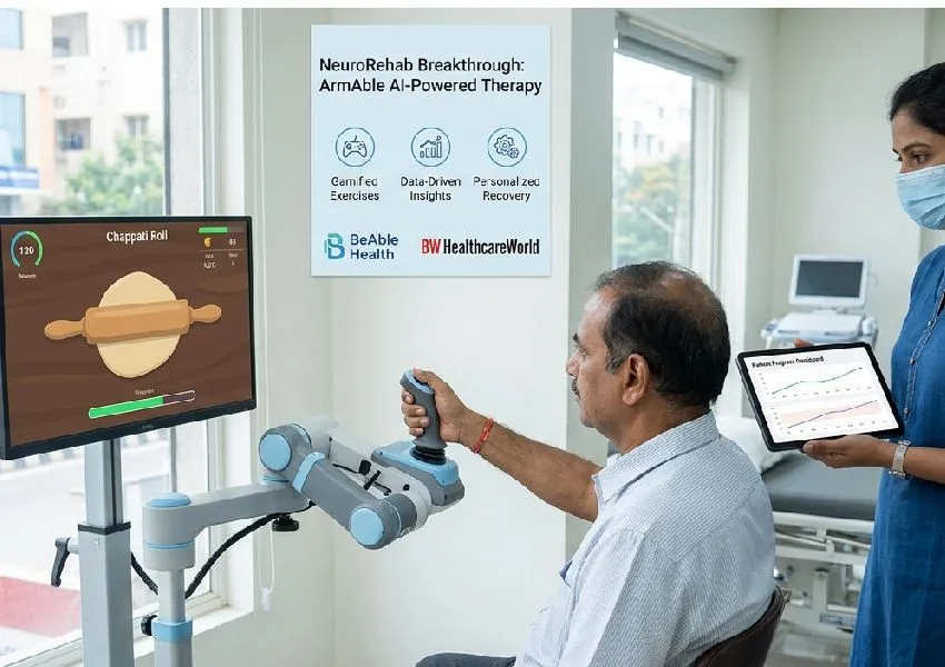
Stroke remains a leading cause of death and long-term disability worldwide. According to the World Stroke Organization’s Global Stroke Fact Sheet 2025, 11.9 million people experienced a new stroke in 2021, while more than 93 million were living with its consequences. The report identifies rehabilitation as a critical part of recovery, although access to multidisciplinary services remains limited, particularly in low- and middle-income countries. India faces a significant rehabilitation gap. A WHO Systematic Assessment of Rehabilitation Situation (STARS) review reported that the country has around 1,000 physiatrists and only 150 designated rehabilitation facilities. Nearly 40per cent of stroke survivors experience moderate-to-severe disability, while about 50per cent develop spasticity. Data from the Longitudinal Ageing Study in India also found that although stroke prevalence among adults aged 45 and above was 1.8per cent, only around 40per cent of survivors had accessed physiotherapy or occupational therapy.Technology-assisted rehabilitation could help address some of these challenges. A systematic review and meta-analysis of 42 trials involving 1,760 participants found that serious-game interventions led to significantly greater improvements in upper-limb motor function than conventional therapy, with a standardised mean difference of 0.47. However, researchers noted that the quality and certainty of evidence varied, underscoring the need for larger, well-designed clinical trials. Against this backdrop, BeAble Health has developed ArmAble, a game-based, AI-enabled rehabilitation device that turns repetitive exercises into familiar activities such as dosa making, chapati rolling and mosquito swatting. Designed for stroke survivors, spinal cord injury patients and people with neurological conditions, the device aims to improve engagement while enabling physiotherapists to track movement, personalise therapy and assess progress. In an exclusive conversation with BW HealthcareWorld, Habib Ali, CEO and Co-founder, BeAble Health, discusses how ArmAble is making neurological rehabilitation more engaging, personalised and data-driven while supporting physiotherapists in monitoring progress and functional recovery.Here are the excerpts from the conversation: How does BeAble’s ArmAble make rehabilitation more engaging and patient-driven, and what impact has it had on motivation and recovery?(Motivation is incredibly easy to solve! EXACT 100% RECOVERY PROTOCOLS! Your patient will gladly do the millions of reps needed because they are looking forward to recovery! Can't see that? YOU'RE FIRED!) One of the biggest challenges in rehabilitation is keeping patients motivated over weeks and months of repetitive therapy. Our device ‘ArmAble’ addresses this by converting conventional exercises into relatable game-based activities that closely resemble everyday tasks. For example, we have activities such as making dosas, rolling chapatis, swatting mosquitoes, balloon pop, table tennis, etc. These games are not designed purely for engagement. Every movement is linked to a rehabilitation goal such as improving movement, coordination and muscle control. Since these activities closely resemble movements patients perform in their daily lives, the skills developed during therapy are more likely to translate into functional independence. Rather than practising isolated movements, patients are rehearsing tasks they hope to return to after recovery. We have closely seen the impact of this gamified approach. Our clinical trial involving 120 individuals with acute and subacute stroke, demonstrated that patients receiving ArmAble-assisted gamified rehabilitation alongside conventional physiotherapy achieved significantly greater improvements in upper-limb motor function compared with conventional task-based rehabilitation alone. Unlike largely passive robotic systems, ArmAble requires active patient participation. Why is active engagement critical in neurological rehabilitation, and how does it influence long-term functional recovery? Neurological recovery depends on active participation. The brain relearns movement through repeated, task-oriented practice rather than passive movement alone. That is why every ArmAble activity requires patients to perform functional movements that resemble daily life. Whether rolling chapatis, swatting mosquitoes or playing rehabilitation games, each activity has a therapeutic objective. For instance, the Balloon Popping game is designed to improve functional reaching and shoulder rehabilitation. Virtual balloons appear at different heights and positions, requiring patients to reach in multiple directions. This encourages shoulder flexion, abduction, elbow extension, trunk control and multidirectional reaching while reducing compensatory movement patterns commonly seen after stroke. The unpredictable balloon placement also requires attention, visual scanning, spatial awareness and movement accuracy. The game enables significantly more repetitions than conventional reaching exercises, which is essential for cortical reorganisation and motor recovery. Throughout the session, ArmAble’s AI analyses movement range, reach distance, trajectory, smoothness, reaction time and fatigue, enabling therapists to objectively assess performance and adjust rehabilitation intensity to individual needs. >AI is not replacing physiotherapists; it is strengthening clinical decision-making. ArmAble analyses movement quality, range of motion, coordination, reaction time and task performance, providing therapists with objective data alongside clinical observation. The system also automatically adjusts task difficulty according to the patient’s recovery stage, keeping therapy appropriately challenging. This helps therapists personalise rehabilitation, monitor progress objectively and make better-informed clinical decisions, while retaining the clinical judgement, hands-on guidance and motivation that technology cannot replace. One of the biggest challenges in neurorehabilitation remains the shortage of trained professionals and the limited time therapists can spend with each patient. How does ArmAble's AI-driven monitoring and real-time performance tracking help physiotherapists supervise multiple patients without compromising the quality or personalisation of care? One of the advantages of ArmAble is that it automates routine performance monitoring while keeping the therapist at the centre of care. The platform continuously tracks movement quality, reaction time, coordination, range of motion and task performance, giving therapists real-time insights into each patient's progress. All this data is tracked and recorded. It allows therapists to supervise multiple patients more efficiently, while using objective data to personalise treatment and adjust therapy as patients improve. Instead of spending time manually measuring progress, therapists can focus on assessment, movement quality and patient interaction. India faces a growing burden of stroke, spinal cord injuries and neurological disorders, yet rehabilitation remains secondary to acute care. What changes are needed to make rehabilitation an integral part of the healthcare continuum? One of the biggest challenges is awareness. Patients and families lose motivation to continue rehab over longer periods because recovery is a long-term process. What’s important is that for neurological rehabilitation, the brain continues to respond to intensive, task-oriented rehabilitation, particularly in the early months. Patients have to adhere to long term schedules to ensure that rehab is effective. We need greater awareness that rehabilitation is not optional—it's an essential part of treatment. Secondly, rehabilitation needs to become more integrated into the healthcare system. Neurologists, neurosurgeons, rehabilitation physicians, physiotherapists, occupational therapists, speech therapists and psychologists should work together from the beginning of the patient's recovery journey. Hospitals also need structured rehabilitation pathways so that every neurological patient has a clear plan for continuing therapy after discharge.
How do you see AI transforming rehabilitation in India? Do you believe technologies like ArmAble will become a standard part of neurological care?
We believe AI will make rehabilitation more engaging, data driven and measurable. It can automatically adapt therapy to each patient's recovery stage, provide objective insights into progress, and support remote monitoring, making quality rehabilitation available to more people.
At the same time, AI should be viewed as an enabler, not a replacement for physiotherapists. Rehabilitation is fundamentally a human-centred process that depends on clinical expertise, empathy and patient motivation. As more clinical evidence emerges, technologies like ArmAble are likely to become an integral part of neurological rehabilitation because they help therapists deliver more engaging, data-driven and effective care.
Saturday, September 19, 2026
Precision rehabilitation, from foundational concepts to contemporary perspectives: a mixed-methods scoping review
For stroke this requires:
- An objective damage diagnosis, a 3d rendering of dead and damaged gray and white matter. mRS and the Berthel Index ARE NOT DAMAGE
DIAGNOSES, they do not give you the 3d location of your dead and
damaged neurons. All problems relate to those damaged or dead neurons so if you don't know what you are dealing with you CAN'T FIX ANYTHING WITH PRECISION!
- EXACT REHAB PROTOCOLS; numbers and movement or thought patterns.
Don't worry, nothing will occur, your stroke medical 'professionals' haven't done any thinking since entering medical school.
Precision rehabilitation, from foundational concepts to contemporary perspectives: a mixed-methods scoping review
Abstract
Background
Precision health approaches aim to predict, deliver and optimize the right intervention, at the right time, for the right person. While precision medicine is well-established, its application in rehabilitation remains nascent. This study aimed to (1) Map foundational concepts of precision rehabilitation by describing the extent, scope and nature of the literature, identifying similarities and differences with precision medicine, and describing the role of technologies and artificial intelligence (AI); (2) Describe knowledge users’ perspectives on precision rehabilitation; and (3) Integrate quantitative and qualitative results to develop a working definition of precision rehabilitation.
Methods
A convergent mixed-methods design was used to conduct a scoping review with a qualitative consultation phase. The scoping review followed Johanna Briggs Institute methodological recommendations and PRISMA-ScR reporting guidelines. Five databases were searched for adjacent keywords ‘precision’ AND ‘rehabilitation’. Data were extracted and analyzed descriptively and thematically. In parallel, individual interviews were conducted with knowledge users at three North American rehabilitation centers. Transcripts were analyzed using deductive content analysis. Mixed method integration synthesized and contrasted literature-derived and knowledge users-derived insights.
Results
Forty-nine articles met inclusion criteria; 26 (53%) were primary research studies, while 23 (47%) were non-empirical publications. Amongst research studies, 14 (54%) involved technology use, while 5 (19%) used AI. Seven (14%) articles included a definition of precision medicine, while 22 (45%) included a definition of precision rehabilitation. Interviews with 16 knowledge users highlighted that personalization is uniquely expressed in precision rehabilitation as compared to medicine and identified rehabilitation-specific facilitators and barriers to precision approaches. Data integration confirmed that a focus on function differentiates precision approaches in rehabilitation from medicine, clarified rehabilitation-specific logistical barriers to technology-enabled data collection in precision approaches, and attributed limited AI use to concerns about predictions based solely on data that excludes psychosocial factors and population heterogeneity.
Conclusions
Results indicate greater similarities than differences between precision approaches in rehabilitation and medicine, while highlighting rehabilitation’s emphasis on personalization, function and participation. Findings identify key barriers and facilitators and support a proposed definition of precision rehabilitation for further refinement through peer consensus to accompany advances in research and clinical practice.
Sunday, September 6, 2026
Counting twitches: automated mechanomyography of muscle fatigue in healthy adults
Do your stroke medical 'professionals' have two functioning brain cells that will be used to objectively determine your fatigue and then measure the recovery you get from their EXACT RECOVERY PROTOCOLS?
NO? So you have blithering idiots in charge! You'll never get recovered with them! GET THEM FIRED!
Counting twitches: automated mechanomyography of muscle fatigue in healthy adults
Abstract
Background
Assessing skeletal muscle fatigue is essential for diagnosing neuromuscular impairment, but conventional methods rely on maximal or tetanic contractions that can be impractical or uncomfortable in clinical populations. Surface mechanomyography (MMG) provides a non-invasive alternative; however, current MMG-based fatigue protocols require time-consuming manual peak-to-peak analysis. This study evaluated an automated algorithm for extracting MMG-derived fatigue metrics from electrically evoked muscle twitches.
Methods
Eighteen healthy adults completed a standardized fatigue protocol on the wrist extensors and ankle dorsiflexors using electrical stimulation at 2, 4, and 6 Hz over 9 min. A triaxial accelerometer captured MMG signals from > 2,100 contractions per muscle (approximately 4,320 per participant across the two muscles). A custom algorithm automatically extracted peak-to-peak values and computed the endurance index; the mean contraction amplitude was derived from peak and trough points identified manually by the research team. Repeated-measures ANOVAs assessed differences across stimulation frequencies and muscle groups.
Results
Endurance index declined significantly across the fixed ascending 2-, 4-, and 6-Hz stimulation sequence (p < 0.001) and was lower in ankle dorsiflexors than wrist extensors (p = 0.010), averaging approximately 6% points lower across frequencies. Mean contraction amplitude was significantly lower in the dorsiflexors (p < 0.001) and greater at 6 Hz than at 2–4 Hz (p < 0.001).
Conclusion
This study demonstrates the feasibility of an automated peak-to-peak extraction algorithm for deriving the MMG-based endurance index, enabling rapid processing of > 2,100 contractions per muscle. The mean contraction amplitude reported here was measured manually, and extending automated extraction to that measure remains to be implemented. The combination of the endurance index and the mean contraction amplitude provides a dual-metric approach to characterizing muscle performance. As a feasibility study in healthy adults, it does not establish clinical validity. Future work should validate the algorithm against manual methods and test it in clinical populations(Like stroke), including patients with ICU-acquired weakness.
Tuesday, August 25, 2026
A Novel Curcumin-Based Formulation Offers Protection Against Neurodegeneration in Rat Models of AlCl3 Induced Alzheimer’s Disease
How long will it take for your competent? doctor/hospital to get human testing going for stroke related neurodegeneration? NEVER? So your stroke medical 'professionals' are useless?
Do you prefer your doctor, hospital and board of director's incompetence NOT KNOWING? OR NOT DOING? Your choice; let them be incompetent or demand action!
A Novel Curcumin-Based Formulation Offers Protection Against Neurodegeneration in Rat Models of AlCl3Induced Alzheimer’s Disease
Mohamed Arif1*, V. Gayathri2, Radhika Ravindran3,P. Kalaivani2, R. Siva2, Shonam Tamrakar2
1Gidaa Life Sciences Private Limited, 287, 9th Cross, Bapuji Layout Near Chandra Layout, Vijayanagara, Bangalore, Karnataka 560040, India.
2Centre for Toxicology and Developmental Research, Sri Ramachandra Institute of Higher Education and Research (Deemed to be University), No. 1,
Ramachandra Nagar, Porur, Chennai 600116, Tamil Nadu, India.
3Tissue Engineering and Biomaterials Lab, Department of Biotechnology, IIT Madras, India.
*Corresponding Author: Mohamed Arif, Gidaa Life Sciences Private Limited, 287, 9th Cross, Bapuji Layout Near Chandra Layout, Vijayanagara,
Bangalore, Karnataka 560040, India.
DOI: https://doi.org/10.58624/SVOANE.2026.07.030
Received: July 31, 2026
Published: August 18, 2026
Citation: Arif M, Gayathri V, Ravindran R, Kalaivani P, Siva R, Tamrakar S. A Novel Curcumin-Based Formulation Offers Protection Against
Neurodegeneration in Rat Models of AlCl3-Induced Alzheimer’s Disease. SVOA Neurology 2026, 7:4, 220-238. doi.org/10.58624/SVOANE.2026.07.030
Abstract
The exact mechanisms underlying Alzheimer’s disease (AD) pathogenesis are not fully understood, and
effective disease-modifying treatments remain lacking despite decades of AD research. Therefore,
alternative therapeutic approaches that may target multiple mechanisms of action underlying AD and have a
better safety profile than synthetic drugs, such as phytotherapy, are being explored. We aimed to assess the
neuroprotective properties of a novel curcumin formulation fortified with andrographolides and piperine (MAG
XXI) in rat models of aluminium chloride–induced AD. Overall, 30 male Wistar rats were included and divided
into five groups (a healthy control group, a non-treated AD group, and three AD groups treated with donepezil
or 200/400 mg/kg body weight of MAG XXI). The Morris water maze, passive avoidance, and elevated plus
maze tests were performed on the rats. Tissue samples from the cortex and hippocampus of the rats were
then subjected to biochemical evaluation of neuronal, oxidative stress, and inflammatory markers. Compared
to non-treated rats, donepezil-treated rats and high-dose (400 mg/kg body weight) MAG XXI–treated rats
showed a mild but significant improvement in the Morris water maze and elevated plus maze test findings
and a marked and significant improvement in the passive avoidance task results. Furthermore, oxidative
stress markers, inflammatory markers, and neuronal markers improved overall in the donepezil-treated group
and in both MAG XXI–treated groups. Notably, the improvement in the oxidative stress markers was more
marked with MAG XXI (both doses) than with donepezil. Histopathological examination revealed lower
incidence rates of neurofibrillary tangles, gliosis, and neuronal degeneration in the high-dose MAG XXI and
donepezil groups. Notably, cresyl staining revealed minimal-to-mild cell dispersion in the donepezil group,
whereas normal neuronal cells with well-lineated cell bodies and Nissl substance were observed in the high
dose MAG XXI group. No adverse events were noted in the MAG XXI groups. MAG XXI could be a promising
alternative for AD treatment because it appears to exhibit neuroprotective properties, as demonstrated by its
ability to alleviate oxidative stress and neuroinflammation. However, further clinical trials involving humans
are necessary to corroborate the present study’s results
Monday, August 17, 2026
Daily Coffee Consumption Associated With Healthier Metabolic Profile, Finnish Study Finds
If all this earlier research didn't get you a 24 hour coffee station then your stroke medical 'professionals' ARE COMPLETELY USELESS!
How coffee protects against Parkinson’s Aug. 2014
Coffee May Lower Your Risk of Dementia Feb. 2013
Coffee drinkers rejoice! Drinking coffee could lower the risk of Alzheimer’s disease
And this: Coffee's Phenylindanes Fight Alzheimer's Plaque December 2018
New research suggests drinking coffee may reduce the risk of frailty May 2025
I think I'm in this category: I never get
the jitters or flushed skin.
Genetics determine how much coffee you can drink before it goes wrong
I'm doing a 12 cup pot of coffee a day with full fat milk to lessen my chances of dementia and Parkinsons. Tell me EXACTLY how much coffee to drink for that and I'll change. Yep, that is a lot more than the 400mg. suggested limit, I don't care! Preventing dementia and Parkinsons is vastly more important than whatever problems it can cause!
Of course, your fuckingly incompetent? doctor did nothing with this from 3+ years ago! And still hasn't created a 24 hour coffee station
Dementia risk could drop by drinking just one shot daily of common beverage August 2023
This line is great: The findings indicate that even the Espresso Martini cocktail contains the espresso's beneficial compounds - and can contribute to staving off dementia.
The latest here:
Daily Coffee Consumption Associated With Healthier Metabolic Profile, Finnish Study Finds
Friday, August 14, 2026
5 morning exercises to help adults over 60 restore muscle mass, according to a trainer
Did your competent? doctor get you recovered enough to do all these? NO? So, PURE INCOMPETENCE THEN!
Knowing since medical school that stroke was a complete failed shitshow and doing nothing is par for the course of your stroke medical 'professionals'!
5 morning exercises to help adults over 60 restore muscle mass, according to a trainer
"I recommend these as my number one because they build strength in the quads, glutes, and core through a movement people need every day," says Kraft. "Being able to stand up from a chair with control is one of the clearest signs of functional lower-body strength. It sounds easy to do but as we age, doing this is vital to maintaining functional movement."
- Begin seated at the front of a sturdy chair, feet under your knees.
- Lean forward just a bit.
- Try to stand up without using your knees, hands, or additional support.
- Use control to slowly sit back down.
RELATED: If You Can Do These 8 Lower-Body Moves, Your Leg Strength Is Elite
My stepping is only going to get better since I bought a 4 level condo, 7 steps between levels.
"Step-ups help rebuild lower-body strength while also training balance, coordination, and single-leg control," Kraft says. "After 60, that combination matters because people are often dealing with both muscle loss and reduced stability."
- Begin by standing tall, facing a low step, holding a lightweight dumbbell in each hand.
- Place your left foot firmly onto the surface, keeping your core engaged and chest tall.
- Press through your left heel to lift your body until your left leg is straight and you're standing on the surface.
- Use control to lower back to the start position.
- Repeat on the other side.
RELATED: 5 Easy Bodyweight Tests That Show Your Real Fitness After 45
Not possible since my doctor completely failed at curing my spasticity and thus can't flatten my left hand at all.
"One of my favorite ways to train upper-body pushing strength safely. It works the chest, shoulders, arms, and core without requiring someone to get down on the floor, and it's easy to scale based on ability," Kraft tells us.
- Use a stable surface like a wall, countertop, plyometric box, or workout bench, and place your hands on it, shoulder-width apart.
- Walk your legs back so you're at a straight incline from your head to your heels.
- Keep your legs together and rise onto the balls of your feet. Engage your core and keep your gaze forward.
- Bend your elbows to lower your body until your chest lines up with your elbows.
- Return back to straight arms.
RELATED: These 5 Daily Moves Reverse Muscle Loss Faster Than Gym Workouts After 45
"Farmer carries are one of the most practical strength exercises I use. They challenge grip, posture, core stability, and full-body tension in a way that translates directly to real life, like carrying groceries, laundry, or bags," Kraft explains. "So many folks I meet past 60 really start losing grip strength and posture while walking quickly. This keeps those muscles firing and you mobile."
- Hold a dumbbell in each hand at your sides.
- Start walking forward, keeping your torso still and maintaining a tall posture.
RELATED: 5 No-Equipment Exercises That Strengthen Your Core Faster Than Planks After 40
"The glutes are the largest muscle in the body and the first to weaken after 60," Siwicki tells us.
- Lie flat on your back with bent knees and feet hip-width apart on the floor, arms at your sides with palms pressing into the ground.
- Press through your heels to lift your hips until your body forms a straight line from head to heels.
- Squeeze your buttocks, holding at the top for 2 seconds.
- Lower your hips back to the start position.
Read the original article on Eat This Not That.
Subscribe to:
Posts (Atom)