Changing stroke rehab and research worldwide now.Time is Brain! trillions and trillions of neurons that DIE each day because there are NO effective hyperacute therapies besides tPA(only 12% effective). I have 523 posts on hyperacute therapy, enough for researchers to spend decades proving them out. These are my personal ideas and blog on stroke rehabilitation and stroke research. Do not attempt any of these without checking with your medical provider. Unless you join me in agitating, when you need these therapies they won't be there.

What this blog is for:

My blog is not to help survivors recover, it is to have the 10 million yearly stroke survivors light fires underneath their doctors, stroke hospitals and stroke researchers to get stroke solved. 100% recovery. The stroke medical world is completely failing at that goal, they don't even have it as a goal. Shortly after getting out of the hospital and getting NO information on the process or protocols of stroke rehabilitation and recovery I started searching on the internet and found that no other survivor received useful information. This is an attempt to cover all stroke rehabilitation information that should be readily available to survivors so they can talk with informed knowledge to their medical staff. It lays out what needs to be done to get stroke survivors closer to 100% recovery. It's quite disgusting that this information is not available from every stroke association and doctors group.

Showing posts with label too good to treat. Show all posts

Thursday, July 30, 2026

Malpractice Risks in Neurology: Lessons From a $4.5 Million Stroke Verdict

 Maybe this will finally get ACTUAL PROTOCOLS IN PLACE and remove the too good to treat bullshit!

I personally think the only way we're ever going to get stroke solved is financial penalties like; $1000 a dead neuron after tPA is delivered or the clot is pulled out. That is the only way we will ever get the neuronal cascade of death solved in the first week!

Malpractice Risks in Neurology: Lessons From a $4.5 Million Stroke Verdict

Medical malpractice claims in neurology often arise from diagnostic errors, particularly missed or delayed identification of conditions such as stroke, intracranial aneurysm, or subdural hematoma, which can result in severe patient harm. As with many medical specialties, communication breakdowns and inadequate documentation are also common contributing factors. The case discussed here highlights several of these issues, including delayed diagnosis as well as deficiencies in communication and documentation.


During an acute stroke, every second counts.

Facts of the Case

Mr B was a 57-year-old White man with no significant medical history. He worked as a master electrician and owned his own company. One morning, he woke up with a headache, noticed a slight weakness in his left arm, and his speech was a bit slurred. While speaking to him, his girlfriend noticed that he seemed confused and had a tremor in his left arm. Concerned, his girlfriend convinced him to go to the emergency department (ED) of the local hospital to get examined.

In the ED, Mr B was seen by Dr E, the attending emergency physician, who recognized that the patient’s symptoms were consistent with a stroke. Dr E ordered a non-contrast head CT at around noon, which showed no acute abnormalities. He assigned the patient a National Institutes of Health (NIH) Stroke Scale score of 2.

Dr N was the on-call neurologist at the hospital that day. Dr E contacted the neurologist after examining the patient to discuss whether to administer tissue plasminogen activator (tPA). After the 2 physicians spoke, it was decided that Mr B was outside the time frame for tPA, and that he was not a candidate for intervention at that point due to his symptoms, duration of onset, and low NIH Stroke Scale score. Instead, Dr E started the patient on aspirin, admitted him for observation, and asked Dr N to evaluate the patient.

Dr N eventually arrived to evaluate the patient several hours after admission. Dr N noted acute gait instability, ataxia, headache, right mouth droop, impaired repetition, and a left arm tremor. The physician ordered a magnetic resonance imaging/magnetic resonance angiography (MRI/MRA) to assess for infarct or vascular occlusion; however, he failed to write ‘stat’ on the order to note the urgency and instead gave it routine priority. Dr N was also unaware that the hospital’s MRI/MRA machine was currently not working and the hospital was waiting for a service call. Hours passed without the tests being performed. In all that time, Dr N did not follow up to find out why the MRI/MRA had not been completed. The rest of the day and night passed without Mr B receiving the MRI/MRA.

Learn about how Lying About Sealed Felony Conviction Cost NP His Job

The next morning, approximately 24 hours after the onset of symptoms, Mr B was found unresponsive, with labored breathing and significant neurologic decline. His NIH Stroke Scale score had risen to 13. A repeat CT revealed a basilar artery thrombus. Mr B was transferred to another hospital, where an MRI revealed acute infarcts in both cerebellar hemispheres, the left mesial temporal lobe, bilateral thalami, and thrombi in the basilar and left posterior cerebral arteries. Although he underwent a mechanical thrombectomy at the second hospital, the intervention came too late to reverse the damage. As a result, the patient suffered “locked-in syndrome,” where he was aware but unable to speak or move for 5 days before he finally died from the damage caused by the severe stroke.

The Trial

After Mr B’s death, his family consulted with a plaintiff’s attorney, who reviewed the medical records and agreed to take the case. The attorney filed a lawsuit against Dr N, alleging that his failure to order the imaging on a ‘stat’ basis, failure to follow up on the order, and failure to send the patient to another hospital with a working MRI machine resulted in Mr B’s severe and life-ending stroke.

Dr N was assigned an attorney from his malpractice insurance company. After several years, the case went to trial. At trial, the defense attorney argued that the severe stroke that occurred the morning after Mr B was admitted was an entirely new and unpredictable event. The plaintiff’s attorney argued that the original stroke did exactly what it was at risk of doing: causing significant harm. The plaintiff’s attorney described the horror of “locked-in syndrome” and elicited testimony from the defense’s expert that he had previously described it as “a terrifying experience for the patient” and “a fate worse than death.”

The testimony lasted for 2 weeks, and the jury deliberated for 5 hours before returning a verdict for the plaintiff and awarding his family over $4.5 million.

Keep Reading | “Don’t Call Us, We’ll Call You”: How MRI Follow-Up Failure Led to Court

Protecting Yourself

We all know that time is of the essence when diagnosing a stroke, which makes it all the more shocking that Dr N did not order the test ‘stat’. Worse still, when the test wasn’t done in the ensuing hours, Dr N did not follow up to find out why. Had the physician known that the MRI machine was down, he could have had the patient transferred to another hospital where the tests could have been conducted. With the information gained from an MRI/MRA, Mr B could have been treated.

The jury’s high monetary award indicated that they were sympathetic to the patient, were swayed by the expert’s description of “locked-in syndrome” as a “fate worse than death,” and found fault with the neurologist for failing to order tests with the proper urgency and to follow up on his own orders.

During an acute stroke, every second counts. Quick diagnosis and treatment are vital to a good outcome. Rapid intervention is essential, and tests and imaging should always be ordered on a stat basis to protect your patient… and yourself.

Learn More: Endocrinologist Sued for Using Own Sperm During IVF

Tuesday, April 14, 2026

Malpractice Risks in Neurology: Lessons From a $4.5 Million Stroke Verdict

 Get the neurologist, CT and MRI scans out of the picture.

Why would you want to do slow CT/MRI scans when much faster methods are available? You do know; 'Time is Brain!'

 The answers are already out there, AND YOU'RE SO FUCKING INCOMPETENT YOU DON'T KNOW THAT!

These people must not have any working brains at all!

Like maybe these fast diagnosis options?

Malpractice Risks in Neurology: Lessons From a $4.5 Million Stroke Verdict

Author’s Note: This periodic feature will focus on legal issues faced by neurologists and other health care practitioners. It will use case studies to illustrate topics such as the elements of medical malpractice, HIPAA, informed consent, delayed diagnosis, and other issues. We hope you will find it informative and useful in your practice.Medical malpractice claims in neurology often arise from diagnostic errors, particularly missed or delayed identification of conditions such as stroke, intracranial aneurysm, or subdural hematoma, which can result in severe patient harm. As with many medical specialties, communication breakdowns and inadequate documentation are also common contributing factors. The case discussed here highlights several of these issues, including delayed diagnosis as well as deficiencies in communication and documentation.


During an acute stroke, every second counts.

Facts of the Case

Mr B was a 57-year-old White man with no significant medical history. He worked as a master electrician and owned his own company. One morning, he woke up with a headache, noticed a slight weakness in his left arm, and his speech was a bit slurred. While speaking to him, his girlfriend noticed that he seemed confused and had a tremor in his left arm. Concerned, his girlfriend convinced him to go to the emergency department (ED) of the local hospital to get examined.

In the ED, Mr B was seen by Dr E, the attending emergency physician, who recognized that the patient’s symptoms were consistent with a stroke. Dr E ordered a non-contrast head CT at around noon, which showed no acute abnormalities. He assigned the patient a National Institutes of Health (NIH) Stroke Scale score of 2.

This 'Too Good to Treat' crapola caused the whole problem, Debunked over a decade ago:

VIDEO: "Too good to treat" stroke patients may benefit from tPA

 February 2015

Dr N was the on-call neurologist at the hospital that day. Dr E contacted the neurologist after examining the patient to discuss whether to administer tissue plasminogen activator (tPA). After the 2 physicians spoke, it was decided that Mr B was outside the time frame for tPA, and that he was not a candidate for intervention at that point due to his symptoms, duration of onset, and low NIH Stroke Scale score. Instead, Dr E started the patient on aspirin, admitted him for observation, and asked Dr N to evaluate the patient.

Dr N eventually arrived to evaluate the patient several hours after admission. Dr N noted acute gait instability, ataxia, headache, right mouth droop, impaired repetition, and a left arm tremor. The physician ordered a magnetic resonance imaging/magnetic resonance angiography (MRI/MRA) to assess for infarct or vascular occlusion; however, he failed to write ‘stat’ on the order to note the urgency and instead gave it routine priority. Dr N was also unaware that the hospital’s MRI/MRA machine was currently not working and the hospital was waiting for a service call. Hours passed without the tests being performed. In all that time, Dr N did not follow up to find out why the MRI/MRA had not been completed. The rest of the day and night passed without Mr B receiving the MRI/MRA.

The next morning, approximately 24 hours after the onset of symptoms, Mr B was found unresponsive, with labored breathing and significant neurologic decline. His NIH Stroke Scale score had risen to 13. A repeat CT revealed a basilar artery thrombus. Mr B was transferred to another hospital, where an MRI revealed acute infarcts in both cerebellar hemispheres, the left mesial temporal lobe, bilateral thalami, and thrombi in the basilar and left posterior cerebral arteries. Although he underwent a mechanical thrombectomy at the second hospital, the intervention came too late to reverse the damage. As a result, the patient suffered “locked-in syndrome,” where he was aware but unable to speak or move for 5 days before he finally died from the damage caused by the severe stroke.

The Trial

After Mr B’s death, his family consulted with a plaintiff’s attorney, who reviewed the medical records and agreed to take the case. The attorney filed a lawsuit against Dr N, alleging that his failure to order the imaging on a ‘stat’ basis, failure to follow up on the order, and failure to send the patient to another hospital with a working MRI machine resulted in Mr B’s severe and life-ending stroke.

Dr N was assigned an attorney from his malpractice insurance company. After several years, the case went to trial. At trial, the defense attorney argued that the severe stroke that occurred the morning after Mr B was admitted was an entirely new and unpredictable event. The plaintiff’s attorney argued that the original stroke did exactly what it was at risk of doing: causing significant harm. The plaintiff’s attorney described the horror of “locked-in syndrome” and elicited testimony from the defense’s expert that he had previously described it as “a terrifying experience for the patient” and “a fate worse than death.”

The testimony lasted for 2 weeks, and the jury deliberated for 5 hours before returning a verdict for the plaintiff and awarding his family over $4.5 million.

Protecting Yourself

We all know that time is of the essence when diagnosing a stroke, which makes it all the more shocking that Dr N did not order the test ‘stat’. Worse still, when the test wasn’t done in the ensuing hours, Dr N did not follow up to find out why. Had the physician known that the MRI machine was down, he could have had the patient transferred to another hospital where the tests could have been conducted. With the information gained from an MRI/MRA, Mr B could have been treated.

The jury’s high monetary award indicated that they were sympathetic to the patient, were swayed by the expert’s description of “locked-in syndrome” as a “fate worse than death,” and found fault with the neurologist for failing to order tests with the proper urgency and to follow up on his own orders.

During an acute stroke, every second counts. Quick diagnosis and treatment are vital to a good outcome. Rapid intervention is essential, and tests and imaging should always be ordered on a stat basis to protect your patient… and yourself.

Wednesday, April 3, 2024

Trends of Intravenous Thrombolysis and Thrombectomy for Low NIHSS Score (<6) Strokes in the United States: A National Inpatient Sample Study

 There are never stroke patients too good to treat. All patients should get to 100% recovery.

Trends of Intravenous Thrombolysis and Thrombectomy for Low NIHSS Score (<6) Strokes in the United States: A National Inpatient Sample Study

Originally publishedhttps://doi.org/10.1161/SVIN.123.001262Stroke: Vascular and Interventional Neurology. 2024;0:e001262

Stroke management has evolved significantly with the use of intravenous thrombolysis (IVT) and endovascular thrombectomy (EVT). However, low‐severity strokes, National Institutes of Health Stroke Scale (NIHSS) score <6, were mostly absent in the main trials that established the efficacy of EVT and IVT. As the indications for thrombectomy expand for larger and older strokes, there is still ambiguity surrounding its use in these low‐severity cases. This study aims to analyze the trends in IVT and EVT usage as well as functional outcome, inpatient death, and intracranial hemorrhage (ICH) among low‐severity strokes in the United States following the landmark 2015 EVT trials. 

METHODS

Anonymized data and materials have been made publicly available at the Healthcare Cost and Utilization Project National Inpatient Sample database and the data from 2016 to 2020 which were utilized can be accessed at www.hcup‐us.ahrq.gov/nisoverview.jsp.1 Our analysis can be made available upon reasonable request. The National Inpatient Sample is the United States’ largest inpatient database, with 7 million annual admissions. Managed by the Healthcare Cost and Utilization Project, it features deidentified hospitalization data based on billing and discharge records. Its discharge weights enable national statistical estimates. We analyzed the NIS database from 2016 to 2020 for patients with stroke with NIHSS score <6 on admission. Functional independence, which entails routine home discharge without assistance, was used as our primary outcome, as this has been shown to correlate with modified Rankin scale score ≤ 2 at 90 days.2 We also analyzed the rates of inpatient death and ICH. We used the ICH variable International Classification of Diseases, Tenth Revision (ICD‐10) code as opposed to ICH as a sequela of cerebrovascular disease. All annual proportions were compared using logistic regression models, with 2016 as the reference year. Statistical analyses were performed accounting for the sampling design of the National Inpatient Sample, with appropriate strata, weights, and clusters according to Healthcare Cost and Utilization Project guidelines.1

RESULTS

Baseline patient characteristics can be viewed in Table S1. Compared with 2016, IVT usage decreased significantly by 2020 (11.32% versus 14.88%; odds ratio [OR], 0.769 [95% CI, 0.676–0.874]; P<0.001), while EVT usage incrementally increased during this time and was significantly higher in 2020 (1.97% versus 1.35%; OR,1.466 [95% CI, 1.012–2.123]; P<0.05). Rates of combined IVT and EVT treatment were 0.37% in 2016 and, while slightly increasing, remained statistically unchanged during the period. There was a significant increase in functional independence in 2017 (49.02% versus 46.42%; OR, 1.110 [95% CI, 1.011–1.219]; P = 0.029), 2018 (48.81% versus 46.42%; OR, 1.101 [95% CI, 1.005–1.204]; P = 0.038), and 2019 (49.37 versus 46.42%; OR, 1.125 [95% CI, 1.030–1.230]; P = 0.009), compared with 2016, and a nearly significantly increase in 2020 (48.54% versus 46.42%; OR, 1.089 [95% CI, 0.996–1.190]; P = 0.06). ICH and inpatient death remained statistically unchanged. Trend lines can be visualized in the Figure and the table of outcomes in Table S2.

Thursday, July 20, 2023

The use of alteplase, although safe, does not offer clear clinical advantages when mild stroke is non-disabling

Did you ask your patients if your definition of non-disabling matches theirs?   I thought not.

There are NO strokes that are too good to treat. Do you ever talk to stroke survivors about leaving them disabled because you did NOTHING?

The use of alteplase, although safe, does not offer clear clinical advantages when mild stroke is non-disabling

Giovanni Merlino1,2*, Lorenzo Nesi2, Pietro Vergobbi2, Marco Domenico Scanni2, Sara Pez2, Alessandro Marziali2, Yan Tereshko2, Giuseppe Sportelli2, Simone Lorenzut1, Francesco Janes1,2, Gian Luigi Gigli3 and Mariarosaria Valente2,3
  • 1Stroke Unit, Department of Head-Neck and Neuroscience, Udine University Hospital, Udine, Italy
  • 2Clinical Neurology, Udine University Hospital, Udine, Italy
  • 3Dipartimento di Area Medica (DAME), University of Udine, Udine, Italy

Introduction: It is unknown whether alteplase is effective and safe in patients with mild acute ischemic stroke (AIS). Determining whether symptoms are “disabling” or not is a crucial factor in the management of these patients. This study aimed to investigate the efficacy and safety of alteplase in patients with mild, non-disabling AIS.

Methods: We included all consecutive patients admitted for AIS at our institution from January 2015 to May 2022 who presented a baseline NIHSS score of 0–5 and fit the criteria to receive intravenous thrombolysis. In order to select only subjects with non-disabling AIS, we excluded patients who scored more than 1 point in the following NIHSS single items: vision, language, neglect, and single limb. Patients who scored at least 1 point in the NIHSS consciousness item were excluded as well. This study is a retrospective analysis of a prospectively collected database.

Results: After the application of the exclusion criteria, we included 319 patients, stratified into patients receiving and not receiving alteplase based on non-disabling symptoms. The two groups were comparable regarding demographic and clinical data. Rates of a 3-month favorable outcome, defined as a 3-month mRS score of 0–1, were similar, being 82.3% and 86.1% in the treated and untreated patients, respectively. Hemorrhagic complications and mortality occurred infrequently and were not affected by alteplase treatment.

Discussion: This observational study suggests that the use of alteplase, although safe, is not associated with a better outcome in highly selected patients with non-disabling AIS.

Sunday, February 19, 2023

Thrombolysis Not Necessary in Mild Nondisabling Stroke: ARAMIS

But you say NOTHING on how you are getting them 100% recovered! There are NO strokes that are too good to treat. Do you ever talk to stroke survivors about leaving them disabled because you did NOTHING?

Thrombolysis Not Necessary in Mild Nondisabling Stroke: ARAMIS

Among Chinese patients with minor nondisabling stroke who presented within 4.5 hours of symptom onset, dual antiplatelet treatment was noninferior to thrombolysis with intravenous alteplase with regard to functional outcome at 90 days in the ARAMIS trial.

The trial was presented by Thanh Nguyen, MD, Boston Medical Center, on February 10 at the International Stroke Conference (ISC) 2023 in Dallas, Texas.

"Given the ease of administration, less intensive monitoring, low cost, and safety profile of dual antiplatelet therapy, the current findings support the use of dual antiplatelet in this population," Nguyen concluded.

Commenting on the ARAMIS trial for theheart.org | Medscape Cardiology, Pooja Khatri, MD, professor of neurology at the University of Cincinnati, Ohio, and lead investigator of the previous PRISMS study of tissue plasminogen activator (tPA) or alteplase in mild stroke, said the results reinforced the current recommendations of giving dual antiplatelet therapy but not alteplase to these patients.

Noting that the standard of care is now to give dual antiplatelet therapy to these patients, Khatri said: "These data reassure that this remains the right way to go."

She added that her take-home message from the study would be: "Keep giving dual antiplatelet therapy, and we may be doing more harm than good with alteplase in this patient population."

Introducing her presentation, Nguyen explained that mild ischemic stroke, defined as having a National Institutes of Health Stroke Scale (NIHSS) score of 5 or less, comprises half of ischemic stroke patients in the US. But the benefit of thrombolysis in patients with minor ischemic stroke that is not disabling is unknown.

A subgroup analysis of one of the major thrombolysis trials (IST-3) found that a higher proportion of patients with mild ischemic stroke that was treated within 3 hours of symptom onset were alive and independent at 6 months if they had been given thrombolysis (84%), compared to 65% in the control group who received standard medical treatment.

This led to the first randomized trial (PRISMS) dedicated to patients with mild nondisabling stroke, which found that alteplase given within 3 hours of symptom onset did not increase the likelihood of a good functional outcome at 90 days in comparison with single-agent aspirin. The study was unfortunately terminated early for administrative reasons, and no definitive conclusions could be drawn on the basis of these results, Nguyen reported.

In 2018, the American Heart Association/American Stroke Association guidelines indicated that for patients who present within 3 hours of symptom onset with mild ischemic stroke that was judged to be nondisabling, thrombolysis with intravenous alteplase could be considered, she noted.

In the meantime, dual antiplatelet therapy was shown to be safe and effective in the POINT and CHANCE trials in patients presenting with minor stroke within 12 or 24 hours, and the CHANCE trial also found a benefit in reducing recurrent stroke that was most effective in the first 2 weeks.

The current ARAMIS trial was therefore conducted to evaluate dual antiplatelet therapy in comparison with thrombolysis for patients with acute minor stroke (NIHSS 5 or less) who presented within 4.5 hours of symptom onset and were without clearly disabling deficit.

The trial was conducted in 38 hospitals in China and included 760 patients (median NIHSS score of 2) who were randomly assigned to receive intravenous alteplase at the standard dose of 0.9 mg/kg, followed by guideline-based antiplatelet treatment, or dual antiplatelet therapy (clopidogrel 300 mg plus 100 mg aspirin loading dose followed by 10 to 14 days of aspirin 100 mg and clopidogrel 75 mg).

The trial was designed to assess noninferiority of dual antiplatelet therapy to alteplase with noninferiority margin of -4.5%.

In the modified intention-to-treat analysis, which included 722 patients, the primary outcome (excellent functional outcome, defined as a Modified Rankin Scale score of 0 or 1 at 90 days) occurred in 93.8% of patients in the dual antiplatelet therapy group and in 91.4% of the alteplase group. This gave a difference of 2.4%, which fell within the limits for noninferiority (P = .0002 for noninferiority test).

"Therefore, this was a positive trial," Nguyen stated.

About 20% of patients crossed over from the dual antiplatelet group to the thrombolysis group, and about 16% of patients crossed over from the thrombolysis group to the dual antiplatelet group. But a per-protocol and an "as treated" analysis showed similar results to the main intention-to-treat analysis.

Secondary outcomes were largely similar between the two groups other than early neurologic deterioration, which was less common in the dual antiplatelet therapy group.

In terms of safety, symptomatic intracranial hemorrhage occurred in 0.3% (1/369) in the dual antiplatelet group and in 0.9% (3/350) in the alteplase group, a nonsignificant difference.

Events of "any bleeding" occurred in more patients in the thrombolysis group (5.4%) than in the dual antiplatelet therapy group (1.6%), and this difference was significant (P = .01).

Subgroup analysis showed a trend toward benefit of alteplase for patients with higher NIHSS score at baseline (NIHSS >3). Otherwise, the other subgroups looked similar to the main results.

Dual Antiplatelet Therapy Now Standard of Care

Nguyen pointed out one limitation of the study ― that dual antiplatelet therapy was updated to standard treatment in this target population in the 2019 AHA/ASA guidelines.

In her discussion of the study, Khatri suggested that the ARAMIS results were what might have been expected.

"Dual antiplatelet therapy is designed to prevent stroke. Even in the POINT trial, dual antiplatelet therapy showed no effect on 90-day functional outcome. It was really about prevention. The PRISMS trial suggested that alteplase was also unlikely to improve 90-day functional outcome in this population of patients with mild and not clearly disabling stroke. So, it is not surprising that dual antiplatelet therapy was noninferior to alteplase for 90-day functional outcome for both those reasons," she explained.

"That being said, while designed as a noninferiority study, it is interesting to note that alteplase again showed no evidence of treatment effect compared to antiplatelet therapy, affirming what was observed in the prematurely terminated PRISMS trial," Khatri added.

In a discussion of the study at an ISC 2023 highlights session, ISC program chair Tudor Jovin, MD, Cooper Neurological Institute, Cherry Hill, New Jersey, said: "This is very important data and it's actually the first completed trial that examines this question."

But, he added, "I think we need to refine our knowledge about what a nondisabling stroke actually is. You could argue that every stroke is disabling. I think we need more clarity on this definition, as in practice, many clinicians still give tPA on account of these mild strokes still being disabling."

The ARAMIS trial was funded by the National Key R&D Program of China and the Science and Technology Project Plan of Liao Ning Province. Nguyen reports research support from Medtronic that was not related to the current study.

International Stroke Conference (ISC) 2023: Abstract LB23. Presented February 10, 2023.

For more from the heart.org | Medscape Cardiology, follow us on Twitter and Facebook.


Friday, November 26, 2021

Acute Ischemic Stroke With Mild Symptoms–To Thrombolyse or Not to Thrombolyse?

Really? doing nothing is an option? Charge them $1000 a dead neuron, that will concentrate the mind. You're supposed to do no harm, dead neurons are considered harm, aren't they?

Acute Ischemic Stroke With Mild Symptoms–To Thrombolyse or Not to Thrombolyse?

 
Julia Ferrari1*, Audrey Reynolds2, Michael Knoflach3 and Marek Sykora1,4
  • 1Department of Neurology, St John's Hospital, Vienna, Austria
  • 2Department of Neurology, St Vincent's University Hospital, Dublin, Ireland
  • 3Department of Neurology, Medical University of Innsbruck, Innsbruck, Austria
  • 4Medical Faculty, Sigmund Freud University Vienna, Vienna, Austria

Management of stroke with minor symptoms may represent a therapeutical dilemma as the hemorrhage risk of acute thrombolytic therapy may eventually outweigh the stroke severity. However, around 30% of patients presenting with minor stroke symptoms are ultimately left with disability. The objective of this review is to evaluate the current literature and evidence regarding the management of minor stroke, with a particular emphasis on the role of IV thrombolysis. Definition of minor stroke, pre-hospital recognition of minor stroke and stroke of unknown onset are discussed together with neuroimaging aspects and existing evidence for IV thrombolysis in minor strokes. Though current guidelines advise against the use of thrombolysis in those without clearly disabling symptoms due to a paucity of evidence, advanced imaging techniques may be able to identify those likely to benefit. Further research on this topic is ongoing.

Introduction

A scenario known to every neurologist: a patient with acute onset mild stroke symptoms is admitted to the hospital. Imaging excludes an intracranial hemorrhage. Should intravenous thrombolysis be given? What are the risks and what are the benefits? It is frequently assumed that for those with mild stroke symptoms, risks of thrombolysis outweigh potential benefits. However, despite having “minor” symptoms, one-third of stroke patients were not functionally independent at 90 days when considered too mild to treat for intravenous thrombolysis (14). The purpose of this review is to provide an update on the acute treatment of patients with minor stroke with a special focus on intravenous thrombolysis.

Definition of Stroke With Mild Symptoms

The definition of a stroke with mild symptoms or minor stroke (MS) is not standardized. Definitions are often based on the National Institutes of Health Stroke Scale (NIHSS) requiring a score ≤ 1 on every item (5) or utilize certain limits, mostly NIHSS ≤ 6 (6). Other definitions include whether symptoms are disabling or non-disabling, e.g., isolated aphasia or a severe distal paresis of the arm will give a low NIHSS score but are very disabling symptoms.

Further questions arise in differentiating minor stroke from a transient ischemic attack (TIA). In the acute phase, it is not possible to tell whether symptoms will persist or resolve spontaneously. The definition of a TIA from the American Heart and the American Stroke Association from 2002, “a transient episode of neurological dysfunction caused by focal brain, spinal cord, or retinal ischemia, without acute infarction” (7) implies the use of an advanced imaging method to differentiate between TIA and minor stroke. This definition will build the basis for the 11th International Classification of Diseases (8). A majority of TIAs are of short duration, and once neurological deficits persist longer than 60 min they resolve in <15% within 24 h (9). Furthermore, only 2% of patients that received placebo in the National Institute of Neurological Disorders and Stroke rt-PA Stroke Study were free of symptoms 24 h later (10).

Those with rapidly improving symptoms are usually excluded from receiving thrombolysis therapy. Rapidly improving symptoms are those which improve spontaneously after presentation, but the definition is ambiguous. However, their outcomes are not predictable, with 30% of those with minor stroke or rapidly improving symptoms not fully functionally independent at hospital discharge (1).

Disabling or Non-Disabling Symptoms?

Determining whether symptoms are “disabling” or not is an important factor in the management of acute MS. A pooled metanalysis of nine trials could show that thrombolysis treatment resulted in a nearly 10% better chance of an excellent functional outcome after 3 months in patients with clearly disabling deficits such as aphasia or hemiparesis (11, 12).

For those with non-disabling symptoms, however, less evidence exists. Only one of these nine trials–the Third International Stroke Trial (IST-3) (13)–did not exclude patients with non-disabling symptoms.

IST-3 found evidence of benefit for thrombolysis for those presenting within 6 h of symptoms of stroke, however the benefit increased with increasing NIHSS and was less beneficial for those with minor stroke symptoms. Out of the 106 patients randomized with NIHSS ≤ 5, 60% showed a favorable outcome after 3 months.

Non-disabling symptoms include transient, fluctuating or persistent symptoms without unilateral motor weakness or language/speech disturbance (e.g., hemi-body sensory symptoms, monocular vision loss, binocular diplopia, hemifield vision loss, dysarthria, dysphagia, or ataxia). The PRISMS trial, a randomized controlled trial (RCT), showed that among patients with a low NIHSS and no disabling deficit, rtPA may not provide a benefit and might increase the risk of symptomatic intracranial hemorrhage (6). A clearly disabling deficit was operationally defined as a deficit that, if unchanged, would prevent the patient from performing basic activities of daily living (i.e., bathing, ambulating, toileting, hygiene, and eating) or returning to work. Judging how disabling a deficit will be in the future is challenging in the hyperacute stroke setting.

A further obstacle to thrombolysis treatment in minor stroke is that patients with minor stroke symptoms do not receive the priority of emergency medical services and in-hospital triage pathways leading to relevant time delays in onset-to-door and door-to-imaging times (14).

Prehospital Recognition of Minor Stroke

The presentation of those with mild symptoms is frequently delayed compared to major stroke as it may not be recognized in the acute phase, leading to undertreatment. Public knowledge of stroke symptoms according to the FAST campaign is only about 70%, with the highest rate found in females and in the older and white population (15). Additionally, the mode of arrival at the hospital plays an important role. Patterns of emergency medical services pre-notification vary across countries. Data from a cohort study in New York showed that patients with minor stroke have longer door to needle times if the mode of arrival was without pre-notification (16).

The clinical significance of posterior circulation symptoms is often not recognized and, therefore, mostly remain undertreated in the acute phase. As in the NIHSS symptoms of the posterior circulation are underrepresented (e.g., vertigo, imbalance of gait), strokes in this territory are more likely to be defined as “minor” if a cut-off NIHSS score is used.

Wake up Stroke and Stroke of Unknown Onset

Those who wake up with stroke were traditionally excluded from revascularization therapies, due to unknown time of onset. Due to circadian rhythms there is diurnal variation in stroke onset, with a higher number occurring in the morning, which may be related to a surge in blood pressure (17). This suggests that the stroke may have occurred shortly before awakening, though the true time of onset is unknown. Modern imaging technologies, such as MRI DWI and FLAIR mismatch and or perfusion imaging, can help identify those who may benefit from thrombolysis or thrombectomy (18). The WAKE-UP trial showed that those with strokes evident from sleep with favorable MRI findings (DWI and FLAIR mismatch) who were treated with IV alteplase had significantly better functional outcomes, though more intracranial hemorrhages, than placebo at 90 days (19). The WAKE-UP trial included patients with all types of stroke severity, but the median NIHSS was of mild to moderate severity (median NIHSS 6, interquartile range 4–9). Analysis of patients with minor stroke has not been reported so far. Penumbral pattern identified using perfusion imaging is another recent radiological paradigm to identify those to benefit from reperfusion in the absence of onset time knowledge. Trials including ECASS4, EPITHET and EXTEND proved positively this concept for wake-up strokes and extended time window (4.5–9 h) thrombolysis (20).

Thrombolysis for wake-up stroke with minor symptoms has not been specifically studied. As mentioned previously, many stroke centers do not perform advanced imaging in those with NIHSS ≤ 6, and may be missing those with mismatch deficits or large vessel occlusions who could potentially benefit from thrombolysis. See also illustrative patient case in Figure 2.

Current Evidence of use of Thrombolytic Agents in Patients With Minor Stroke

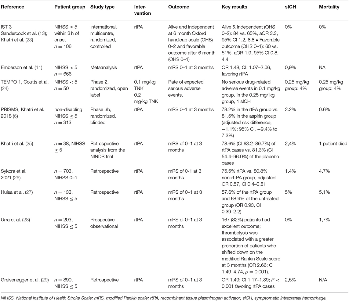
Current guidelines and recommendations state that for patients with acute minor disabling ischemic stroke of <4.5 h duration, intravenous thrombolysis with recombinant alteplase is recommended/ may be reasonable (21, 22). RCTs and observational studies addressing this topic so far showed promising results with a good functional outcome and a low complication rate (Table 1).

TABLE 1
www.frontiersin.org

Table 1. Randomized controlled trials and observational trials on thrombolysis in minor stroke.

For patients with acute minor non-disabling ischemic stroke of <4.5 h duration, no intravenous thrombolysis is recommended. One exception may be patients with non-disabling symptoms and a large vessel occlusion. However, many acute stroke centers do not perform angiography for those with NIHSS <6 as part of their internal protocol, and many centers do not have access to advanced imaging such as CT perfusion. Therefore, an unknown proportion of stroke with minor symptoms who have large vessel occlusions amenable to intervention are being missed. TEMPO 1, a case series of 50 patients with mild symptoms and intracranial vessel occlusion, which showed that administration of tenecteplase-tissue-type plasminogen activator in minor stroke with intracranial occlusion is feasible and safe (24). Wang et al. found that intravenous thrombolysis benefits though with mild stroke symptoms (NIHSS ≤ 5) and large artery atherosclerosis, though not those who had a tandem proximal intracranial occlusion and cervical internal artery lesion (complete occlusion or severe stenosis ≥ 90%) (30). They found that LAA-type patients (as defined by TOAST criteria) had significantly favorable outcomes after treatment with thrombolysis compared to untreated patients, however no such benefits were observed in other stroke subtypes, such as cardioembolic, small vessel occlusion and undetermined. This suggests that CT or MR angiography might be helpful to choose patients for thrombolysis that present with stroke with minor symptoms.

Alteplase or Tenecteplase in Patients With Minor Stroke

In recent years, the recombinant plasminogen activator tenecteplase is increasingly competing with the gold standard alteplase. The first publication of the EXTEND IA TNK study showed that higher perfusion rates and better clinical results can be achieved with tenecteplase in the 0.25 mg/kg dose than with alteplase in patients with an acute ischemic stroke (31). Tenecteplase was used as so-called bridging thrombolysis in the 4.5 h time window until the mechanical thrombectomy was performed. In addition, tenecteplase has advantages in handling, as it can be administered as single intravenous bolus and does not require a continuous infusion over 1 h, as alteplase does. The results of the EXTEND TNK study prompted the authors of the US guideline and the European Stroke Organization (ESO) to include tenecteplase in their recommendation as an alternative fibrinolytic (AHA/ASA Class IIb recommendation), although the AHA/ASA recommendation can also be considered to the 0.4 mg/kg dose for patients with less severe neurological impairments and if there are no large vessel occlusions (Level of Evidence: IIb) (22).

The second part of the EXTEND TNK study was recently published (32) which evaluated different doses of tenecteplase. The higher dose of tenecteplase (0.4 mg/kg) did not have any disadvantages in terms of safety: there were 16 and 22 death in the high and low dose groups, respectively. Symptomatic intracerebral hemorrhages 36 h after thrombolysis were numerically more frequent in the high dose group (7 vs. 2 patients), but four bleeding events in this group were associated with wire perforations during the endovascular procedure and were therefore not attributable to thrombolysis directly. The authors of the study report that the latter results are in contrast to an earlier study with the 0.40 mg/kg dose that was terminated prematurely for safety reasons, as some patients developed symptomatic intracranial hemorrhage. As a limitation, Campbell and colleagues point out that the study may not have been powered to reveal differences in efficacy. There was no restriction on clinical severity using NIHSS scores in these trials, but showed that probably a higher perfusion rate can be achieved with tenecteplase in patients with vessel occlusions. TEMPO 2 is an ongoing multicentre prospective randomized open label blinded-endpoint (PROBE) controlled trial of thrombolysis with low dose TEnecteplase vs. standard of care in Minor ischemic stroke with Proven acute symptomatic Occlusion (33). The hypothesis is that patients with mild (NIHSS < = 5) or even non-disabling symptoms due to identifiable vessel occlusion will benefit from IVT as compared to standard antiplatelet therapy. Results are expected in 2024. In summary, currently no evidence exists that tenecteplase should be preferred to alteplase in acute treatment of minor stroke patients, though further research is ongoing.


Thursday, February 11, 2021

Intravenous thrombolysis in stroke with admission NIHSS score 0 or 1

 So we still know nothing about treating mild strokes and until we do your doctors will just assume you are too good to treat and let your neurons die. The job of your doctor is to prevent disability not just stand by watching neurons die.  Contact researchers and get appropriate research going. Not doing so is an indictment of the complete stroke hospital starting at the top with the board of directors.

Tuesday, November 10, 2020

Safety and Hospital Costs Averted Using a Rapid Outpatient Management Strategy for Transient Ischemic Attack and Minor Strokes: The RAVEN Clinic

This is absolutely appalling, suggesting that TIAs and minor strokes are too good to treat because of cost concerns.  You better hope this doesn't happen to you because the damage won't magically disappear.


Safety and Hospital Costs Averted Using a Rapid Outpatient Management Strategy for Transient Ischemic Attack and Minor Strokes: The RAVEN Clinic

Show all authors First Published November 5, 2020 Research Article 

Patients presenting to emergency departments (ED) with transient ischemic attack and minor strokes (TIAMS) are often admitted for evaluation, though experience in other countries have suggested that an expedited outpatient care models may be a safe alternative. We hypothesized that a rapid access clinic for select TIAMS was feasible and would avert hospitalization costs.

This retrospective analysis included patients presenting to our institution’s ED with TIAMS and NIHSS ≤5 in calendar year 2017. We referred low-risk patients with TIAMS to a Rapid Access Vascular Evaluation-Neurology (RAVEN) clinic within 24 hours of ED discharge. We identified admitted patients who met RAVEN criteria at ED presentation. Rates of follow-up to the RAVEN clinic were recorded. Financial data collected included total hospital costs and time spent in the ED, as well hospital length of stay for admitted patients with low-risk TIAMS.

In 2017, 149 patients were referred to RAVEN clinic and 50 patients were admitted. Of the RAVEN patients 99 (94%) appeared as scheduled. None had clinical changes between ED discharge and clinical evaluation. One patient required hospitalization at the RAVEN evaluation. When compared to RAVEN patients, admitted patients had significantly higher $7,719 (SD 354) total hospital costs and were hospitalized for 2 days on average. Overall, the RAVEN strategy averted approximately $764,000 in hospitalization costs and 208 hospital bed-days in accounting year 2017.(But what the hell happened to those you didn't treat? Did they magically recover?)

For select patients presenting with TIAMS without disabling deficits, a rapid outpatient evaluation may be feasible while averting significant total hospital costs and preserving inpatient hospital beds.(My god, your focus is completely wrong, get the hell out of the stroke business and take your cost containment someplace that doesn't affect lives.)

Access Options