Changing stroke rehab and research worldwide now.Time is Brain! trillions and trillions of neurons that DIE each day because there are NO effective hyperacute therapies besides tPA(only 12% effective). I have 523 posts on hyperacute therapy, enough for researchers to spend decades proving them out. These are my personal ideas and blog on stroke rehabilitation and stroke research. Do not attempt any of these without checking with your medical provider. Unless you join me in agitating, when you need these therapies they won't be there.

What this blog is for:

My blog is not to help survivors recover, it is to have the 10 million yearly stroke survivors light fires underneath their doctors, stroke hospitals and stroke researchers to get stroke solved. 100% recovery. The stroke medical world is completely failing at that goal, they don't even have it as a goal. Shortly after getting out of the hospital and getting NO information on the process or protocols of stroke rehabilitation and recovery I started searching on the internet and found that no other survivor received useful information. This is an attempt to cover all stroke rehabilitation information that should be readily available to survivors so they can talk with informed knowledge to their medical staff. It lays out what needs to be done to get stroke survivors closer to 100% recovery. It's quite disgusting that this information is not available from every stroke association and doctors group.

Showing posts with label Sphenopalatine Ganglion. Show all posts

Tuesday, November 23, 2021

Stimulating the Facial Nerve to Treat Ischemic Stroke: A Systematic Review

Interesting, but are these other methods to increase blood flow better? 

Your doctor should be doing something from one of these. 

Or is it more important to deliver more oxygen to your brain?

Possible solutions: Obviously not vetted coming from me. Don't do them. 

Normobaric oxygen (10)

How to Improve Your Brain Function with An Oxygen Concentrator April 2018 

Or is it more important to increase the loading ability of red blood cells to carry more oxygen? 

Like this?

University of Glasgow Study Demonstrates the Ability of Oxycyte® to Supply Oxygen to Critical Penumbral Tissue in Acute Ischemic Stroke  August 2012

Or like this?

chronic cannabis users have higher cerebral blood flow and extract more oxygen from brain blood flow than nonusers. August 2017  

The latest here:

Stimulating the Facial Nerve to Treat Ischemic Stroke: A Systematic Review

 
Turner S. Baker1,2*†, Justin Robeny1,2†, Danna Cruz2,3, Alexis Bruhat1,2, Alfred-Marc Iloreta4, Anthony Costa1,2 and Thomas James Oxley1,2
  • 1Department of Neurosurgery, Icahn School of Medicine at Mount Sinai, New York, NY, United States
  • 2Sinai BioDesign, Icahn School of Medicine at Mount Sinai, New York, NY, United States
  • 3The Grove School of Engineering, The City College of New York, New York, NY, United States
  • 4Department of Otolaryngology, Icahn School of Medicine at Mount Sinai, New York, NY, United States

Acute ischemic stroke (AIS) is a common devastating disease that has increased yearly in absolute number of cases since 1990. While mechanical thrombectomy and tissue plasminogen activator (tPA) have proven to be effective treatments, their window-of-efficacy time is very short, leaving many patients with no viable treatment option. Over recent years there has been a growing interest in stimulating the facial nerves or ganglions to treat AIS. Pre-clinical studies have consistently demonstrated an increase in collateral blood flow (CBF) following ganglion stimulation, with positive indications in infarct size and neurological scores. Extensive human trials have focused on trans-oral electrical stimulation of the sphenopalatine ganglion, but have suffered from operational limitations and non-significant clinical findings. Regardless, the potential of ganglion stimulation to treat AIS or elongate the window-of-efficacy for current stroke treatments remains extremely promising. This review aims to summarize results from recent trial publications, highlight current innovations, and discuss future directions for the field. Importantly, this review comes after the release of four important clinical trials that were published in mid 2019.

Introduction

Acute Ischemic Stroke

Stroke is the leading cause of disability and the fifth leading cause of death in the United States (US). Approximately 795,000 people experience a new or recurrent stroke each year (1). Acute ischemic stroke (AIS) occurs when an obstruction within a blood vessel decreases cerebral blood flow, depriving nerve cells of oxygen and leading to severe metabolic failure and neural death (2–4). Immediately following stroke, a section of the brain referred to as the ischemic core is subject to extreme hypoxia, leading to irreversible brain damage (5). The area surrounding the ischemic core, the ischemic penumbra, is severely hypoperfused and non-functioning, yet can regain functionality if blood flow is restored to the area (5, 6). This recovery is highly time-dependent, as the penumbra rapidly evolves into the ischemic core (6, 7). The recovery of the penumbra has been demonstrated to have a significant effect on clinical outcomes; Meretoja et al. showed that for every 20-min reduction in time to reperfusion increases the average disability-free life span by 3 months (8).

Current Treatments: Endovascular Thrombectomy and Tissue Plasminogen Activator

Management for AIS relies on rapid treatment times to avoid penumbra evolution. The goal of modern stroke treatment facilities is to reperfuse the ischemic area via endovascular thrombectomy, mechanically removing the blood clot with catheter-based devices (9). The faster the patient achieves reperfusion, the more likely the patient will have excellent neurological outcomes at 90 days (10). The second treatment paradigm for AIS is the use of intravenous (IV) tissue plasminogen activator (tPA), which acts to chemically break down clots (11–14). IV-tPA is commonly used prior to patient transport for thrombectomy.

Both endovascular thrombectomy and IV-tPA suffer from a limited window-of-efficacy. Currently, stroke guidelines list the acceptable window of treatment for mechanical thrombectomy at 24 h (15, 16), and a recommended IV-tPA door-to-treatment time of 60 min (10, 17, 18). These windows are frequently missed, with fewer than a third of patients in the U.S. treated within the IV-tPA 60 min window (19). Mandatory neuroimaging, presence of a highly-trained neurointerventionalist, and hospital transfer times all reduce event-to-treatment times and result in reductions of successful functional outcomes following recanalization (16, 20, 21).

Thrombectomy is a well-established treatment (15, 16, 22), but patients routinely fail to achieve functional independence (mRS ≤ 2) due to extensive event-to-treatment times (15, 16). There is a clear need for innovative approaches to extend the window-of-efficacy for endovascular thrombectomy and IV-tPA.

Time Is Brain: Inhibiting the Evolution of the Ischemic Penumbra

Researchers have recently sought to find new approaches to arrest the evolution of the ischemic penumbra and keep this susceptible region from becoming irreversibly damaged. Current approaches aim to either enhance oxygen delivery to the penumbra or reduce tissue oxygen demand (6). In addition to door-to-treatment time limitations, many patients also become ineligible for mechanical thrombectomy due to large ischemic core volumes. Inhibiting the evolution of the penumbra may help buy time by limiting core volume growth from reaching recommended exclusionary levels (23, 24). Inhibition of penumbra evolution could also be highly beneficial when combined with IV-tPA, potentially increasing its effectiveness and elongating its window-of-efficacy (6).

Increased Collateral Blood Circulation Through Ganglion Stimulation

Facial nerve-induced vasodilation of the cerebral arteries is an emerging therapeutic target that seeks to increase collateral blood flow in ischemic brain tissue, improve oxygen availability, and improve patient functional outcomes after stroke. Collateral circulation refers to alternative, pre-existing vascular pathways that deliver blood to target tissue when the primary vessel is occluded (25). Imaging of the brain and vessels has shown that collateral blood flow can preserve brain tissue for hours after major arteries to the brain are blocked (26). Figure 1 illustrates how facial nerve-induced increased collateral blood flow has the ability to ameliorate clot-induced tissue death by limiting ischemic penumbra evolution.
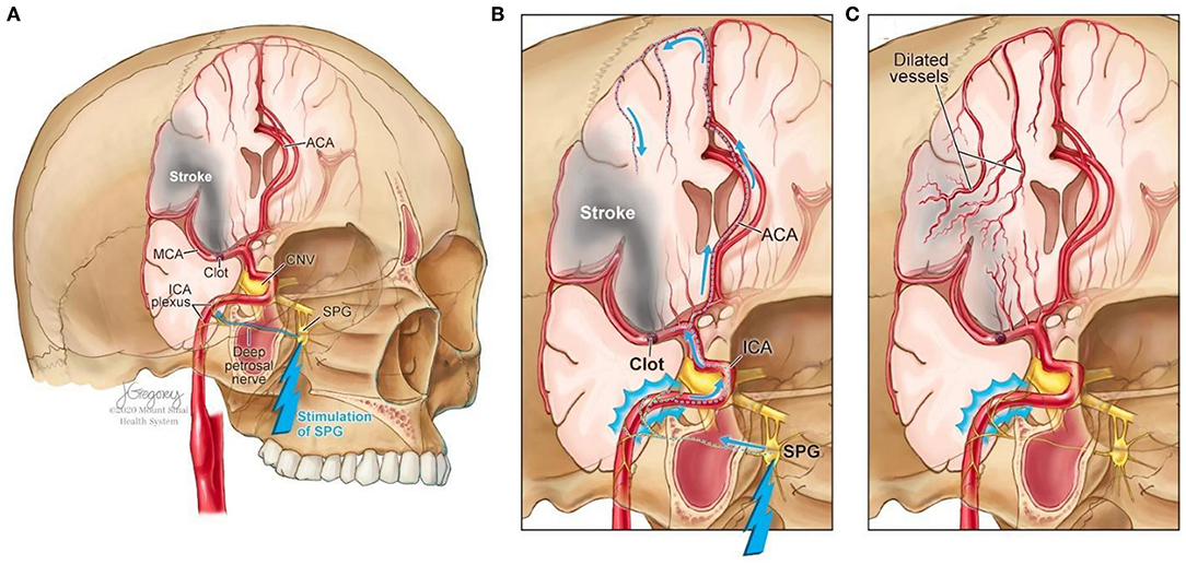
FIGURE 1
www.frontiersin.org

Figure 1. Increased collateral blood circulation by sphenopalatine ganglion (SPG) stimulation. (A) Anatomical overview of stroke affected brain region before SPG stimulation. (B) The SPG contains parasympathetic fibers that synapse in the ganglion and innervate the internal carotid artery (ICA) through the deep petrosal nerve. (C) SPG stimulation induces vasodilation of blood vessels in the anterior circulation, increasing blood flow to the affected area limiting the evolution of the penumbra and reducing infarct volume.

The sphenopalatine ganglion (SPG), also known as the pterygopalatine ganglion, is the largest and most superior ganglion of the sympathetic and parasympathetic nervous system and contains the largest collection of neurons in the calvarium outside of the brain (27). Humans have two SPGs, on each side of the midface, located within the viscerocranium in a space called the pterygopalatine fossa. This fossa has direct connections to the middle cranial fossa, nasal cavity, orbit, infratemporal fossa and oral cavity (28, 29). The SPG contains parasympathetic fibers that synapse in the ganglion and innervate the internal carotid artery (ICA) through the deep petrosal nerve (30). Electrical stimulation of the SPG activates the fibers, releasing several neurotransmitters such as acetylcholine, vasoactive intestinal polypeptide, peptide histidine isoleucine, and nitrous oxide that play a role in inducing vasodilation of blood vessels in the anterior circulation (26, 31).

The geniculate ganglion is a small collection of somatosensory and gustatory ganglion cells (32) located within the temporal bone along the axis of the ear canal. Similar to the SPG, the geniculate ganglion has parasympathetic connections to cerebral arteries and has also been demonstrated to increase cerebral blood flow (33, 34). Its curved shape allows for greater susceptibility to be activated location makes it easier to access through non-invasive routes (33, 35).

More at link.

Sunday, July 7, 2019

Sphenopalatine Ganglion Stimulation to Augment Cerebral Blood Flow

The takeaway from this is that your doctor and hospital have the responsibility  to followup, contact researchers and get further trials done. No followup, contact the board of directors and have those doctors, directors and president responsible fired.  Why not do this for all stroke patients? Better cerebral blood flow would probably save more of the penumbra from dying.

Sphenopalatine Ganglion Stimulation to Augment Cerebral Blood Flow

A Randomized, Sham-Controlled Trial
and The ImpACT-24A Investigators include the Site Principal Investigators, DSMB members, and the Site Co-Investigators listed
Originally publishedhttps://doi.org/10.1161/STROKEAHA.118.024582Stroke. ;0

Background and Purpose—

Many patients with acute ischemic stroke are not eligible for thrombolysis or mechanical reperfusion therapies due to contraindications, inaccessible vascular occlusions, late presentation, or large infarct core. Sphenopalatine ganglion (SPG) stimulation to enhance collateral flow and stabilize the blood-brain barrier offers an alternative, potentially more widely deliverable, therapy.

Methods—

In a randomized, sham-controlled, double-masked trial at 41 centers in 7 countries, patients with anterior circulation ischemic stroke not treated with reperfusion therapies within 24 hours of onset were randomly allocated to active SPG stimulation or sham control. The primary efficacy outcome was improvement beyond expectations on the modified Rankin Scale of global disability at 90 days (sliding dichotomy), assessed in the modified intention-to-treat population. The initial planned sample size was 660 patients, but the trial was stopped early when technical improvements in device placement occurred, so that analysis of accumulated experience could be conducted to inform a successor trial.

Results—

Among 303 enrolled patients, 253 received at least one active SPG or sham stimulation, constituting the modified intention-to-treat population (153 SPG stimulation and 100 sham control). Age was median 73 years (interquartile range, 64–79), 52.6% were female, deficit severity on the National Institutes of Health Stroke Scale was median 11 (interquartile range, 9–15), and time from last known well median 18.6 hours (interquartile range, 14.5–22.5). For the primary outcome, improved 3-month disability beyond expectations, rates in the SPG versus sham treatment groups were 49.7% versus 40.0%; odds ratio, 1.48 (95% CI, 0.89–2.47); P=0.13. A significant treatment interaction with stroke location (cortical versus noncortical) was noted, P=0.04. In the 87 patients with confirmed cortical involvement, rates of improvement beyond expectations were 50.0% versus 27.0%; odds ratio, 2.70 (95% CI, 1.08–6.73); P=0.03. Similar response patterns were observed for all prespecified secondary efficacy outcomes. No differences in mortality or serious adverse event safety end points were observed.

Conclusions—

SPG stimulation within 24 hours of onset is safe in acute ischemic stroke. SPG stimulation was not shown to statistically significantly improve 3-month disability above expectations, though favorable outcomes were nominally higher with SPG stimulation. Beneficial effects may distinctively be conferred in patients with confirmed cortical involvement. The results of this study need to be confirmed in a larger pivotal study.

Clinical Trial Registration—

URL: https://www.clinicaltrials.gov. Unique identifier: NCT03767192.

Wednesday, May 29, 2019

SPG Stimulation 'Likely' of Benefit for Some Acute Strokes

So WHOM is going to followup to see if this treatment as a standalone immediately post tPA or thrombectomy would benefit stroke survivors? Or is nobody thinking that far ahead and prefer to stay with the failed status quo of stroke? I'm guessing that absolutely no followup will be done because we don't have two functioning neurons in stroke leadership.

SPG Stimulation 'Likely' of Benefit for Some Acute Strokes


Trial narrowly misses endpoint in confirmed cortical involvement group

  • by Staff Writer, MedPage Today
Acute ischaemic stroke patients ineligible for thrombolytic therapy and with confirmed cortical involvement (CCI) may derive benefit from an injectable neurostimulator implant that works on the sphenopalatine ganglion (SPG), a sham-controlled randomized trial found.
In the modified intent-to-treat population, the proportion of patients whose disability level was better than anticipated at 90 days was 49% in the SPG stimulation group versus 45% in the sham group (OR 1.14, P=0.31), reported Jeffrey Saver, MD, of the University of California Comprehensive Stroke Center in Los Angeles, and colleagues.
In the CCI population, 50% of the intervention group had better than expected outcomes compared with 40% of the control group (OR 1.48, P=0.0258), which narrowly missed statistical significance (P<0.025 required), as described in The Lancet.
The research was also presented at the European Stroke Organisation Conference in Milan.
"Although the improvement in 90-day functional outcome among patients with imaging evidence of cortical involvement at presentation was not significant in this trial alone, study findings indicate the use of sphenopalatine ganglion stimulation is likely to be beneficial, on the basis of consistent effects across the primary and all secondary endpoints, and of the presence of an inverted U-shaped dose-response curve," Saver's group wrote.
When assessing the U-shaped dose-response relationship between the primary outcome and SPG stimulation intensity in the CCI group, the proportion with favorable outcomes went up from 40% to 70% at low-midrange intensity and declined back to 40% during high-intensity stimulation (P=0.0034).
"Pooled analysis of all completed trials indicated that, among patients with imaging evidence of cortical involvement at presentation, sphenopalatine ganglion stimulation improves functional outcome," the researchers concluded.
IMPACT-24B (Implant Augmenting Cerebral Blood Flow Trial-24B) was a pivotal, international, double-blind study that randomized 1,078 patients with anterior-circulation acute ischaemic stroke, who were not receiving reperfusion treatment, to SPG stimulation (n=481) or sham treatment (n=519). Median patient age was about 70 years and 51% were women. Among these, 52% of patients had CCI -- 244 in the intervention group and 276 in the sham group. No differences in serious adverse events or mortality were seen between the two groups.
Patients were eligible if they had radiological and clinical evidence of acute ischemic stroke in the anterior cerebral circulation, had National Institutes of Health Stroke Scale scores between 7 and 18, and were able to start treatment 8 to 24 hours from stroke onset. Age requirements were 40-85 for women and 40-80 years for men.
Exclusion criteria included pregnancy, oral cavity conditions that might hinder device implantation, uncontrolled high blood pressure, bilateral stroke, bleeding propensity, radiological evidence of intracranial haemorrhage, and others.
Results of the present investigation fall into line with previous pilot trial (IMPACT-24A), which also looked at SPG stimulation within 1 day of stroke onset.
"Additional studies are planned or underway in patients receiving intravenous thrombolytic therapy and endovascular mechanical thrombectomy, to assess whether adding sphenopalatine ganglion stimulation improves outcomes in these patients as well," the authors wrote.
The study was funded by BrainsGate.
Saver disclosed relationships with BrainsGate.

Saturday, May 25, 2019

An injectable implant to stimulate the sphenopalatine ganglion for treatment of acute ischaemic stroke up to 24 h from onset (ImpACT-24B): an international, randomised, double-blind, sham-controlled, pivotal trial

Well then if it works that well your job isn't done until you have written a stroke protocol on this. AND distributed it to every stroke doctor and hospital in the world. OR IF YOU'D RATHER, distribute it to every one of the 10 million yearly stroke survivors. Your choice,

DOING NOTHING IS NOT AN OPTION.


An injectable implant to stimulate the sphenopalatine ganglion for treatment of acute ischaemic stroke up to 24 h from onset (ImpACT-24B): an international, randomised, double-blind, sham-controlled, pivotal trial


Summary

Background

Sphenopalatine ganglion stimulation increased cerebral collateral blood flow, stabilised the blood–brain barrier, and reduced infarct size, in preclinical models of acute ischaemic stroke, and showed potential benefit in a pilot randomised trial in humans. The pivotal ImpACT-24B trial aimed to determine whether sphenopalatine ganglion stimulation 8–24 h after acute ischaemic stroke improved functional outcome.

Methods

ImpACT-24B is a randomised, double-blind, sham-controlled, pivotal trial done at 73 centres in 18 countries. It included patients (men aged 40–80 years and women aged 40–85 years) with anterior-circulation acute ischaemic stroke, not undergoing reperfusion therapy. Enrolled patients were randomly assigned via web-based randomisation to receive active sphenopalatine ganglion stimulation (intervention group) or sham stimulation (sham-control group) 8–24 h after stroke onset. Patients, clinical care providers, and all outcome assessors were masked to treatment allocation. The primary efficacy endpoint was the difference between active and sham groups in the proportion of patients whose 3-month level of disability improved above expectations. This endpoint was evaluated in the modified intention-to-treat (mITT) population (defined as all patients who received one active or sham treatment session) and the population with confirmed cortical involvement (CCI) and was analysed using the Hochberg multi-step procedure (significance in both populations if p<0·05 in both, and in one population if p<0·025 in that one). Safety endpoints at 3 months were all serious adverse events (SAEs), SAEs related to implant placement or removal, SAEs related to stimulation, neurological deterioration, and mortality. All patients who underwent an attempted sphenopalatine ganglion stimulator or sham stimulator placement procedure were included in the safety analysis. This trial is registered with ClinicalTrials.gov, number NCT00826059.

Findings

Between June 10, 2011, and March 7, 2018, 1078 patients were enrolled and randomly assigned to either the intervention or the sham-control group. 1000 patients received at least one session of sphenopalatine ganglion stimulation or sham stimulation and entered the mITT population (481 [48%] received sphenopalatine ganglion stimulation, 519 [52%] were sham controls), among whom 520 (52%) patients had CCI on imaging. The proportion of patients in the mITT population whose 3-month disability level was better than expected was 49% (234/481) in the intervention group versus 45% (236/519) in the sham-control group (odds ratio 1·14, 95% CI 0·89–1·46; p=0·31). In the CCI population, the proportion was 50% (121/244) in the intervention group versus 40% (110/276) in the sham-control group (1·48, 1·05–2·10; p=0·0258). There was an inverse U-shaped dose–response relationship between attained sphenopalatine ganglion stimulation intensity and the primary outcome in the CCI population: the proportion with favourable outcome increased from 40% to 70% at low–midrange intensity and decreased back to 40% at high intensity stimulation (p=0·0034). There were no differences in mortality or SAEs between the intervention group (n=536) and the sham-control group (n=519) in the safety population.

Interpretation

Sphenopalatine ganglion stimulation is safe for patients with acute ischaemic stroke 8–24 h after onset, who are ineligible for thrombolytic therapy. Although not reaching significance(Will this be your excuse for doing nothing?), the trial's results support that, among patients with imaging evidence of cortical involvement at presentation, sphenopalatine ganglion stimulation is likely to improve functional outcome.

Funding

BrainsGate Ltd.
To read this article in full you will need to make a payment


Thursday, February 14, 2019

Stimulation of the Spenopalatine Ganglion Improves Outcomes in Patients With Acute Ischaemic Stroke

Good, then write up a protocol and distribute this to every stroke hospital in the world. OR, figure out a way to get this to everyone of the 10 million yearly stroke survivors. Your choice, DOING NOTHING IS NOT AN OPTION. 

Find out what this is here, for training your doctor:

Sphenopalatine Ganglion Stimulation


Stimulation of the Spenopalatine Ganglion Improves Outcomes in Patients With Acute Ischaemic Stroke

By Alex Morrisson
HONOLULU -- February 11, 2019 -- Stimulation of the sphenopalatine ganglion (SPG) with a device appears to improve functional outcomes among a subset of patients diagnosed with acute ischaemic stroke, according to a study presented here at the 2019 International Stroke Conference(ISC).
Stimulation of the SPG has been associated with increased cerebral collateral blood flow, stabilisation of the blood-brain barrier, and reduced infarct size.
In analysing pooled data from the ImpACT 24A and ImpACT 24B trials, 48.9% of the 634 patients who had SPG stimulation had outcomes considered better than expected on a sliding dichotomy assessment compared with 44.6% of the 619 patients who received sham stimulation (odds ratio = 1.19; 95% confidence interval, 0.95-1.48; P = .13) -- a non-significant finding.
However, when researchers scrutinised outcomes in patients who had a conformed cortical involvement stroke, there was a significant positive finding. Of the 294 people treated with stimulation, 34% achieved better than expected outcomes -- achieving a 0-2 score on the modified Rankin Scale -- compared with 26% of the 313 treated with sham (P = .005).
“SPG stimulation in these patients was safe,” said Ashfaq Shuaib, MD, University of Alberta, Edmonton, Alberta. “There was no difference in adverse events, and placement was fast and robust.”
“This meta-analysis further supports that in patients with acute ischaemic stroke with confirmed cortical infarcts, SPG stimulation started within 24 hours reduced post-stroke disability over the entire outcome range and increases the proportion of patients who are alive and independent 3 months after stroke,” he said.
Patients had an average National Institutes of Health Stroke Scale score of 12, more than 80% were diagnosed with hypertension, and about 25% were diagnosed with diabetes. The primary endpoint was improvement on the modified Rankin Scale at 90 days using a sliding dichotomy, assessed in the modified intention-to-treat populations and in those with confirmed cortical involvement. In the confirmed cortical involvement group, about 31% had atrial fibrillation while in the intention to treat population, about 25% had atrial fibrillation.
[Presentation title: Spenopalatine Ganglion Stimulation for the Treatment of Acute Ischemic Stroke: Pooled Meta-Analysis of the Impact 24A and Impact 24B Trials. Abstract LB12]