Use the labels in the right column to find what you want. Or you can go thru them one by one, there are only 34,224 posts. Searching is done in the search box in upper left corner. I blog on anything to do with stroke. DO NOT DO ANYTHING SUGGESTED HERE AS I AM NOT MEDICALLY TRAINED, YOUR DOCTOR IS, LISTEN TO THEM. BUT I BET THEY DON'T KNOW HOW TO GET YOU 100% RECOVERED. I DON'T EITHER BUT HAVE PLENTY OF QUESTIONS FOR YOUR DOCTOR TO ANSWER.
Changing stroke rehab and research worldwide now.Time is Brain!trillions and trillions of neuronsthatDIEeach day because there areNOeffective hyperacute therapies besides tPA(only 12% effective). I have 523 posts on hyperacute therapy, enough for researchers to spend decades proving them out. These are my personal ideas and blog on stroke rehabilitation and stroke research. Do not attempt any of these without checking with your medical provider. Unless you join me in agitating, when you need these therapies they won't be there.
What this blog is for:
My blog is not to help survivors recover, it is to have the 10 million yearly stroke survivors light fires underneath their doctors, stroke hospitals and stroke researchers to get stroke solved. 100% recovery. The stroke medical world is completely failing at that goal, they don't even have it as a goal. Shortly after getting out of the hospital and getting NO information on the process or protocols of stroke rehabilitation and recovery I started searching on the internet and found that no other survivor received useful information. This is an attempt to cover all stroke rehabilitation information that should be readily available to survivors so they can talk with informed knowledge to their medical staff. It lays out what needs to be done to get stroke survivors closer to 100% recovery. It's quite disgusting that this information is not available from every stroke association and doctors group.
Two University of Texas at Arlington undergraduates have won a national invention award for developing a lightweight, wearable soft robotic wrist exoskeleton designed to help stroke survivors regain wrist function.
Suyog Neupane, a senior majoring in electrical engineering, and Kritazya Upreti, a senior majoring in computer science, earned first place in the university-level division of the 2026 National Academy of Inventors GenSpiration Prize Competition in Los Angeles. The competition brought together student inventors from universities across the United States, and UTA was one of only three finalist teams.
The students were inspired by the global impact of stroke, which affects millions of people each year. Loss of wrist and hand function is one of the most common consequences of stroke, making everyday activities such as eating, writing and turning a doorknob difficult or impossible.
"Competing against such talented innovators was both an incredible honor and an inspiring experience," Upreti said. "Seeing our hard work and innovation recognized on such a large stage was truly rewarding."
Their invention is a soft pneumatic wrist exoskeleton powered by air pressure rather than heavy motors or rigid mechanical components. Its key innovation is that it can produce two degrees of wrist motion simultaneously—flexion/extension and radial/ulnar—using a single compact soft actuator, improving portability while reducing mechanical complexity.
"Most devices currently available for wrist rehabilitation are either too heavy, bulky or difficult to operate," Upreti said. "One of our main motivations was to develop a solution that could provide multiple wrist motions using a single actuator."
From left to right: Suyog Neupane, Kritazya Upreti and Inderjeet Singh pose with their invention. (UT Arlington)
The undergraduate team was mentored by Inderjeet Singh, a research scientist at the University of Texas at Arlington Research Institute. He emphasized that the design is grounded in modern rehabilitation robotics principles, where effective recovery depends on providing controlled, repeatable and task-specific movement training. He added that supporting multiple wrist movements together is critical because natural hand function depends on them working in coordination, making that capability essential for meaningful rehabilitation.
The exoskeleton is designed to support patients throughout different stages of stroke recovery. For individuals with severe motor impairments, it can assist with the repetitive wrist movements commonly prescribed during rehabilitation. As patients regain strength and mobility, the device transitionts to assisting voluntary wrist movements, enabling users to perform therapeutic exercises and promoting active participation in the recovery process.
Related: Do more with less strain: UTA’s robotic arm
The award-winning project was the result of a collaborative effort within the Biomedical Technologies Lab at UTARI. Along with mentoring the student inventors, Dr. Singh and UTARI research scientist Veysel Erel led the project's research and development. UTARI research scientists Allison Palomino and Sahil Mangaonkar contributed to device fabrication and electronics development, while UTARI research scientist Alex Jamieson provided valuable support in refining and finalizing the overall system. Together, the multidisciplinary team transformed an innovative concept into a nationally recognized technology.
The exoskeleton can be used to help with many tasks. (UT Arlington)
Beyond stroke rehabilitation, the researchers see broader applications for the device. Individuals whose occupations involve repetitive wrist movements—such as assembly-line workers and heavy-equipment operators—could potentially use the exoskeleton to reduce fatigue, minimize strain, and lower the risk of work-related musculoskeletal injuries.
"What means the most to us is the potential for our technology to make a positive impact on the lives of stroke survivors," Neupane said. "The journey was both challenging and exciting, filled with moments of trial and error that pushed us to think more creatively and innovate."
About The University of Texas at Arlington (UTA)
The University of Texas at Arlington is a growing public research university in the heart of Dallas-Fort Worth. With a student body of over 42,700, UTA is the second-largest institution in the University of Texas System, offering more than 180 undergraduate and graduate degree programs. Recognized as a Carnegie R-1 university, UTA stands among the nation’s top 5% of institutions for research activity. UTA and its 300,000 alumni generate an annual economic impact of $28.8 billion for the state. The University has received the Innovation and Economic Prosperity designation from the Association of Public and Land Grant Universities and has earned recognition for its focus on student access and success, considered key drivers to economic growth and social progress for North Texas and beyond.
Oh jeez, are you that fucking stupid that tracking wrist movements does ANYTHING AT ALL to get survivors recovered? And you're using a healthy subject to suggest stroke recovery! My God, I'd fire everyone here!
Newswise — For survivors of strokes, which afflict nearly 800,000
Americans each year, regaining fine motor skills like writing and using
utensils is critical for recovering independence and quality of life.
But getting intensive, frequent rehabilitation therapy can be
challenging and expensive.
Now, researchers at NYU Tandon School
of Engineering are developing a new technology that could allow stroke
patients to undergo rehabilitation exercises at home by tracking their
wrist movements through a simple setup: a smartphone strapped to the
forearm and a low-cost gaming controller called the Novint Falcon.
The
Novint Falcon, a desktop robot typically used for video games, can
guide users through specific arm motions and track the trajectory of its
controller. But it cannot directly measure the angle of the user's
wrist, which is essential data for therapists providing remote
rehabilitation.
"Patients would strap their phone to their forearm and
manipulate this robot," said Maurizio Porfiri, NYU Tandon Institute
Professor and director of its Center for Urban Science + Progress
(CUSP), who is the paper’s senior author. "Data from the phone's
inertial sensors can then be combined with the robot's measurements
through machine learning to infer the patient's wrist angle."
The
researchers collected data from a healthy subject performing tasks with
the Falcon while wearing motion sensors on the forearm and hand to
capture the true wrist angle. They then trained an algorithm to predict
the wrist angles based on the sensor data and Falcon controller
movements.
The resulting algorithm could predict wrist angles with
over 90% accuracy, a promising initial step toward enabling remote
therapy with real-time feedback in the absence of an in-person
therapist.
"This technology could allow patients to undergo
rehabilitation exercises at home while providing detailed data to
therapists remotely assessing their progress," Roni Barak Ventura, the
paper’s lead author who was an NYU Tandon postdoctoral fellow at the
time of the study. "It's a low-cost, user-friendly approach to
increasing access to crucial post-stroke care."
The researchers
plan to further refine the algorithm using data from more subjects.
Ultimately, they hope the system could help stroke survivors stick to
intensive rehab regimens from the comfort of their homes.
"The
ability to do rehabilitation exercises at home with automatic tracking
could dramatically improve quality of life for stroke patients," said
Barak Ventura. "This portable, affordable technology has great potential
for making a difficult recovery process much more accessible."
This study adds to NYU Tandon’s body of work that aims to improve stroke recovery. In 2022, Researchers from NYU Tandon began collaborating with the FDA
to design a regulatory science tool based on biomarkers to objectively
assess the efficacy of rehabilitation devices for post-stroke motor
recovery and guide their optimal usage. A study
from earlier this year unveiled advances in technology that uses
implanted brain electrodes to recreate the speaking voice of someone who
has lost speech ability, which can be an outcome from stroke.
In
addition to Porfiri and Barak Ventura, the study’s authors are Angelo
Catalano, who earned an MS from NYU Tandon in 2024, and Rayan Succar, an
NYU Tandon PhD candidate. The study was funded by grants from the
National Science Foundation.
In
the last two decades robot training in neuromotor rehabilitation was
mainly focused on shoulder-elbow movements. Few devices were designed
and clinically tested for training coordinated movements of the wrist,
which are crucial for achieving even the basic level of motor competence
that is necessary for carrying out ADLs (activities of daily life).
Moreover, most systems of robot therapy use point-to-point reaching
movements which tend to emphasize the pathological tendency of stroke
patients to break down goal-directed movements into a number of jerky
sub-movements. For this reason we designed a wrist robot with a range of
motion comparable to that of normal subjects and implemented a
self-adapting training protocol for tracking smoothly moving targets in
order to facilitate the emergence of smoothness in the motor control
patterns and maximize the recovery of the normal RoM (range of motion)
of the different DoFs (degrees of Freedom).
Methods
The
IIT-wrist robot is a 3 DoFs light exoskeleton device, with direct-drive
of each DoF and a human-like range of motion for Flexion/Extension
(FE), Abduction/Adduction (AA) and Pronation/Supination (PS). Subjects
were asked to track a variable-frequency oscillating target using only
one wrist DoF at time, in such a way to carry out a progressive
splinting therapy. The RoM of each DoF was angularly scanned in a
staircase-like fashion, from the "easier" to the "more difficult"
angular position. An Adaptive Controller evaluated online performance
parameters and modulated both the assistance and the difficulty of the
task in order to facilitate smoother and more precise motor command
patterns.
Results
Three
stroke subjects volunteered to participate in a preliminary test
session aimed at verify the acceptability of the device and the
feasibility of the designed protocol. All of them were able to perform
the required task. The wrist active RoM of motion was evaluated for each
patient at the beginning and at the end of the test therapy session and
the results suggest a positive trend.
Conclusion
The
positive outcomes of the preliminary tests motivate the planning of a
clinical trial and provide experimental evidence for defining
appropriate inclusion/exclusion criteria.
Background
Decreased
wrist range of motion (ROM) (flexion and/or extension,
abduction/adduction or pronation/supination) after trauma or surgery can
be a challenging problem. Physical therapy, orthoses, and additional
surgical interventions may not restore the desired functionality even
after an intensive rehabilitation program. Therapists spend a
considerable amount of practice time in differential diagnosis of these
losses and selecting appropriate intervention strategies to restore
passive and active motion in concordance with the pathology and to
prevent loss of range of motion after injury.
While the regular
treatment for wrist stiffness is physical therapy or surgery,
researchers are looking for an alternative and more efficient and
automatic procedure by means of robotic applications.
Several systems for wrist rehabilitation have been developed in research centres and universities, for example RiceWrist [1]; MIME [2]; IMT3 [3], HWARD [4]; the Okayama University pneumatic manipulator [5], and the devices overviewed in [6–9]. The majority are also used for rehabilitation in health centres and hospitals, often coupled with MIT-MANUS [10], ARMIN [11], MIME, HapticMaster [12] and wire-based device from Rosati et. al. [13]
for rehabilitation of proximal limb. Robot assisted therapy are
primarily based on goal-directed point-to-point movement involving
multiple DoFs [14];
main purpose is increasing the ROM of the paretic limb in order to
regain motor abilities for the Activities of Daily Living (ADL).
Contrarily regular physical therapy of wrist rehabilitation consists in a
splinting treatment for each single DoF at time, and there have been
many studies that look at the splints' effectiveness and what type of
splint would be best [15, 16].
Static progressive splinting is a time-honored concept, for more than
20 years, clinicians have recognized the effectiveness of static
progressive splints to improve passive range of motion (PROM). Splint
designers then sought a means to improve the technique with components
that offer infinitely adjustable joint torque control and are easy to
apply, lightweight, low-profile, and reasonably priced.
Dynamic splints use some additional component (springs, wires, rubber bands) to mobilize contracted joints [17–19].
This dynamic pull functions to provide a controlled gentle force to the
soft tissue over long periods of time, which encourages tissue
remodeling without tearing. The issues that make dynamic or static
progressive splinting technically difficult include determining how much
force to use, how to apply the force, how long to apply the force, and
how to prevent added injury to the area. Things could change if the
dynamic splinting is delivered using devices which are able to modulate
torque delivering and space the range of motion.
Therefore we
intend to approach the robotic therapy for wrist rehabilitation using a
continuous dynamic splinting of each single DoF but contrarily to the
regular progressive splinting we want also to highlight the voluntary
component of movement. A performance adaptive control strategy has been
developed, with the purpose of providing variable assistance by means of
a general training paradigm for stroke patients.
Methods
Apparatus: the wrist device
The Wrist-Robot [20],
herewith reported, has been developed at the Italian Institute of
Technology with three main requirements: 1) back-drivability of the 3
DoFs (Degree of Freedom), in order to assure a smooth haptic interaction
between the robot and the patient; 2) mechanical and electronic
modularity, in order to facilitate the future integration into a haptic
bimanual arm-wrist-hand system with up to 12 DoFs; 3) scalable software
architecture. The Wrist Robot is intended to provide kinesthetic
feedback during the training of motor skills or rehabilitation of
reaching movements. Motivations for application of robot therapy in
rehabilitation of neurological patients come from experimental studies
about the practice-induced plastic reorganization of the brain in humans
and animal models [21, 22].
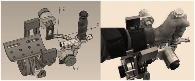
The robot (figure 1) is a 3 DOFs exoskeleton: F/E (Flexion/Extension); Ad/Ab (Adduction/Abduction); P/S (Pronation/Supina-tion).
Figure 1
3DoF Wrist Device. It has 3 DOFs: F/E, P/S, Ad/Ab. One motor is used for F/E and P/S; two motors for Ad/Ab.
The
chosen class of mechanical solutions is based on a serial structure,
with direct drive by the motors: one motor for pronation/supination, one
motor for flexion/extension and two parallel coupled motors for
abduction/adduction that allow to balance the pronosupination rotation
during motion.
The problem of measurement of arm position is thus
reduced to the solution of the device kinematics, with no further
transformations required, allowing to actuate the robot to control
feedback to a specific human joint, for example to constrain the forearm
rotation during wrist rehabilitation, without affecting other joints.
The corresponding rotation axes meet at a single point as shown in figure 1.
The subjects hold a handle connected to the robot and their forearms are constrained by velcros®
to a rigid holder in such a way that the biomechanical rotation axes
are as close as possible to the robot ones. Unavoidable small joints
misalignments are partially reduced by means of a sliding connection
between the handle and the robot and the forearm can be moved vertically
in order to fit the rotation axis of the pronation/supination DoF. In
order to minimize the effect of occasional compensatory shoulder/trunk
movements during training exercises, the body is firmly strapped to a
robust chair and the chair is positioned in such a way to have the elbow
flexed about 90 deg and the hand pointing to the centre of a 21" CD
screen, in correspondence with the neutral anatomical orientation of the
hand.
Having in mind the general requirements of robot therapy [22, 23], we identified the following design specifications:
1.
sufficient level of the torque at the handle (tab. I)
2.
large workspace
low friction and direct drive motors enhance
the back-driveability of the manipulandum, thus simplifying its control
without needing a closed loop force control scheme. The mechanical range
of motion (ROM) is as follows: F/E = -70° ↔ +70°; Ad/Ab = -35° ↔ +35°; P/S = -80° ↔ +80°. These values approximately match the ROM of a typical human subject (Table 1).
Each
DOF is measured by means of a high-resolution encoder (2048 bits/rev)
and is actuated by one or two brushless motors, in a direct-drive,
back-drivable connection, providing the continuous torque values
reported in table 1.
The control architecture integrates the wrist controller with a
bi-dimensional visual virtual reality environment (VR) for showing to
the subjects the actual joint rotation transformation of the hand, the
corresponding target direction and two performance indicators defined in
the following. The software environment is based on Simulink® and RT-Lab®.
The control architecture includes three nested control loops: 1) an
inner loop, running at 7 kHz, used by the motor servos; 2) an
intermediate loop, running at 1 kHz, for the low level control; 3) a
slower loop, running at 100 Hz, for implementing the VR environment and
the user interface. The mechanical structure of the wrist robot was
designed in such a way to allow a simple and immediate mounting for
patients' forearm.
Are you going to do followup research that will create protocols that deliver wrist and hand recovery? WHY NOT? If not, I'd have you all fired for incompetence! We have to remove a lot of dead wood in stroke, there must be some suitable researchers out there actually trying to solve stroke to 100% recovery, but they are not sticking their necks out for fear of the incompetent leadership in stroke.
Laziness? Incompetence? Or just don't care? NO leadership? NO strategy? Not my job? Not my Problem?
Seventy-five
percent of stroke survivors, caregivers, and health care professionals
(HCP) believe current therapy practices are insufficient, specifically
calling out the upper extremity as an area where innovation is needed to
develop highly usable prosthetics/orthotics for the stroke population. A
promising method for controlling upper extremity technologies is to
infer movement intention non-invasively from surface electromyography
(EMG). However, existing technologies are often limited to research
settings and struggle to meet user needs.
Approach
To address these limitations, we have developed the NeuroLife®
EMG System, an investigational device which consists of a wearable
forearm sleeve with 150 embedded electrodes and associated hardware and
software to record and decode surface EMG. Here, we demonstrate accurate
decoding of 12 functional hand, wrist, and forearm movements in chronic
stroke survivors, including multiple types of grasps from participants
with varying levels of impairment. We also collected usability data to
assess how the system meets user needs to inform future design
considerations.
Main results
Our
decoding algorithm trained on historical- and within-session data
produced an overall accuracy of 77.1 ± 5.6% across 12 movements and rest
in stroke participants. For individuals with severe hand impairment, we
demonstrate the ability to decode a subset of two fundamental movements
and rest at 85.4 ± 6.4% accuracy. In online scenarios, two stroke
survivors achieved 91.34 ± 1.53% across three movements and rest,
highlighting the potential as a control mechanism for assistive
technologies. Feedback from stroke survivors who tested the system
indicates that the sleeve’s design meets various user needs, including
being comfortable, portable, and lightweight. The sleeve is in a form
factor such that it can be used at home without an expert technician and
can be worn for multiple hours without discomfort.
Significance
The
NeuroLife EMG System represents a platform technology to record and
decode high-resolution EMG for the real-time control of assistive
devices in a form factor designed to meet user needs. The NeuroLife EMG
System is currently limited by U.S. federal law to investigational use.
Introduction
Stroke is a leading cause of long-term disability in the United States, affecting more than 800,000 people per year [1].
Unilateral paralysis (hemiparesis) affects up to 80% of stroke
survivors, leaving many to struggle with activities of daily living
(ADLs) including the ability to manipulate objects such as doors,
utensils, and clothing due to decreased upper-extremity muscle
coordination and weakness [2].
Restoration of hand and arm function to improve independence and
overall quality of life is a top priority for stroke survivors and
caregivers [3].
Intensive physical rehabilitation is the current gold standard for
improving motor function after stroke. Unfortunately, 75% of stroke
survivors, caregivers, and health care providers report that current
upper extremity training practice is insufficient [4].
The development of user-centric neurotechnologies to restore motor
function in stroke survivors could address these unmet clinical needs
through a range of different mechanisms, such as improving motivation,
enhancing neuroplasticity in damaged sensorimotor networks, and enabling
at-home therapy.
Assistive technologies (AT) hold potential to restore hand function and independence to individuals with paralysis [5].
ATs, including exoskeletons and functional electrical stimulation
(FES), can assist with opening the hand and also evoke grips strong
enough to hold and manipulate objects [6].
Additionally, these systems have been used therapeutically during
rehabilitation to strengthen damaged neural connections to restore
function [7]. A wide variety of mechanisms to control ATs have been investigated including voice [8], switch [9], position sensors [10], electroencephalography (EEG) [11], electrocorticography (ECoG) [12], intracortical microelectrode arrays (MEA) [13], and electromyography (EMG) [14].
Unfortunately, no single system has simultaneously delivered an
intuitive, user-friendly system with a high degree-of-freedom (DoF)
control for practical use in real-world settings [4].
Recent
advances in portable, high-density EMG-based (HDEMG) systems have the
potential to overcome several of these barriers and deliver an intuitive
and entirely non-invasive AT control solution [15, 16]. While various EMG-based ATs exist, including the commercially available MyoPro Orthosis [15], most of these systems use a small number of electrodes and rely on threshold-based triggering [14].
Consequently, these systems have limited DoF control which constrains
their practical use. Conversely, HDEMG systems consisting of dozens of
electrodes and leveraging machine learning approaches to infer complex
movement intention can provide high DoF control, significantly expanding
functional use cases as well as increasing the proportion of the stroke
population that could benefit from these technologies [16,17,18,19].
Currently, HDEMG systems are primarily research systems and are not
optimized for usability, including being difficult to set up, requiring
manual placement of electrodes, and being non-portable and bulky, which
can hinder the successful translation of technologies [4].
To address these limitations, we developed the NeuroLife®
EMG System to decode complex forearm motor intention in chronic stroke
survivors while simultaneously addressing end user needs. The EMG system
was designed to be used as a control device for various end effectors,
such as FES systems and exoskeletons. Additionally, the system was
specifically designed to meet user needs in domains previously
identified as high-value for stroke survivors: donning/doffing
simplicity, device setup and initialization, portability, robustness,
comfortability, size and weight, and intuitive usage [4].
The sleeve is a wearable garment consisting of up to 150 embedded
electrodes that measure muscle activity in the forearm to decode the
user’s motor intention. A single zipper on one edge of the sleeve allows
for a simplified and streamlined donning and doffing by the user and/or
a caregiver. The sleeve design facilitates an intuitive setup process
as embedded electrodes that span the entire forearm are consistently
placed, eliminating the need for manual electrode placement on specific
muscles. The lightweight stretchable fabric, similar to a compression
sleeve, was chosen to enhance comfort for long-term use. The sleeve
connects to backend Intan hardware housed in a lightweight, 8 × 10″
signal acquisition module appropriate for tabletop upper-extremity
rehabilitation. Overall, these design features help address critical
usability factors for ATs [4].
In
this work, we demonstrate that our EMG system can extract task-specific
myoelectric activity at high temporal and spatial resolution to resolve
individual movements. Based on EMG data collected from seven
individuals with upper extremity hemiparesis due to stroke, trained
neural network machine learning models can accurately decode muscle
activity in the forearm to infer movement intention, even in the absence
of overt motion. We demonstrate the viability of this technique for
online decoding, as two subjects used the system for closed-loop control
of a virtual hand. This online demonstration is a promising step
towards using HDEMG sleeves for high DoF control of ATs based on motor
intention. Finally, we present usability data collected from study
participants that highlight the user-centric design of the sleeve. These
data will be used to inform future developments to deliver an effective
EMG-based neural interface that meets end user needs.
In
the last two decades robot training in neuromotor rehabilitation was
mainly focused on shoulder-elbow movements. Few devices were designed
and clinically tested for training coordinated movements of the wrist,
which are crucial for achieving even the basic level of motor competence
that is necessary for carrying out ADLs (activities of daily life).
Moreover, most systems of robot therapy use point-to-point reaching
movements which tend to emphasize the pathological tendency of stroke
patients to break down goal-directed movements into a number of jerky
sub-movements. For this reason we designed a wrist robot with a range of
motion comparable to that of normal subjects and implemented a
self-adapting training protocol for tracking smoothly moving targets in
order to facilitate the emergence of smoothness in the motor control
patterns and maximize the recovery of the normal RoM (range of motion)
of the different DoFs (degrees of Freedom).
Methods
The
IIT-wrist robot is a 3 DoFs light exoskeleton device, with direct-drive
of each DoF and a human-like range of motion for Flexion/Extension
(FE), Abduction/Adduction (AA) and Pronation/Supination (PS). Subjects
were asked to track a variable-frequency oscillating target using only
one wrist DoF at time, in such a way to carry out a progressive
splinting therapy. The RoM of each DoF was angularly scanned in a
staircase-like fashion, from the "easier" to the "more difficult"
angular position. An Adaptive Controller evaluated online performance
parameters and modulated both the assistance and the difficulty of the
task in order to facilitate smoother and more precise motor command
patterns.
Results
Three
stroke subjects volunteered to participate in a preliminary test
session aimed at verify the acceptability of the device and the
feasibility of the designed protocol. All of them were able to perform
the required task. The wrist active RoM of motion was evaluated for each
patient at the beginning and at the end of the test therapy session and
the results suggest a positive trend.
Conclusion
The
positive outcomes of the preliminary tests motivate the planning of a
clinical trial and provide experimental evidence for defining
appropriate inclusion/exclusion criteria.
Background
Decreased
wrist range of motion (ROM) (flexion and/or extension,
abduction/adduction or pronation/supination) after trauma or surgery can
be a challenging problem. Physical therapy, orthoses, and additional
surgical interventions may not restore the desired functionality even
after an intensive rehabilitation program. Therapists spend a
considerable amount of practice time in differential diagnosis of these
losses and selecting appropriate intervention strategies to restore
passive and active motion in concordance with the pathology and to
prevent loss of range of motion after injury.
While the regular
treatment for wrist stiffness is physical therapy or surgery,
researchers are looking for an alternative and more efficient and
automatic procedure by means of robotic applications.
Several systems for wrist rehabilitation have been developed in research centres and universities, for example RiceWrist [1]; MIME [2]; IMT3 [3], HWARD [4]; the Okayama University pneumatic manipulator [5], and the devices overviewed in [6–9]. The majority are also used for rehabilitation in health centres and hospitals, often coupled with MIT-MANUS [10], ARMIN [11], MIME, HapticMaster [12] and wire-based device from Rosati et. al. [13]
for rehabilitation of proximal limb. Robot assisted therapy are
primarily based on goal-directed point-to-point movement involving
multiple DoFs [14];
main purpose is increasing the ROM of the paretic limb in order to
regain motor abilities for the Activities of Daily Living (ADL).
Contrarily regular physical therapy of wrist rehabilitation consists in a
splinting treatment for each single DoF at time, and there have been
many studies that look at the splints' effectiveness and what type of
splint would be best [15, 16].
Static progressive splinting is a time-honored concept, for more than
20 years, clinicians have recognized the effectiveness of static
progressive splints to improve passive range of motion (PROM). Splint
designers then sought a means to improve the technique with components
that offer infinitely adjustable joint torque control and are easy to
apply, lightweight, low-profile, and reasonably priced.
Dynamic splints use some additional component (springs, wires, rubber bands) to mobilize contracted joints [17–19].
This dynamic pull functions to provide a controlled gentle force to the
soft tissue over long periods of time, which encourages tissue
remodeling without tearing. The issues that make dynamic or static
progressive splinting technically difficult include determining how much
force to use, how to apply the force, how long to apply the force, and
how to prevent added injury to the area. Things could change if the
dynamic splinting is delivered using devices which are able to modulate
torque delivering and space the range of motion.
Therefore we
intend to approach the robotic therapy for wrist rehabilitation using a
continuous dynamic splinting of each single DoF but contrarily to the
regular progressive splinting we want also to highlight the voluntary
component of movement. A performance adaptive control strategy has been
developed, with the purpose of providing variable assistance by means of
a general training paradigm for stroke patients.
Methods
Apparatus: the wrist device
The Wrist-Robot [20],
herewith reported, has been developed at the Italian Institute of
Technology with three main requirements: 1) back-drivability of the 3
DoFs (Degree of Freedom), in order to assure a smooth haptic interaction
between the robot and the patient; 2) mechanical and electronic
modularity, in order to facilitate the future integration into a haptic
bimanual arm-wrist-hand system with up to 12 DoFs; 3) scalable software
architecture. The Wrist Robot is intended to provide kinesthetic
feedback during the training of motor skills or rehabilitation of
reaching movements. Motivations for application of robot therapy in
rehabilitation of neurological patients come from experimental studies
about the practice-induced plastic reorganization of the brain in humans
and animal models [21, 22].
The robot (figure 1) is a 3 DOFs exoskeleton: F/E (Flexion/Extension); Ad/Ab (Adduction/Abduction); P/S (Pronation/Supina-tion).
Figure 1
3DoF Wrist Device. It has 3 DOFs: F/E, P/S, Ad/Ab. One motor is used for F/E and P/S; two motors for Ad/Ab.
The
chosen class of mechanical solutions is based on a serial structure,
with direct drive by the motors: one motor for pronation/supination, one
motor for flexion/extension and two parallel coupled motors for
abduction/adduction that allow to balance the pronosupination rotation
during motion.
The problem of measurement of arm position is thus
reduced to the solution of the device kinematics, with no further
transformations required, allowing to actuate the robot to control
feedback to a specific human joint, for example to constrain the forearm
rotation during wrist rehabilitation, without affecting other joints.
The corresponding rotation axes meet at a single point as shown in figure 1.
The subjects hold a handle connected to the robot and their forearms are constrained by velcros®
to a rigid holder in such a way that the biomechanical rotation axes
are as close as possible to the robot ones. Unavoidable small joints
misalignments are partially reduced by means of a sliding connection
between the handle and the robot and the forearm can be moved vertically
in order to fit the rotation axis of the pronation/supination DoF. In
order to minimize the effect of occasional compensatory shoulder/trunk
movements during training exercises, the body is firmly strapped to a
robust chair and the chair is positioned in such a way to have the elbow
flexed about 90 deg and the hand pointing to the centre of a 21" CD
screen, in correspondence with the neutral anatomical orientation of the
hand.
Having in mind the general requirements of robot therapy [22, 23], we identified the following design specifications:
1.
sufficient level of the torque at the handle (tab. I)
2.
large workspace
low friction and direct drive motors enhance
the back-driveability of the manipulandum, thus simplifying its control
without needing a closed loop force control scheme. The mechanical range
of motion (ROM) is as follows: F/E = -70° ↔ +70°; Ad/Ab = -35° ↔ +35°; P/S = -80° ↔ +80°. These values approximately match the ROM of a typical human subject (Table 1).
Each
DOF is measured by means of a high-resolution encoder (2048 bits/rev)
and is actuated by one or two brushless motors, in a direct-drive,
back-drivable connection, providing the continuous torque values
reported in table 1.
The control architecture integrates the wrist controller with a
bi-dimensional visual virtual reality environment (VR) for showing to
the subjects the actual joint rotation transformation of the hand, the
corresponding target direction and two performance indicators defined in
the following. The software environment is based on Simulink® and RT-Lab®.
The control architecture includes three nested control loops: 1) an
inner loop, running at 7 kHz, used by the motor servos; 2) an
intermediate loop, running at 1 kHz, for the low level control; 3) a
slower loop, running at 100 Hz, for implementing the VR environment and
the user interface. The mechanical structure of the wrist robot was
designed in such a way to allow a simple and immediate mounting for
patients' forearm.
Task
The
task is mono-dimensional tracking of a sinusoidally moving target,
using one DOF at a time: F/E, Ad/Ab or P/S, respectively; this approach
is consistent with the dynamic splinting paradigm which is primarily
used to regain the passive ROM after trauma or surgical intervention;
the subject aims to move the handle to track the harmonic motion of the
target using his/her active ROM; the robot gently intervenes if the
subject is not able to actively cover the required angular displacement.
Three different experiments were then carried out for the three
different DoFs of the wrist. For each experiment, there was one active
DoF, which received controlled assistance by the robot, while the two
other DoFs were hold by the robot in a small neighbourhood of the
neutral position [24–26].
In
order to make the task interesting and challenging at the same time,
the level of difficulty was managed by the controller modulating two
parameters as a function of the performance: a) frequency of the target
motion; b) level of the robot assistance. The controller implementation
is discussed and illustrated in the next section.
Controller architecture
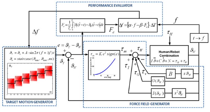
The
general control architecture consists of three blocks: 1) target motion
generator; 2) force filed generator; 3) performance evaluator.
Figure 2 shows (on the left) the control scheme named "Target Motion generator" and exemplifies a segment of the oscillatory pattern that span the entire ROM in a progressive manner. The Target Motion Generator
is characterized by the following set of equations that are sampled at 1
kHz by the inner control loop and they will be explained in present
section.
Figure 2
Controller diagram. The "assist-as-needed" force parabolic term continuously inputs torque τmwhen errors are present during the
tracking task. The input torque to the robot/hand system is the sum of
different contributions of a viscous field τv, a gravity τGand inertia τIcompensation. τHis the torque applied by the subjects wrist.
Here ϑWstands for the joint angular rotation of anyone of the three DoFs of the robot: F/E, Ab/Ad, P/S (figure 2). In particular, ϑTis the time-varying target angular position, characterized by an harmonic motion with frequency f, amplitude A, and bias or offset ϑo(eq. 1).
(1)
The bias is moved in a staircase manner (eq. 2), in order to progressively span the whole ROM of each DoF (ϑmin ↔ ϑmax) by means of ns steps (ns = 11 in our experiments).
(2)
Each step of the staircase has a duration
of 40s plus a 4s rest interval, during which the harmonic motion of the
target is stopped as well as the attractive force. For each DoF, the
ROM is scanned by the staircase starting from the "easier" to the "more
difficult" angular position, taking into account the specific
pathological conditions of the treated subjects. In this feasibility
study the sequence was, for all the patients, from Flexion to Extension,
from Adduction to Abduction, and from Pronation to Supination,
respectively. The sequence is ordered "from easy to difficult"
considering the hypertonic trend in the range of motion for each trained
DoF: 1) the offset angle steps from the easy (more natural and less
hypertonic) to the difficult (less natural) joint configuration; 2) the oscillation is modulated from slow (easy) to quick (difficult) frequency.
Table 2
shows the amplitude of the target oscillations and the range of values
of the angular offset/bias: such range is divided into 11 parts
corresponding to the steps of the staircase. Therefore each step
amplitude is different for the different three spaced ROMs. Thus, the
subjects are progressively trained in a limited workspace but the
gradual change of the offset angle allows them to experience the whole
ROM for each single DoF (as a progressive splinting). The initial
position was chosen taking into consideration the specific pathological
conditions; i.e. subjects train each Dof starting form the less
hypertonic portion of each ROM to gradually space the whole workspace.
Table 2 Growth and decay coefficients of Eq. 9 for each DOF and amplitude oscillation and max/min ROM for each Dof
Eq. 3 identifies the tracking error for each time instant (ϑwis the current angular position of the wrist DoF) which is input in the "Force Field Generator" and the "Performance Evaluator".
(3)
The assistive torque provided by the motor is computed in the "Force Field Generator" according to eq. 4 and then transformed into the corresponding current drive.
(4)
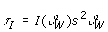
The actual delivered torque τwis the sum of different control efforts that consider assistance τm(eq. 5), gravity compensation τG(eq. 6), inertia compensation τr(eq. 7) and a viscous field τv(eq. 8) in order to stabilize by a damping effect the unwanted oscillation at the end effector.
(5)
(6)
(7)
(8)
The different contribution of the force field generator is shown in figure 2 (right).
The assistive control law τmconsists of a non linear elastic field with a
parabolic profile (eq. 5). This non linear characteristic was chosen
according to the principle of minimal assistance [27] or also assist as needed [28]:
assistance forces/torques should be kept as low as possible in order to
promote the emergence of voluntary control. In fact, the chosen pattern
of assistance has a less-than-linear increase for small errors, thus
facilitating the emergence of active un-aided control at the end of
training; for large errors, which are likely to occur at the beginning
of training, the assistance grows more than linearly in order to speed
up the learning process. The same concept of minimal assistance is used
for selecting, in an individual-specific manner, the gain K:
it is chosen as the minimum value capable to induce the initiation of
movements of the paretic wrist and it was chosen by experimentally
observing the active voluntary movements of the participating subjects
before starting the rehabilitation protocol.
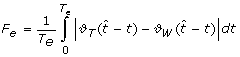
The "Performance Evaluator" computes intermittently the average angular error given by eq. 3 in a time window (Te= 2 s):
(9)
where is the time instant at which the current oscillation terminates or also the zero-crossing of the ϑT-ϑWwaveform.
The "Performance Evaluator" modulates the "difficulty" of the tracking task, i.e. the oscillation frequency f = 1/ΔT, by changing it in a smooth way at the end of each complete oscillation cycle according to the following equation:
(10)
The equation contains two terms: a raising term with a coefficient α and a decaying term depending on the average angular error Femultiplied by the decay coefficient β. For clarity sake figure 2
shown the entire controller scheme highlighting the different blocks of
the controller. There are also two saturation levels that keep the task
in a suitable range of difficulty: we chose the range 0.1-1.0 Hz
empirically, looking at the performance of the unimpaired subjects. Also
the values of α and β for each DoF were experimentally chosen, in order to balance the conflicting requirements of readiness and smoothness and provide a symmetric counterbalance of decaying and raising contributions: these values are listed in table 2.
During the performance of an exercise, when eq. 2 switches the offset ϑofrom one step to the next one, the initial value of eq. 10
is reset to the minimum value of frequency (0.1 Hz). Therefore, the
initial target oscillation will be very slow and will smoothly speed-up
as a function of the tracking accuracy e = ϑT- ϑW, until the end of the step (40s).
Virtual Reality environment
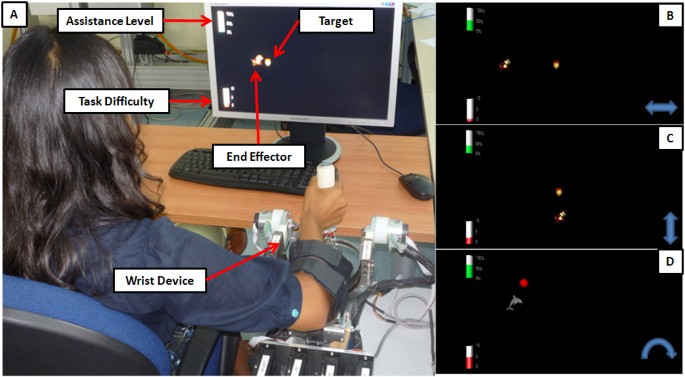
The VR process displays on the screen the trajectory of the target and the wrist angular position (figure 3). The target and the wrist positions are represented graphically as 'pleasant' images: a dolphin chasing a ball or a squirrel hunting an acorn.
The target path on the PC screen is horizontal in the F/E experiment,
vertical in the Ab/Ad experiment, and a circular segment in the P/S
experiment.
Figure 3
Virtual reality environment in the therapy session.
A) Experimental set-up in the P/S case: the dolphin chasing the ball.
The two bars on the left of the screen display two performance
indicators. B) F/E excercise; D) Ab/Ad excercise; D) P/S exercise.
We
wanted to strengthen the effectiveness of the system in monitoring
wrist use while providing encouragement and reminders throughout a
therapy session [29].
Hence
we also display, on the left side of the screen, the instantaneous
levels of the two performance indicators by means of height-modulated
bars: 1) the level of assistance and 2) the frequency of oscillation.
The patients were instructed to minimize the height of the former one
while maximizing the height of the latter. This kind of intuitive
performance feedback was easily understood by the patients and well
appreciated by them.
Subjects
Three
stroke subjects volunteered to participate in this preliminary study.
The recruitment was among the outpatients of the ART Rehabilitation and
Educational Centre (Genoa, Italy), and based the following inclusion
criteria: 1) diagnosis of a single, unilateral stroke verified by brain
imaging; 2) sufficient cognitive and language abilities to understand
and follow instructions; 3) chronic condition (at least 1 year after
stroke). Table 3
summarizes the anagraphic data (age, sex) and the clinical state
(etiology, disease duration, affected side, Fugl Meyer and Ashworth
scores) collected at the ART Rehabilitation and Educational Centre
(Genoa, Italy). The research conforms to the ethical standards laid down
in the 1964 Declaration of Helsinki, which protects research subjects.
Each subject signed a consent form that conforms to these guidelines.
The robot training sessions were carried out at the Human Behaviour Lab
of IIT (Genoa, Italy), under the supervision of an experienced
physiotherapist of the ART Rehabilitation and Educational Center.
The following parameters were estimated for each DoF:
Max frequency: the maximal frequency that the subject is able to reach, in the possible range 0.1-1 Hz;
Mean assistive torque: the average torque delivered to the patient during the rehabilitation protocol for each DoF
ROM achieved in the single step;
Mean speed.
Moreover we estimated:
The ROM in the whole session (minimum-maximum degree of movement in the entire exercise).
The active voluntary ROM of the subject holding
the passive inactivated device, before and after the exercise in order
to compare if the rehabilitation protocol would provide fast benefits
even after one therapy session.
Results
Although the clinical states of the three subjects are rather different, as reported in table 3,
all of them were able to carry out the proposed exercises in a
consistent way, with different performance profiles considering the
performance adaptive nature of the controller architecture. For clarity
sake, in the present preliminary/feasibility study, the following
figures will refer to subject S3, who is the most severely affected and
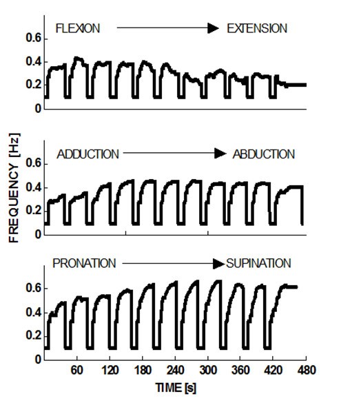
therefore the worst case in the experienced population. Figure 4 shows the evolution of the frequency of the moving target for each DoF, while the ϑoposition scans through the 11 values that are
uniformly placed in the corresponding ROM: 40s for each step + 4s of
rest between one step and the next one. For each step, the peak value of
the frequency depends on the position in the workspace of each DoF and
on the specific pathological condition of each patient: the figure shows
that S3 has higher difficulty in extension than flexion, in adduction
than abduction, and in pronation than supination.
Figure 4
Course of the target frequency when the offset position steps through the ROM.
At the beginning of each step the frequency is reset to its minimum
value (0.1 Hz); the maximum possible value is 1 Hz. Subject S3.
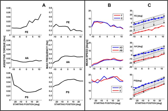
Figure 5A
summarizes the trend of the peak frequency at the different steps
comparing it with the corresponding evolution of the assistive torque
provided by the robot. It appears that the two sets of curves provide
compatible and complementary messages as regards the overall performance
of S3: he reaches peak frequency at about full flexion and mid-range of
abduction/adduction and prono/supination; in the same areas the
assistance torque reaches local minima, highlighting the fact that
higher performance is obtained when a higher capability of voluntary
motion is present needing a lower level of assistance.
Figure 5
complementary analysis between assistive torque and maximum frequency reached during tracking (subject S3).
(A) Left panel: Maximal target frequency reached for the different DOFs
during the 40s steps, identified by the starting position in the ROM
with respect to the neutral position. Right panel: Mean value of the
assistive torque (in 10-3Nm) during the corresponding steps.(B) Mean
tracking speed, for the different DOFs, in the different 40s steps,
identified by the starting position in the ROM with respect to the
neutral position. Gray and black curves correspond to the opposing parts
of the movements (F vs. E, Ad vs. Ab, P vs. S). (C) For each value of
the offset rotation and each DOF, the graphs show the ROM of the robot
(shaded band) and the ROM of subject S3 (black curves). X-axis
identified the spammed ROM for the exercised Dof; positive and negative
value are referred respectively to F/E, Ab/Ad and P/S while zero is the
neutral position. Y-axis is the amplitude oscillation reached by the
target (shaded band) and by the subject.
The information provided by figures 4 and 5A
is complemented by the measurement of the Active ROM (voluntary
capability of moving) for each type of movement of the wrist DoFs. These
measurements were carried out at the beginning and at the end of the
training session, by using the same wrist robot in order to normalize
the intrinsic constraints (biomechanical and neurological) as well as
the constraints determined by the robot. In the measurement, only one
DoF at a time was allowed to move freely (with no assistive control
applied), while the two remaining DoF were hold by the robot in the
approximated neutral positions. Table 4
summarizes the measurements before starting the protocol. Shaded cells
correspond to the more impaired movements for each subject: 1) all of
them lack mobility in Extension rather than Flexion; 2) S1 has a higher
deficiency in Abduction that in Adduction, while S2 and S3 have the
opposite impairment; 3) S1 shows a higher deficit in Supination whereas
S2 and S3 are worse in Pronation.
Table 4 Active Range of motion of the subjects pre and post treatment
A
similar kind of pattern, i.e. asymmetry of performance for easier vs.
more difficult movement directions, can be shown as regards the maximal
values of frequency reached by the target (table 5).
Table 5 Maximal frequency reached and average assistive torque
We
can also observe that minimal frequency values correspond to the
position in which subjects have a reduced range of motion. Moreover,
table 5
shows that maximal assistive joint torque is generally provided on the
side of the movement of each DoF where the subject is more defective.
The
performance of the subjects can also be investigated by comparing the
mean speed of the two opposite movements for each DoF in relation with
each offset step of the staircase (Figure 5B: F vs. E, Ad vs. Ab, and P vs. S).
We
can observe that, for each DoF, the speed curves for the opposing
rotations are quite similar in spite of the fact that there is a
significant asymmetry in the ROM, as shown in tables 4
before and after threatment. This suggests that the training protocol
is effective in two main ways, by inducing at the same time the patient
to behave in a more functional and physiological way:
1)
exercising movements that are more difficult
for him/her, given his specific pathological condition, for example
Extension vs. Flexion;
2)
moderating the predominance of pathology-aided behaviours that would enhance Flexion vs. Extension etc.
At last, figure 5C
compares, for each DoF, the ROM of the robot target motions (shaded
grey band is the amplitude of the target oscillation at different
starting position on each DoF workspace) with the actual ROM (bold lines
with markers for the two directions of each Dof) exhibited by patient
S3 in relation with each offset position. It appears that generally the
maximal joint rotation achieved by the patient is asymmetric in the two
opposing directions of each DoF (P vs S, F vs. E, Ad vs. Ab) and this is
reflected in the pattern of values stored in table 4
of the active range of motion measured by the uncontrolled device at
the beginning of protocol. i.e. In spite of the assistance, the subject
S3 does not succeed in following the harmonic motion of the target
represented by the shaded grey band; he systematically undershoots
extension (blue line) and overshoots flexion (red line), whereas the
performance is closer to physiological conditions for the two other
DoFs.
On the other hand, table 4
reports the active range of motion (uncontrolled device) measured at
the end of the training session and the comparison between the part of
the table 4
shows a clear increase and symmetrisation before and after the
treatment; this result suggests that using robot to generate mobilising
splints might be useful to modify the joint stiffness, and reducing
hypetonia; even if the total ROM is reduced the symmetry noticeably
increases; it is possible the passive component due to hyper tonicity
before the splinting added a bias to each joint drifting from the
anatomical neutral position.
In the lights of these considerations
however we present a preliminary study on the feasibility of using a
performance adaptive control strategy combined with a dynamic splinting;
in order to strengthen the effectiveness of the proposed approach a
wider clinical protocol with higher number of subjects and therapy
session is needed.
Discussion
Although
it has been shown in a number of studies that robots can decrease motor
impairment after stroke with certain advantages, less emphasis to date
has been put on robotic developments for the hand and on corresponding
preliminary clinical studies. A notable exception is the work by
Takahashi et al. [4]
who reported the use of the pneumatic-actuated HWARD wrist robot with
13 patients. The main difference of HWARD with respect to the Wrist
robot (here with reported) is related to the wrist movements: HWARD can
only operate with F/E whereas Wrist Robot can operate equally well with
Ab/Ad and P/S.
In this preliminary experiment investigating
patients, only one joint DoF was exercised at a time. The procedure
simulated as much as possible the use of splints widely used in clinical
applications. However, there is no hardware or software limitation to
design 2D and 3D experiments, which indeed are planned and will be
carried out in the near future.
We wish to emphasize that our
control system is based of a principle of minimal assistance that
focuses on the initiation of the movement; on the contrary most of the
other rehabilitation robots, focuses on the termination phase (goal
directed movements), by forcing the patient to complete the movements if
he/she is unable to achieve the target. We also plan to integrate in
the robot an active finger F/E unit, by means of a motorized handle [30]
to study the impact of single-DoF rehabilitation protocol on
cylindrical grasping and compare the effectiveness of different
rehabilitation strategies that include distal and/or proximal limb.
The
results reported in this single-session study show that the proposed
adaptive control strategy is robust, in terms of patient response, is
well accepted by the subjects and the control architecture is capable to
smoothly adapt to the specific impairments of the patients without
needing a fine customization of the controller gains for each subject;
this controller robustness allows to introduce the system in the
clinical application providing a user friendly interface for users and
patients, and to deliver an automatic execution of the therapy sessions.
Conclusion
The
results of the presented preliminary work shows that robotic therapy
may improve motivations in patients and provide tangible results even in
a short term experience. The technological approach with the use of
customized devices may strengthen the potentials of the regular physical
therapy in delivering assistance and training. The proposed controller
strategy is simply based on an automation of the well established
methodology of dynamic splinting; this kind of approach can result
familiar to the medical staff allowing technology to progressively take
part to the emerging and increasing needs of rehabilitation, without
shocking the entrenched application of regular therapy. It remains to be
investigated, as we plan to do in a systematic clinical trial, to which
extent a suitable protocol can induce permanent improvements in the
neural control of wrist movements, necessary for any attempt to achieve
functional gains in the activities of daily life.









 is the time instant at which the current oscillation terminates or also the zero-crossing of the ϑ
T
-ϑ
W
waveform.
is the time instant at which the current oscillation terminates or also the zero-crossing of the ϑ
T
-ϑ
W
waveform.