Changing stroke rehab and research worldwide now.Time is Brain! trillions and trillions of neurons that DIE each day because there are NO effective hyperacute therapies besides tPA(only 12% effective). I have 523 posts on hyperacute therapy, enough for researchers to spend decades proving them out. These are my personal ideas and blog on stroke rehabilitation and stroke research. Do not attempt any of these without checking with your medical provider. Unless you join me in agitating, when you need these therapies they won't be there.

What this blog is for:

My blog is not to help survivors recover, it is to have the 10 million yearly stroke survivors light fires underneath their doctors, stroke hospitals and stroke researchers to get stroke solved. 100% recovery. The stroke medical world is completely failing at that goal, they don't even have it as a goal. Shortly after getting out of the hospital and getting NO information on the process or protocols of stroke rehabilitation and recovery I started searching on the internet and found that no other survivor received useful information. This is an attempt to cover all stroke rehabilitation information that should be readily available to survivors so they can talk with informed knowledge to their medical staff. It lays out what needs to be done to get stroke survivors closer to 100% recovery. It's quite disgusting that this information is not available from every stroke association and doctors group.

Showing posts with label when the hell?. Show all posts

Saturday, October 5, 2024

Motor Cortex Activation During Treatment May Predict Therapeutic Gains in Paretic Hand Function After Stroke

 It is vastly more important to DELIVER HAND FUNCTION than predict it! When the hell will stroke leadership get a strategy going to DELIVER 100% RECOVERY? I'm guessing never since survivors are not in charge.

Yeah, Bruce Dobkin is a superstar stroke researcher but I think even he is not actually solving stroke to get survivors recovered.  Hope he has 100% stroke recovery solved before he becomes the 1 in 4 per WHO that has a stroke!

  • Dr. Bruce H. Dobkin (15 posts to December 2011)
  • Motor Cortex Activation During Treatment May Predict Therapeutic Gains in Paretic Hand Function After Stroke

    Abstract

    Background and Purpose— Functional brain imaging after stroke offers insight into motor network adaptations. This exploratory study examined whether motor cortical activation captured during arm-focused therapy can predict paretic hand functional gains.
    Methods— Eight hemiparetic patients had serial functional MRI (fMRI) while performing a pinch task before, midway, and after 2 weeks of constraint-induced therapy. The Wolf Motor Function Test (WMFT) was performed before and after intervention.
    Results— There was a linear reduction in ipsilateral (contralesional) primary motor (M1) activation (voxel counts) across time. The midpoint M1 Laterality Index anticipated post-therapeutic change in time to perform the WMFT. The change in ipsilateral M1 voxel count (pre- to mid-) correlated with the change in mean WMFT time (pre- to post-).
    Conclusions— The relationship between brain activation during treatment and functional gains suggests a use for serial fMRI in predicting the success and optimal duration for a focused therapeutic intervention.
    Functional MRI (fMRI) has revealed reorganization in the primary and secondary motor cortices during poststroke recovery and after therapeutic interventions.1 Few studies have explored the evolution of brain activation in relation to behavioral gains in a “one-to-one” correspondence during a specific rehabilitation intervention.2 This exploratory study examined whether the brain activation midway through a 2-week arm-focused intervention might capture adaptations induced by the initial week of training and, in turn, could be used to anticipate post-therapeutic behavioral changes in paretic hand function. If so, this brain–behavior correspondence may offer guidance to determine an optimal duration for task-specific therapy.2

    Subjects and Methods

    Eight patients with hemiparetic stroke (Fugl-Meyer [FM] motor score 33 to 62) participated. Inclusion criteria were >3 months after stroke, ability to perform the fMRI task, and a minimum of 10° of voluntary wrist and finger extension. Lesions varied in location, but all spared the hand motor representation (M1). No alternative therapy group was studied. Seven healthy volunteers were scanned twice to test the reproducibility of fMRI activation.

    Physical Therapy and Functional Measure

    All patients received constraint-induced therapy for 2 weeks as defined for the EXCITE trial.3 The Wolf Motor Function Test (WMFT)4 was performed before and after intervention. The behavioral outcome measure consisted of 6 dexterity items from the full 15-item WMFT (Lift Can; Lift Pencil; Lift Paper Clip; Stack Checkers; Flip Cards; Turn Key in Lock) that most directly captured fine motor control. The change in mean WMFT (mWMFT) time for the 6-item subset was correlated with that for the 15-item test (r=0.98), indicating reliability and validity for the subset. The pre-mWFT–post-mWFMT (absolute time) difference was used as a proxy for functional change in motor skill.

    fMRI Acquisition

    fMRI acquisition parameters were described previously.5 fMRI sessions were performed before intervention, midintervention, and after intervention, each with 4 30-s bouts of repetitive pinch alternating with 5 30-s rest periods. The pinch apparatus included a vertical plastic tube connected to a pressure transducer. The task required tube compression with the index and middle fingers against the thumb, creating enough pressure to match 50% of maximum, viewed through goggles as a target line, and paced by auditory cues at 75% maximum rate. These parameters were maintained constant across the 3 sessions. Practice before each fMRI session minimized unwanted movements and deviations from consistent task performance.

    Data Analysis

    fMRI data were analyzed as described previously.5,6 Volumes related to head motion (>2 mm), and associated movements (visually identified from videotape) were excluded. Z statistic images were thresholded at Z>3.1, and significant clusters were defined atP<0.01 (corrected for multiple comparisons). Regions of interest (ROIs) were set in bilateral M1 and dorsal premotor (PMd) areas. Percentage signal change (% SC) and voxel counts (VCs) within each ROI were measured and a Laterality Index [LI=(contralateral−ipsilateral)/(contralateral+ipsilateral)] (contralateral and ipsilateral activation to the hand movement. LI ranges from −1 [all ipsilateral activation] to 1 [all contralateral activation]) was calculated using VC for each ROI. Linear Mixed Model was used for intersession comparisons of fMRI variables (% SC, VC), pinch pressure, and rate, separately. Individual linear regression analyses were performed between LI, VC (M1 and PMd; independent variable) pre-, mid-, and post- and the post-pre–mWMFT time difference (dependent variable). Pearson correlation coefficient analysis was used to assess the relationship between changes in fMRI measures and changes in mWMFT time. Preintervention fMRI from patients 5 and 6 was technically unusable.

    More at link.

    Thursday, June 13, 2024

    Robot-like exoskeleton helps Tampa stroke patient walk again

    But will it get you back to 100% walking recovery while in the hospital?  If not, what is the EXACT followup rehab that will get you there? Don't have that and no plans to accomplish that? Then you aren't a functioning stroke hospital. Stroke survivors want 100% recovery! When the hell will you start delivering that?

     Robot-like exoskeleton helps Tampa stroke patient walk again

    TAMPA, Fla. — A robot-like device is helping stabilize patients at Tampa General Hospital and help them to walk again.

    Daily therapy means stroke patients and those suffering from spinal cord injuries or neurological conditions can work toward taking more steps each session.


    What You Need To Know

    • A robot-like device is helping stabilize Tampa General Hospital patients enough for them to walk again

    • Patrick McCreery suffered a stroke in April and is now using the Ekso Bionics exoskeleton as part of his treatment

    • The frame is customized with each patient’s measurements and level of assistance for their specific needs

    Patrick McCreery suffered a stroke in April, which caused him to lose function on the right side of his body. The 73-year-old has been using the Ekso Bionics exoskeleton as part of his treatment for about a month.

    The frame is customized with each patient’s measurements and level of assistance for their specific needs.

    “You take for granted the ability to walk until you no longer have the ability,” McCreery said. “So it feels really good.”

    With a chuckle, McCreery says his grandchildren have given him the nickname “RoboPop.”

    “I can control the speed, the step length, the step height,” said Manuel Garcia, Doctor of Physical Therapy at Tampa General Hospital’s Rehabilitation Center. “We can actually increase the speed on it as well, which is very important for muscle memory and neurological re-function of the brain.”

    Rehabilitation experts say this approach to treatment gets patients walking faster than with traditional treatments or technology.

    “We’re able to get patients up sooner, so getting upright is usually a big barrier because the weakness is very significant after a stroke,” said Dr. Rigoberto Nunez, who serves as vice chief of Physical Medicine and Rehabilitation at Tampa General Hospital.

    Garcia said the device also can help accelerate the patient’s progress.

    “The unique part about this is that we’re able to actually take about 1,000 steps, or up to 1,000 steps or more(That's not what needs to be measured; you ask the patient how close they feel to 100% recovery without the device!), compared to other walking devices or other walking treatments where we can only get up to 40 or 50 steps at a time, or per session,”  he said.

    McCreery said he is astounded by the progress he has made using the exoskeleton.

    “It is very incredible actually, if you think about it, to be able to walk when you're not supposed to,” said McCreery, who took more than 800 steps during his session.

    Video at link.

    Robot-like exoskeleton helps Tampa stroke patient walk again
    Robot-like exoskeleton helps Tampa stroke patient walk again
    Robot-like exoskeleton helps Tampa stroke patient walk again
    Robot-like exoskeleton helps Tampa stroke patient walk again

    Saturday, May 11, 2024

    Intravenous Alteplase in Patients With Minor Acute Ischemic Stroke — Where is the Limit?

     Survivors don't want 'better'. They want 100% recovery! WHEN THE HELL ARE YOU GOING TO DO THE WORK TO GET THERE? Maybe after you are the 1 in 4 per WHO that has a stroke?  Perhaps you might want to start researching those solutions now, while you still can.

    Intravenous Alteplase in Patients With Minor Acute Ischemic Stroke — Where is the Limit?

    Originally published 10.1161/blog.20240426.989508

    Zhang Y, Lv T, Nguyen TN, Wu S, Li Z, Bai X, Chen D, Zhao C, Lin W, Chen S, Sui Y. Intravenous Alteplase Versus Best Medical Therapy for Patients With Minor Stroke: A Systematic Review and Meta-Analysis. Stroke. 2024;55:883–892.

    Intravenous thrombolysis (IVT) is probably the most effective weapon for stroke physicians in acute ischemic stroke to achieve a better functional outcome. Guidelines and treatment recommendations evolved from a strict NIHSS >3 points regime to a more liberal indication, namely the use in case of disabling symptoms, regardless of NIHSS-scoring. Guidelines do not recommend alteplase thrombolysis for patients with mild non-disabling symptoms (NIHSS 0–5).

    At least half of acute ischemic stroke patients present with minor or mild symptoms at admission, but research showed that 29% of patients with minor stroke had a non-excellent outcome (mRS 2-6) after three months.1 Reasons for this are most likely progressive strokes or recurrent strokes. Most major trials excluded patients with mild or minor symptoms; therefore, safety and efficacy of IVT in these patients is not entirely clear. Moreover, the exploration of effects on specific subgroups in previous meta-analyses is not sufficient to draw final conclusions.


    Zhang et al. conducted a systematic review and meta-analysis on comparison of IVT with best medical therapy (BMT) in patients with minor stroke to find an answer to the question of IVT or no IVT in these patients.2

    The inclusion criteria for this study were patients with minor ischemic stroke (NIHSS score, 0–5) who could receive thrombolysis within 4.5 hours after onset of stroke; IVT with alteplase; comparison: BMT (including dual antiplatelet therapy [DAPT] or single use); and reporting of functional outcomes and any safety outcomes in randomized controlled trials (RCTs) and observational studies. Studies with patients who received mechanical thrombectomy or bridging therapy were excluded. The primary outcome was excellent functional outcome at 90 days (mRS 0-1). Secondary outcomes included favorable functional outcome at 90 days (mRS 0-2), mortality, early neurological deterioration, recurrent stroke, and recurrent ischemic stroke. Safety outcomes included symptomatic intracranial hemorrhage (sICH) and hemorrhagic transformation. Of 5393 publications found until August 2023, 3 RCTs and 17 observational studies were included.

    Rates of excellent functional outcome (mRS 0-1) showed no significant difference comparing IVT and BMT (2252/2717, 82.89% versus 3295/4073, 80.90%, p=0.274). The pooled estimated effect on mRS score of 0-1 at 90 days was consistent in each predefined subgroup including age >80 years, disabling symptoms, comparing different antiplatelet regimes, large vessel occlusion, thrombolytic time window of 0 to 3 hours or 0 to 4.5 hours, baseline NIHSS score of 0-3 or 0-5 and premorbid mRS scores of 0 and 0-2. No differences were disclosed between subgroups.

    Also, rates of favorable functional outcome (mRS 0-2) showed no significant difference (1790/1960, 91.33% versus 2878/3188, 90.28%, p=0.141). Mortality and recurrent stroke or recurrent ischemic stroke at 90 days also showed no difference comparing IVT and BMT. The study revealed a higher rate of early neurological deterioration, sICH, and hemorrhagic transformation in patients treated with IVT.

    This meta-analysis showed no difference between IVT and BMT in patients with minor ischemic stroke (NIHSS score ≤5) regarding excellent or good functional outcome at 90 days. IVT was associated with an increased risk of safety outcomes, such as early neurological deterioration, sICH, and hemorrhagic transformation, while BMT was similar to IVT in preventing recurrent stroke and recurrent ischemic stroke. All in all, functional outcome of patients with nondisabling strokes is not improved by intravenous alteplase, and a small number of patients may actually be harmed by thrombolysis.

    These results are subject to use of alteplase as thrombolytic therapy agent. No RCTs on other intravenous thrombolytics such as tenecteplase are available in patients with minor stroke. The TEMPO-2 trial could give more insight, but it is subject to patients with mild deficits and large vessel occlusion. The TRUST trial will assess urokinase in comparison to antiplatelet agents for the management of minor ischemic stroke and is ongoing. Limitations include the lack of a universal recognized definition of disabling symptoms and inconsistencies among the included studies in terms of the time of treatment initiation, baseline NIHSS score, definitions of minor stroke, and dosing paradigms of antiplatelets.

    Stroke physicians often see spontaneous neurological improvement in patients with mild symptoms; therefore, these results seem reassuring. The critical question is, what makes a mild/minor stroke a non-disabling stroke? Stroke physicians want to rapidly administer IVT, but in cases like these, we should carefully weigh the risks and benefits and consider the patients’ life circumstances to evaluate if the suspected stroke is causing mild, truly non-disabling symptoms.3 For example, a vocalist with mild dysarthria might consider this a disabling symptom, and a waiter might consider mild paralysis of the non-dominant hand as disabling. It is, therefore, of uttermost importance to consider the whole picture and to carefully assess the risk for sICH like considering age and comorbidities in a setting where time is the enemy.

    There are several questions which remain to be answered. First, the arrival of new thrombolytic agents for acute stroke reperfusion therapy, i.e., tenecteplase, might exploit other new RCTs to address the question if IVT with other substances — which are endowed with an increased half-life compared to alteplase — might reduce the risk of progressive stroke and show a beneficial effect on functional outcomes in acute minor ischemic stroke.

    Second, most patients who receive IVT in case of mild symptoms in observational studies might be seen as being at a formally higher risk of neurological worsening and, therefore, received IVT despite minor symptoms. These scenarios could include cervical artery dissection, instable plaques, or intracranial stenosis — therefore, one has to raise the question of a selection bias in favor of IVT in these patients.

    Third, one has to keep in mind the patients who undergo endovascular treatment and achieve a good outcome. The effect of endovascular treatment on functional outcomes in high-risk patients with minor stroke might narrow the treatment effect of IVT on functional outcomes.

    Finally, future research might explore other beneficial effects of IVT and reperfusion in general besides short-term functional outcome, like cognitive function and poststroke dementia.

    To conclude, IVT with alteplase seems not to be beneficial in non-disabling stroke to ameliorate functional outcome in the short-term. The arrival of tenecteplase in clinical practice, specific subgroups with potential benefit, and the exploration of other outcomes besides functional outcome after 3 months warrant further investigation of IVT in minor acute ischemic stroke.

    Thursday, May 2, 2024

    Stroke Recovery Tips May 2024

    Actually survivors don't want lazy 'tips'! They want 100% recovery protocols! WHEN THE HELL WILL YOU DO THAT?

    This tyranny of low expectations needs to stop. I'd suggest screaming at your stroke medical 'professionals' for not solving stroke! Maybe you can get them motivated so your children and grandchildren will recover from their strokes.

     Stroke Recovery Tips May 2024


    Tuesday, April 23, 2024

    Endovascular therapy improved ischemic stroke outcomes at 1 year

     You blithering idiots actually think 'improves' is what survivors want? They want 100% recovery; nothing less! WHEN THE HELL WILL YOU START TRYING TO GET THERE?

    I'd have you all fired for incompetence!

    Endovascular therapy improved ischemic stroke outcomes at 1 year 

    Key takeaways:

    • The study examined 352 individuals with stroke randomized to EVT and medical care or medical care alone.
    • EVT intervention led to improved functional independence and better ambulation.

    DENVER — In patients with ischemic stroke, treatment with endovascular therapy with medical care led to better functional outcomes at 1 year compared with medical care alone. according to a speaker.

    “In 2023, we had several trials that were groundbreaking in our field, that showed significant benefit for endovascular therapy for patients who present with large core ischemic stroke,” Amanda Opaskar, MD, a vascular neurologist and assistant professor of neurology at Case Western Reserve University, said during her presentation at the American Academy of Neurology annual meeting.

    Ischemic Stroke
    Recent research has determined benefits of endovascular therapy for ischemic stroke outcomes at 1 year. Image: Adobe Stock

    “The question was how do these patients recover over that longer term, first year.”

    Opaskar and colleagues sought to evaluate safety and efficacy of endovascular therapy (EVT) for ischemic stroke in the SELECT2 trial using clinical outcomes at 1-year follow-up. SELECT2 was a phase 3, international, multicenter, prospective, randomized, open-label trial with blinded endpoint assessment.

    It included 352 individuals diagnosed with a large ischemic core on non-contrast CT (ASPECTS 3-5) scan, perfusion or MRI who received either EVT and best medical management (n = 178; median age 66 years; 71% female) or best medical management only (n = 174; median age 67 years; 74% female).

    The study’s primary outcome was modified Rankin Scale (mRS) score at 1-year follow-up, evaluated using generalized odds ratio (GOR), with secondary outcomes including functional independence (0 to 2 mRS score), independent ambulation (mRS 0 to 3) and quality of life scores at 1-year follow-up.

    A total of 93% (329 of 352) of enrollees completed the follow-up, with 23% of those who received EVT recording an mRS score of 0 to 2 compared with 8% of those who received medical care only.

    Thrombectomy significantly improved the 1-year mRS score distribution (WMW probability of superiority = 0.59; 95% CI, 0.53-0.64; GOR = 1.43; 95% CI, 1.14-1.78), functional independence (EVT: 24% vs MM: 6%, RR = 3.17; 95% CI, 1.73-5.79) and independent ambulation (EVT: 37% vs MM: 18%, RR =1.85; 95% CI, 1.3-2.63) compared with medical care alone.

    Data additionally showed EVT led to better QoL scores in functional independence, mobility, ambulation, sociability, cognition and depression compared with medical care alone.

    “It is important to note we think about the potential recovery of these patients both in clinical trial design as well as when we are counseling (them),” Opaskar said. “That their recovery extends not just in 3 months, but it goes longer, up to a full year.”

    Sources/Disclosures

    Collapse

    Source:

    Opaskar A, et al. Long-term outcomes and EVT treatment effect from randomized controlled trial of endovascular thrombectomy for large ischemic strokes (SELECT2). Presented at: American Academy of Neurology annual meeting; April 13-18, 2024; Denver.

    Disclosures: Opaskar reports no relevant financial disclosures.

    Tuesday, July 18, 2023

    FUNCTIONAL MAGNETIC RESONANCE IMAGING (FMRI) AS A PREDICTIVE TOOL FOR REHABILITATION OUTCOMES IN STROKE PATIENTS

    When the hell are you people going to actually deliver EXACT RECOVERY PROTOCOLS? Instead of this crapola of predicting failure to recover?


    Tuesday, August 16, 2022

    Uruguay adopts pioneering plan for treating patients with strokes

     So they have access to 'care'. Survivors want results which means 100% recovery. WHEN THE HELL ARE YOU GOING TO GET THERE? Not anytime soon with the WSO not leading anything.

    Uruguay adopts pioneering plan for treating patients with strokes

    All cases of stroke will be admitted through a single, universal and free emergency door for treatment,” the MSP explained All cases of stroke will be admitted through a single, universal and free emergency door for treatment,” the MSP explained

    Uruguayan President Luis Lacalle Pou last week signed the decree whereby his country would join the Global Stroke Alliance, after an international conference held in Sao Paulo, Brazil.

    Under the new scheme, Uruguay's Health Ministry (MSP) guarantees for universal and free emergency care in case of strokes.

    Now, “any user of the National Integrated Health System who presents symptoms of having a stroke must be transferred to the closest or most accessible thrombolysis center in the place where he/she is, regardless of the health provider to which he/she is affiliated. In other words, all cases of stroke will be admitted through a single, universal and free emergency door for treatment,” the MSP explained.

    Also resulting from this year's meeting, Uruguay will be holding the 2023 edition in Punta del Este in November. “It is an honor for Uruguay to have been designated today, on the basis of its progress in stroke, for the first time as the venue for the Latin American and Caribbean meeting of the Global Stroke Alliance, in conjunction with the Ibero-American Society of Cerebrovascular Disease,” Health Minister Daniel Salinas pointed out.

    The goal of the World Stroke Organization (WSO) is to change the line of work in the approach to preventing cardiovascular pathologies and strokes. During the Sau Paulo event, the organization and Brazilian Health authorities praised Uruguay's “innovative and egalitarian” plan.

    In 2020 the MSP created the brain health program, the first and only one in the Americas and the third worldwide, together with Poland and Norway, which positions the country as an outstanding reference in the subject at a global level, with achievements such as the creation of a national stroke protocol, which brings together national guidelines for the approach to acute ischemic stroke; the incorporation in December 2021 of financial coverage through the National Resources Fund of mechanical thrombectomy for the treatment of hyperacute ischemic stroke; the training of 3,000 healthcare professionals for the best approach to this type of episodes; and now the single, universal availability of treatment.

    Sunday, July 10, 2022

    Rehabilitation of Motor Function after Stroke: A Multiple Systematic Review Focused on Techniques to Stimulate Upper Extremity Recovery

    We don't need your tyranny of low expectations; 'promising outcomes'.  Survivors want 100% recovery!  WHEN THE HELL ARE YOU GOING TO GET THERE?

    I'd have you all fired for incompetence!

    Rehabilitation of Motor Function after Stroke: A Multiple Systematic Review Focused on Techniques to Stimulate Upper Extremity Recovery

    Samar M. Hatem1,2,3*, Geoffroy Saussez2, Margaux della Faille2, Vincent Prist4, Xue Zhang5, Delphine Dispa2,6 and Yannick Bleyenheuft2
    • 1Physical and Rehabilitation Medicine, Brugmann University Hospital, Brussels, Belgium
    • 2Systems and Cognitive Neuroscience, Institute of Neuroscience, Université Catholique de Louvain, Brussels, Belgium
    • 3Faculty of Medicine and Pharmacy, Faculty of Physical Education and Physiotherapy, Vrije Universiteit Brussel, Brussels, Belgium
    • 4Physical and Rehabilitation Medicine, Centre Hospitalier de l'Ardenne, Libramont, Belgium
    • 5Movement Control and Neuroplasticity Research Group, Motor Control Laboratory, Department of Kinesiology, Katholieke Universiteit Leuven, Leuven, Belgium
    • 6Physical Medicine and Rehabilitation, Cliniques Universitaires Saint-Luc, Université Catholique de Louvain, Brussels, Belgium

    Stroke is one of the leading causes for disability worldwide. Motor function deficits due to stroke affect the patients' mobility, their limitation in daily life activities, their participation in society and their odds of returning to professional activities. All of these factors contribute to a low overall quality of life. Rehabilitation training is the most effective way to reduce motor impairments in stroke patients. This multiple systematic review focuses both on standard treatment methods and on innovating rehabilitation techniques used to promote upper extremity motor function in stroke patients. A total number of 5712 publications on stroke rehabilitation was systematically reviewed for relevance and quality with regards to upper extremity motor outcome. This procedure yielded 270 publications corresponding to the inclusion criteria of the systematic review. Recent technology-based interventions in stroke rehabilitation including non-invasive brain stimulation, robot-assisted training, and virtual reality immersion are addressed. Finally, a decisional tree based on evidence from the literature and characteristics of stroke patients is proposed. At present, the stroke rehabilitation field faces the challenge to tailor evidence-based treatment strategies to the needs of the individual stroke patient. Interventions can be combined in order to achieve the maximal motor function recovery for each patient. Though the efficacy of some interventions may be under debate, motor skill learning, and some new technological approaches give promising outcome prognosis in stroke motor rehabilitation.

    Introduction

    The World Health Organization (WHO) estimates that stroke events in EU countries are likely to increase by 30% between 2000 and 2025 (Truelsen et al., 2006). The most common deficit after stroke is hemiparesis of the contralateral upper limb, with more than 80% of stroke patients experiencing this condition acutely and more than 40% chronically (Cramer et al., 1997). Common manifestations of upper extremity motor impairment include muscle weakness or contracture, changes in muscle tone, joint laxity, and impaired motor control. These impairments induce disabilities in common activities such as reaching, picking up objects, and holding onto objects (for a review on precision grip deficits, see Bleyenheuft and Gordon, 2014).

    Motor paresis of the upper extremity may be associated with other neurological manifestations that affect the recovery of motor function and thus require focused therapeutic intervention. Deficits in somatic sensations (body senses such as touch, temperature, pain, and proprioception) after stroke are common with prevalence rates variously reported to be 11–85% (Carey et al., 1993; Yekutiel, 2000; Hunter, 2002). Functionally, the motor problems resulting from sensory deficits after stroke can be summarized as (1) impaired detection of sensory information, (2) disturbed motor tasks performance requiring somatosensory information, and (3) diminished upper extremity rehabilitation outcomes (Hunter, 2002). Sensation is essential for safety even if there is adequate motor recovery (Yekutiel, 2000). Also, up to 50% of patients experience pain of the upper extremity during the first year after stroke, especially shoulder pain and complex regional pain syndrome-type I (CRPS-type I), which may impede adequate early rehabilitation (Jönsson et al., 2006; Kocabas et al., 2007; Sackley et al., 2008; Lundström et al., 2009). Furthermore, joint subluxation and muscle contractures can lead to nociceptive musculoskeletal pain (de Oliveira et al., 2012). Among other complications of stroke the neglect syndrome (Ringman et al., 2004) and spasticity (Sommerfeld et al., 2004; Welmer et al., 2010) affect motor and functional outcomes.

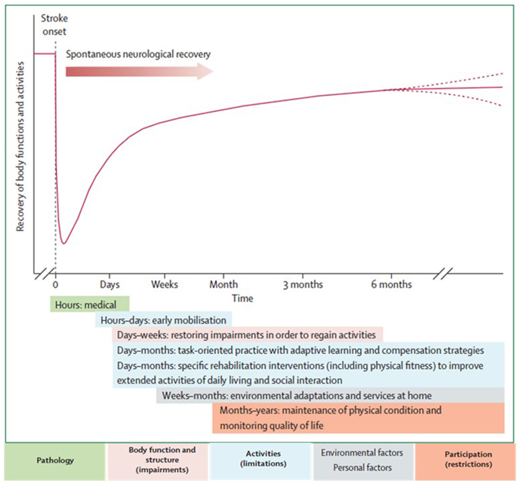
    The neurological recovery after stroke displays a nonlinear, logarithmic pattern (Figure 1; Kwakkel et al., 2006; Langhorne et al., 2011). The greater part of recovery is reported to take place in the first 3 months following stroke (Wade et al., 1983). However, there is evidence that recovery is not limited to this time period; hand and upper extremity recovery has been reported many years after stroke (Carey et al., 1993; Yekutiel and Guttman, 1993). Improvement probably occurs through a complex combination of spontaneous and learning-dependent processes including: restitution, substitution, and compensation (Kwakkel et al., 2004; Langhorne et al., 2011). Until the third month after stroke onset, a variable spontaneous neurological recovery can be considered a confounder of rehabilitation intervention (Kwakkel et al., 2006). In the past, the observation of spontaneous recovery after stroke has misled some authors to believe that recovery of upper extremity function is intrinsic and that little can be done by therapists to influence it (Wade et al., 1983; Heller et al., 1987). Progresses in functional outcome appearing after 3 months seem largely dependent on learning adaptation strategies (Kwakkel et al., 2004). Evidence suggests that neurological repair through brain reorganization supporting true recovery or, alternatively through compensation, may also take place in the subacute and chronic phase after stroke (Krakauer, 2006).

    FIGURE 1
    www.frontiersin.org

    Figure 1. Hypothetical pattern of recovery after stroke with timing of intervention strategies. The neurological recovery after stroke displays a nonlinear, logarithmic pattern. The greater part of recovery is reported to take place in the first three months following stroke. Rehabilitation interventions targeting at improving a stroke patients' performance should be implemented according to the phase of neurological recovery. Reprinted from Langhorne et al. (2011), Copyright [2011] by Elsevier. Reprinted with permission.

    Functional imaging of stroke recovery corroborates this temporal pattern of activation shifts. Shortly after stroke, an initial contralesional shift of activation toward the “unaffected” hemisphere is observed, followed by the activation of learning-related brain structures (including the cerebellum, basal ganglia, and frontal cortices) (Hikosaka et al., 1998; Lehéricy et al., 2005). Finally, two activation patterns are described depending on the degree of recovery (related to the amount of remaining fibers in the impaired corticospinal tract), either a perilesional (refocusing), or a distributed recruitment pattern (Feydy et al., 2002; Ween, 2008). Rehme et al. (2012) confirmed this last assumption and concluded that a good functional outcome relies on the recruitment of the original functional network rather than on contralesional activity. The meta-analysis by Richards et al. (2008) concluded that brain activations increase within the lesioned hemisphere after an upper extremity rehabilitation program. Brain plasticity including reorganization and compensation processes is the base for neurological recovery, as described above, however the exact pathophysiological mechanisms underlying rehabilitation's efficacy remain unclear (Eliassen et al., 2008).

    Stroke recovery is heterogeneous in terms of functional outcome. Patients with mild to moderate upper extremity paresis in acute phase have a good prognosis for functional recovery, as 71% of these patients achieve at least some dexterity at 6 months after stroke (Nijland et al., 2010). The prognosis in severely affected patients is poor with about 60% failing to achieve some dexterity at 6 months after stroke (Kwakkel et al., 2003; van Kuijk et al., 2009). Finally, only 5% of patients who initially experienced complete paralysis achieve functional use of their arm. Upper extremity impairments chronically affect the functional independence and satisfaction in 50–70% of all stroke patients. (Bonita and Beaglehole, 1988).

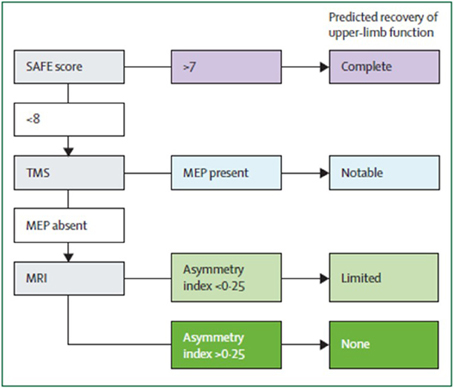
    Algorithms have been developed to predict motor function recovery after stroke (Stinear et al., 2007). Predictor variables include age, sex, lesion site, initial motor impairment, motor-evoked potentials, and somatosensory-evoked potentials. Initial measures of upper extremity impairment and function were found to be the most significant predictors of upper extremity recovery (Coupar et al., 2012). Findings so far suggest that the first assessments should be quick and simple, such as bedside tests of motor impairment, with progression to more complex tests if uncertainty remains (Figure 2). Later tests can include neurophysiological assessments and neuroimagery of the motor system integrity.

    FIGURE 2
    www.frontiersin.org

    Figure 2. Suggested sequence of tests to predict the recovery of motor function in patients with subacute stroke (weeks after stroke). Although this particular algorithm requires validation, it illustrates a potentially efficient progression from simple to more complex predictive measures. SAFE, sum of muscle force on shoulder abduction and finger extension according to Medical Research Council muscle grades at 72 h after stroke; TMS, transcranial magnetic stimulation; MEP, motor evoked potentials in the affected upper limb; Asymmetry index, asymmetry index of fractional anisotropy in the posterior limbs of the internal capsules measured with diffusion-weighted MRI. From Stinear et al. (2014).

    Interdisciplinary complex rehabilitation interventions represent the mainstay of post-stroke care (Langhorne and Legg, 2003; Langhorne et al., 2011). Stroke rehabilitation aims at providing all possible means to recover lost function and to increase the autonomy of stroke patients taking into account the remaining impairments and disabilities. Carr and Shepherd (2011) suggested that poor upper extremity recovery may be due to the direct impact of the stroke itself as well as to insufficient, inadequate or inappropriate therapeutic interventions. Little information is available, however, to describe what best represents “optimum treatment” (Ballinger et al., 1999). From a theoretical point of view, a stroke rehabilitation program for upper extremity motor impairment should include global motor rehabilitation, electrical brain stimulation, hemispheric subspecialization in motor activities, and multisensory interaction (Johansson, 2011). A recent Cochrane review focussing on the recovery of function and mobility in stroke patients reported the potential benefit of rehabilitation therapy on motor impairments and disabilities, compared with no treatment, in function of the time since stroke (Pollock et al., 2014). While these type of systematic reviews and meta-analyses are very powerful, they only take into account rehabilitation techniques that already have been reported in other systematic reviews and may thus ignore rehabilitation approaches that pertain to the routine clinical setting. Furthermore, in most systematic reviews only randomized controlled trials are reported.

    The purpose of the present manuscript was to undertake a systematic review for each of the neurorehabilitation techniques that may be useful in promoting upper extremity motor recovery. The search terms and inclusion criteria of reported trials have been chosen as large as possible in order to detect pertinent information on rehabilitation methods that are currently used in clinical practice, but are uncommonly discussed in systematic reviews (examples: music therapy, motor skill learning, isokinetic muscle strengthening, paired associative stimulation, theta burst stimulation). In some cases, routine clinical treatments that have not been investigated in a randomized controlled way, are still included in the present systematic review if the trial demonstrated sufficient quality evidence. The scientific evidence of each stroke rehabilitation intervention is discussed and presented with a practical recommendation for clinicians working in the field of neurorehabilitation. A decisional tree according to the patient's characteristics is proposed based on scientific evidence available for the different interventions.

    More at link.

    Wednesday, May 11, 2022

    Exosomes Derived From Mesenchymal Stem Cells: Novel Effects in the Treatment of Ischemic Stroke

     When the hell will we get EXACT REHAB PROTOCOLS from exosome research? Specifics names only!

    Exosomes Derived From Mesenchymal Stem Cells: Novel Effects in the Treatment of Ischemic Stroke

    Yu Xiong1, Jianping Song2,3,4,5,6,7, Xinyue Huang1, Zhigang Pan1, Roland Goldbrunner8, Lampis Stavrinou9, Shu Lin10,11*, Weipeng Hu1*, Feng Zheng1* and Pantelis Stavrinou8,12
    • 1Department of Neurosurgery, The Second Affiliated Hospital, Fujian Medical University, Quanzhou, China
    • 2Department of Neurosurgery, Shanghai Medical College, Huashan Hospital, Fudan University, Shanghai, China
    • 3National Center for Neurological Disorders, Shanghai, China
    • 4Neurosurgical Institute of Fudan University, Shanghai, China
    • 5Shanghai Clinical Medical Center of Neurosurgery, Shanghai, China
    • 6State Key Laboratory of Medical Neurobiology, MOE Frontiers Center for Brain Science, Institutes of Brain Science, Fudan University, Shanghai, China
    • 7Department of Neurosurgery, National Regional Medical Center, Fudan University Huashan Hospital Fujian Campus, The First Affiliated Hospital Binhai Campus, Fujian Medical University, Fuzhou, China
    • 8Department of Neurosurgery, Faculty of Medicine and University Hospital, Center for Neurosurgery, University of Cologne, Cologne, Germany
    • 92nd Department of Neurosurgery, Athens Medical School, “Attikon” University Hospital, National and Kapodistrian University, Athens, Greece
    • 10Centre of Neurological and Metabolic Research, The Second Affiliated Hospital of Fujian Medical University, Quanzhou, China
    • 11Diabetes and Metabolism Division, Garvan Institute of Medical Research, Sydney, NSW, Australia
    • 12Department of Neurosurgery, Metropolitan Hospital, Athens, Greece

    Ischemic stroke is defined as an infarction in the brain, caused by impaired cerebral blood supply, leading to local brain tissue ischemia, hypoxic necrosis, and corresponding neurological deficits. At present, revascularization strategies in patients with acute ischemic stroke include intravenous thrombolysis and mechanical endovascular treatment. However, due to the short treatment time window (<4.5 h) and method restrictions, clinical research is focused on new methods to treat ischemic stroke. Exosomes are nano-sized biovesicles produced in the endosomal compartment of most eukaryotic cells, containing DNA, complex RNA, and protein (30–150 nm). They are released into surrounding extracellular fluid upon fusion between multivesicular bodies and the plasma membrane. Exosomes have the characteristics of low immunogenicity, good innate stability, high transmission efficiency, and the ability to cross the blood–brain barrier, making them potential therapeutic modalities for the treatment of ischemic stroke. The seed sequence of miRNA secreted by exosomes is base-paired with complementary mRNA to improve the microenvironment of ischemic tissue, thereby regulating downstream signal transduction activities. With exosome research still in the theoretical and experimental stages, this review aims to shed light on the potential of exosomes derived from mesenchymal stem cells in the treatment of ischemic stroke.

    More at link.