Changing stroke rehab and research worldwide now.Time is Brain! trillions and trillions of neurons that DIE each day because there are NO effective hyperacute therapies besides tPA(only 12% effective). I have 523 posts on hyperacute therapy, enough for researchers to spend decades proving them out. These are my personal ideas and blog on stroke rehabilitation and stroke research. Do not attempt any of these without checking with your medical provider. Unless you join me in agitating, when you need these therapies they won't be there.

What this blog is for:

My blog is not to help survivors recover, it is to have the 10 million yearly stroke survivors light fires underneath their doctors, stroke hospitals and stroke researchers to get stroke solved. 100% recovery. The stroke medical world is completely failing at that goal, they don't even have it as a goal. Shortly after getting out of the hospital and getting NO information on the process or protocols of stroke rehabilitation and recovery I started searching on the internet and found that no other survivor received useful information. This is an attempt to cover all stroke rehabilitation information that should be readily available to survivors so they can talk with informed knowledge to their medical staff. It lays out what needs to be done to get stroke survivors closer to 100% recovery. It's quite disgusting that this information is not available from every stroke association and doctors group.

Showing posts with label surface electromyography. Show all posts

Thursday, August 8, 2024

Neuromuscular conditions in post-stroke ankle-foot dysfunction reflected by surface electromyography

If you can't finish the job and provide EXACT PROTOCOLS to fix the problems identified then you don't belong in stroke! Survivors do want to get recovered, is everyone in your facility that blitheringly stupid you can't see and provide that? This was completely useless.

Neuromuscular conditions in post-stroke ankle-foot dysfunction reflected by surface electromyography

Abstract

Background

Rating scales and linear indices of surface electromyography (sEMG) cannot quantify all neuromuscular conditions associated with ankle-foot dysfunction in hemiplegic patients. This study aimed to reveal potential neuromuscular conditions of ankle-foot dysfunction in hemiplegic patients by nonlinear network indices of sEMG.

Methods

Fourteen male patients with hemiplegia and 10 age- and sex-matched healthy male adults were recruited and tested in static standing position. The characteristics of the root mean square (RMS), median frequency (MF), and three nonlinear indices, the clustering coefficient (C), the average shortest path length (L), and the degree centrality (DC), of eight groups of muscles in bilateral calves were observed.

Results

Compared to those of the control group, the RMS of the medial gastrocnemius (MG), flexor digitorum longus (FDL), and extensor digitorum longus (EDL) on the affected side were significantly lower (P < 0.05), and the RMS of the tibial anterior (TA) and EDL on the unaffected side were significantly higher (P < 0.05). The MF of the EDL on the affected side was significantly higher than that on the control side (P < 0.05). The C of the unaffected side was significantly higher than that of the control group, whereas the L was lower (P < 0.05). Compared to those of the control group, the DC of the TA, EDL, and soleus (SOL) on the unaffected sides were higher (P < 0.05), and the DC of the MG on the affected sides was lower (P < 0.05).

Conclusion

The change trends and clinical significance of these three network indices, including C, L, and DC, are not in line with those of the traditional linear indices, the RMS and the MF. The C and L may reflect the degree of synchronous activation of muscles during a certain motor task. The DC might be able to quantitatively assess the degree of muscle involvement and reflect the degree of involvement of a single muscle. Linear and nonlinear indices may reveal more neuromuscular conditions in hemiplegic ankle-foot dysfunction from different aspects.

Trial registration

ChiCTR2100055090.

Thursday, February 22, 2024

Research Team Using Deep-Learning Algorithms to Improve Stroke Rehabilitation

 Improving stroke rehab is so fucking simple! Stop the 5 causes of the neuronal cascade of death in the first days, thus saving hundreds of millions to billions of neurons from dying. With not nearly as many dead neurons to recover from; the existing stroke rehab might work a lot better! You're going about this wrong, rehab is unlikely to get much better, so preventing neuronal death makes a lot more sense. Or don't you have any sense?

Research Team Using Deep-Learning Algorithms to Improve Stroke Rehabilitation

By Dave DeFusco

A Katz School-led research team is building a framework for improved stroke rehabilitation that can take surface electromyography (sEMG) signals from the activity of arm muscles, address the signal inconsistencies using novel processing methods and predict gestures using deep-learning algorithms.

“By addressing non-stationarity of sEMG signals using efficient signal decomposition algorithms, this research helps improve gesture classification accuracy by providing better support to stroke subjects,” said Dr. Sai Praveen Kadiyala, a postdoctoral research fellow in the Katz School's M.S. in Artificial Intelligence. “The research is also helping to build optimal exoskeletons that can offer effective stroke rehabilitation.”

Dr. Ashwin Satyanarayana, an associate professor at CUNY

Dr. Kadiyala and colleagues from Yeshiva University, University of Maryland Baltimore County (UMBC) and City University of New York (CUNY) presented their work in December at the IEEE Engineering in Medicine and Biology Society (EMBS) International Conference on Data Science and Engineering in Healthcare, Medicine and Biology in Malta.

In their study, “Novel Hand Gesture Classification Based on Empirical Fourier Decomposition of sEMG Signals,” published in IEEE Xplore, the researchers compared two signal processing techniques. One is called Empirical Fourier Decomposition (EFD), which breaks down electrical signals into parts, allowing researchers to extract valuable information about the underlying dynamics and structure of the signals. The other is the Empirical Wavelet Transform (EWT), which decomposes a signal into a set of wavelets that captures transient features and localized changes in a frequency within a signal.

Dr. Ramana Vinjamuri of the University of Maryland Baltimore County

The researchers found when using EFD to decompose a signal, it required a lower number of frequency components compared to EWT to retain a significant portion of the original data’s information. Specifically, to capture 90% of the information from the original data, the EFD approach needed 5.96% fewer components compared to EWT for the original dataset.

“EFD is a versatile technique that can be applied to various types of non-stationary signals, which vary with time, for tasks such as feature extraction, classification and pattern recognition,” said Ke Chen, a 2022 Katz School graduate and co-author of the study.

The researchers validated their approach using the Non-Invasive Adaptive Prosthetics database, or NinaPro, which is publicly available and commonly used in research related to sEMG signal processing and analysis. The database consists of 40 subjects but to avoid bias, 10 subjects were randomly selected and all 17 gestures corresponding to finger and wrist movements for these subjects were considered.

Ke Chen, Katz School Class of '22

“The dataset comprises recordings from multiple subjects, typically individuals with intact hand function, as well as those with upper-limb impairments,” said Dr. Ramana Vinjamuri, associate professor of computer science and electrical engineering at UMBC. “This diversity allowed us to study a wide range of hand gestures across different populations.”

EFD is particularly useful for analyzing signals with non-stationarity characteristics, such as EMG signals, which exhibit changes over time. In the context of EMG signals, non-stationarity can arise due to factors such as changes in muscle activity, fatigue, electrode shift, unwanted movements and environmental noise. These changes can affect the frequency content, amplitude and overall pattern of an EMG signal, making it challenging to analyze and interpret accurately.

“Addressing the non-stationarity of EMG signals is crucial for developing effective signal processing techniques and classification algorithms,” said Dr. Ashwin Satyanarayana, an associate professor at CUNY, “especially in applications such as gesture recognition, prosthetics control and rehabilitation.”

Wednesday, October 14, 2020

“It's All Sort of Cool and Interesting…but What Do I Do With It?” A Qualitative Study of Stroke Survivors' Perceptions of Surface Electromyography

 If you wanted to do something useful you would measure survivors perceptions of their recovery. I'm sure you would get an earful unless you have already brainwashed them into accepting the tyranny of low expectations.

“It's All Sort of Cool and Interesting…but What Do I Do With It?” A Qualitative Study of Stroke Survivors' Perceptions of Surface Electromyography


  • 1Department of Rehabilitation Medicine, University of Washington, Seattle, WA, United States
  • 2Department of Mechanical Engineering, University of Washington, Seattle, WA, United States

Background: Stroke is one of the most common neurologic injuries worldwide. Over decades, evidence-based neurorehabilitation research and advancements in wireless, wearable sensor design have supported the deployment of technologies to facilitate recovery after stroke. Surface electromyography (sEMG) is one such technology, however, clinical application remains limited. To understand this translational practice gap and improve clinical uptake, it is essential to include stakeholder voices in an analysis of neurorehabilitation practice, the acceptability of current sEMG technologies, and facilitators and barriers to sEMG use in the clinic and the community. The purpose of this study was to foreground the perspectives of stroke survivors to gain a better understanding of their experiences in neurorehabilitation, the technologies they have used during their recovery, and their opinions of lab-designed and commercially-available sEMG systems.

Methods: A qualitative, phenomenological study was completed. In-depth, semi-structured interviews were conducted with eight stroke survivors (age range 49–78 years, 6 months to 12 years post-stroke) and two caregivers from a large metropolitan region. A demonstration of four sEMG systems was provided to gather perceptions of sensor design, features and function, and user interface. Interviews were audio-recorded, transcribed verbatim, and coded for analysis using constant comparison until data saturation was reached.

Results: Three themes emerged from the data: (1) “Surface EMG has potential….but…” highlights the recognition of sEMG as a valuable tool but reveals a lack of understanding and need for clear meaning from the data; (2) “Tracking incremental progress over days or years is important” highlights the persistence of hope and potential benefit of sEMG in detecting small changes that may inform neurorehabilitation practice and policy; and (3) “Neurorehabilitation technology is cumbersome” highlights the tension between optimizing therapy time and trying new technologies, managing cost, logistics and set-up, and desired technology features.

Conclusion: Further translation of sEMG technology for neurorehabilitation holds promise for stroke survivors, but sEMG system design and user interface needs refinement. The process of using sEMG technology and products must be simple and provide meaningful insight to recovery. Including stroke survivors directly in translational efforts is essential to improve uptake in clinical environments.

Introduction

Over the past decades, there has been a prolific amount of research and development of technology to enhance both the understanding of neurologic injuries and the application of evidence-based neurorehabilitation interventions. Surface electromyography (sEMG) is one such technology that has undergone rapid advancement in development, but has yet to reach its full translational potential to help drive neurorehabilitation and maximize recovery. Understanding this translational gap must consider multiple factors across a complex landscape of healthcare provision, especially given the public/private healthcare model in the United States. Successful deployment of sEMG in clinical environments relies on an interaction of system design, funding, translational research findings, clinician training, and user acceptance, among many other factors. While user acceptance of neurorehabilitation technology is just a small piece of a much larger puzzle, it is an essential one, and a more explicit understanding of the perceptions and experiences of individuals with neurologic injury, such as stroke, is warranted to better understand the barriers, facilitators, and untapped potential of sEMG technology in clinical neurorehabilitation,

Stroke is one of the most common neurologic injuries worldwide (1, 2). Recent global statistics estimate nearly 14 million new instances of stroke annually; stroke related healthcare costs in the US alone have topped $750 billion annually and are projected to increase as a result of the aging population (3, 4). Further, the psychosocial and functional impacts of stroke are also significant, leading to stress, isolation, and potential comorbid health conditions (5, 6). While neurorehabilitation is a central feature of recovery for individuals with stroke, outcomes can be disparate and long-term impairment is common, further influenced by the extent to which stroke survivors have the geographic, financial, healthcare, and socio-emotional resources to maximize recovery following their injury (1). It is because of this significant impact of stroke at both individual and institutional levels that the field of neurorehabilitation must engage in a deeper exploration of the translation of advanced healthcare technologies into clinical settings to enhance our knowledge and provision of care during recovery from neurologic injuries.

Surface EMG today is used in research and clinical environments across a wide variety of physiological and engineering applications relating to rehabilitation, sport performance, occupational performance, and beyond (7). More specific to neurorehabilitation, foundational literature in the mid-twentieth century described sEMG as a useful tool to characterize neuromuscular patterns, demonstrated the relative contribution of different muscles in functional movement, and in some cases, assisted in prognosis of recovery following neurologic injury (8, 9). Across many subsequent decades, researchers have used sEMG to examine factors in participants with and without neurologic impairments such as interlimb coordination, muscle activation and co-activation patterns, response to biofeedback, and most recently, as a tool to determine treatment appropriateness and costs in stroke survivors with gait impairments (7, 1014). Despite these advances, a significant body of literature supporting the use of sEMG, and the establishment of expert guidelines for sEMG implementation through SENIAM (Surface EMG Non-Invasive Assessment of Muscles), a lack of clinical translation of sEMG technology has also been recognized by researchers (7, 1518).

One potential reason for the slow clinical uptake of sEMG and related neurorehabilitation technologies may be the paucity of perspectives in research from clinicians as providers of sEMG assessment or intervention, and individuals with neurologic conditions and their caregivers as recipients of sEMG assessment or intervention. Considering sEMG alongside other neurorehabilitation technologies more broadly, the literature is lacking a clear picture of how and how often these technologies are used in clinics across the US, and how technology users and their caregivers respond to the design, logistics of use, and output of the devices. However, user and caregiver perspectives are a key untapped resource in the design and implementation of rehabilitation technologies such as sEMG, and have the potential to richly contextualize the barriers and facilitators that affect technology acceptance and use. For example, within the broader realm of neurorehabilitation technology, Alt Murphy et al. (19) recently published a qualitative analysis of participant responses to a novel wearable sensor garment to monitor physiologic and movement parameters for individuals with stroke, Parkinson's Disease, or Epilepsy. The authors reported that responses to the upper body garment was acceptable, but participants noted challenges with fit and comfort and felt uncertain about consistent monitoring and privacy (19). Another study noted similar comfort issues with wearable sensors, but highlighted that despite the discomfort, participants preferred the automated data tracking features of the sensors compared to more time-intensive activities such as completing activity or symptom diaries (20).

Additional qualitative work with stroke survivors and clinicians has also explored perspectives and experiences of the rehabilitation process itself, as well as technologies such as virtual reality, gaming, robotic exoskeletons, or other wearable devices, but little work has focused specifically on sEMG (2129). One study included gaming as part of a structured, enriched rehabilitation environment, which garnered positive responses from participants who noted increased motivation to move as well as friendly competition between other participants on the unit (29). Perceptions of virtual reality systems varied, with one study reporting low rates of side effects but high rates of perceived exertion by stroke survivors (21), and another describing how users felt enjoyment and motivation using a novel technology they would not otherwise have had access to, but felt that the experiences with virtual reality did not translate into improved functional carryover (23). Many studies have examined robotic applications for stroke rehabilitation, but very few have included survivor perspectives. Those that have describe user priorities of cost, better movement quality, endurance, practicality, and appropriate training and support, but also highlight technology acceptance issues as a potential barrier for clinical or home use (3033). One set of studies investigated the preliminary use of sEMG as a control mechanism for a gaming system in chronic stroke survivors, finding significant pre and post intervention sEMG changes, and qualitative outcomes which indicated most participants would recommend neurogaming to others for enjoyment, despite a lack of reported functional carryover (26, 34). Our recent work has explored rehabilitation clinicians' perspectives of the use of sEMG in practice with individuals with neurologic conditions, who noted the potential benefits of objective recovery tracking, muscle training, and patient motivation, but also acknowledged barriers to sEMG use such as time, training, and access to funds and technical support for sEMG equipment (35).

The literature notes that the introduction of novel healthcare technologies into existing clinical practices can be challenging, as the process often disrupts engrained care routines (36). Resistance to new technology integration, as well as distinct ways of evaluating the utility of technology from professional and lay perspectives are common (37). This has consequences for both healthcare providers as well as patients. For example, healthcare providers have noted translational difficulties, including challenges with clearly communicating results to patients and using technology outputs to meaningfully guide treatment decisions. Patients have expressed uncertainty about the purpose of technology as a part of their care, and a failure to receive meaningful results from their providers (37). Applied to rehabilitation, it is reasonable to expect that there may be similar challenges when considering the implementation of sEMG technology, especially considering the introduction of a high-tech, objective, instrumented assessment tool juxtaposed with clinical standards that typically involve low-tech, subjective, scaled tools such as manual muscle testing or dynamometry. Experiences such as these underscore that clinician training, communication about technology intent, impact, and translational capacity to assist in healthcare decision-making are important factors to consider in improving uptake of technology in clinical settings.

The purpose of this early-stage study was to foreground the perspectives of stroke survivors and gain a better understanding of their experiences in neurorehabilitation, the technologies they have used during their recovery, and their introductory perceptions of one lab-designed prototype and three commercially available sEMG systems. Centering these perspectives is critical to understanding the barriers and untapped potential of sEMG and other neurorehabilitation technologies that may support the recovery of individuals with neurologic injuries. This qualitative work complements and builds upon past milestones in sEMG research across rehabilitation and engineering fields. It offers a preliminary look at baseline user perspectives to inform more robust research in the future, and provides a unique opportunity to leverage user-centered perspectives to support potential innovations in sEMG design, implementation, and outcomes.


Sunday, October 11, 2020

Muscle Activity After Stroke: Perspectives on Deploying Surface Electromyography in Acute Care

What crapola. 'Monitoring'. Nothing here is going to get any survivor recovered. Useless. Oh we will monitor you, but we have NO STROKE PROTOCOLS to get you recovered. Hope you are happy with our services.

Muscle Activity After Stroke: Perspectives on Deploying Surface Electromyography in Acute Care

  • 1Department of Mechanical Engineering, University of Washington, Seattle, WA, United States
  • 2Department of Rehabilitation Medicine, University of Washington, Seattle, WA, United States

After a stroke, clinicians and patients struggle to determine if and when muscle activity and movement will return. Surface electromyography (EMG) provides a non-invasive window into the nervous system that can be used to monitor muscle activity, but is rarely used in acute care. In this perspective paper, we share our experiences deploying EMG in the clinic to monitor stroke survivors. Our experiences have demonstrated that deploying EMG in acute care is both feasible and useful. We found that current technology can be used to comfortably and non-obtrusively monitor muscle activity, even for patients with no detectable muscle activity by traditional clinical assessments. Monitoring with EMG may help clinicians quantify muscle activity, track recovery, and inform rehabilitation. With further research, we perceive opportunities in using EMG to inform prognosis, enable biofeedback training, and provide metrics necessary for supporting and justifying care. To leverage these opportunities, we have identified important technical challenges and clinical barriers that need to be addressed. Affordable wireless EMG system that can provide high-quality data with comfortable, secure interfaces that can be worn for extended periods are needed. Data from these systems need to be quickly and automatically processed to create round-ready results that can be easily interpreted and used by the clinical team. We believe these challenges can be addressed by integrating and improving current methods and technology. Deploying EMG in the clinic can open new pathways to understanding and improving muscle activity and recovery for individuals with neurologic injury in acute care and beyond.

Introduction

Every brain injury is unique—making individualized evaluations especially important for diagnosis and prognosis. For individuals who have had a stroke, impaired movement is one of the most persistent and disabling sequela, severely limiting participation, and quality of life (13). Many individuals initially have limited or no ability to move their limbs after stroke. However, determining when and if an individual will regain movement is challenging (46). Surface electromyography (EMG) provides a non-invasive window to observe neuromotor activity. By monitoring activity and observing resulting movements, we can evaluate the integrity of neuromotor pathways (7). The initial weeks after stroke are viewed as a critical period of neural plasticity and recovery (8), yet EMG is rarely deployed during this time.

In acute care, function-based clinical exams remain the standard for evaluating and monitoring muscle activity and movement. The Manual Muscle Test (MMT) and NIH Stroke Scale (NIHSS) are among the most common evaluation measures used in the United States. These measures are often performed daily in the hospital to track recovery and document outcomes for insurance purposes. Clinicians conduct these measures by asking individuals to attempt to voluntarily move specific body parts, assigning an ordinal score based upon observed movement or muscle activity felt by palpation (912). Members of the care team can conduct these exams quickly, but they are coarse measures that provide limited insight into the extent of injury or prognosis, especially for individuals with language barriers, receptive aphasia, neglect, or other impairments that limit ability to follow instructions. The Fugl-Meyer Assessment (FMA) expands the repertoire of movements to evaluate synergistic or other inappropriate muscle activity (13, 14). While the FMA has shown promise for predicting recovery and future function (15), it is not often used in the clinic due to the time and training required. Like the MMT and NIH Stroke Scale, it also has limited utility for individuals with impaired voluntary movement or difficulty following instructions. An ideal assessment tool to monitor muscle activity and movement after stroke would provide deeper insight into the quantity and quality of movement, while requiring minimal time to execute.

In the 1950s, clinicians like Thomas Twitchell deployed EMG to monitor muscle activity (1619), but today EMG is mainly confined to research settings. Twitchell's detailed observations of EMG recordings from stroke survivors in acute care remain some of our most detailed descriptions of early muscle activity after stroke. Twitchell would not recognize today's sophisticated EMG systems (20). Large sensors and tangles of wires have been replaced by sleek, small packages that wirelessly transmit data from dry electrodes that make it easier to target and isolate activity from individual muscles. Material selection and electrode design continue to improve, such that EMG sensors can even be worn for multiple days with minimal impact on signal quality or skin health (2123). One of the largest changes has come in our processing and analytic ability. We have replaced the chart recorders that Twitchell used with systems that easily capture and analyze recordings (24, 25). EMG sensors can also be integrated with other sensors, such as inertial measurement units (IMUs) that provide concurrent measurements of movement.

Despite all of the opportunities provided by this advancement, the translation of EMG to clinical care has been a slow process. In this paper, we share our team's perspective translating EMG into the clinic through a multidisciplinary collaboration between engineers and clinicians. Over the past two years, we have monitored muscle activity with adult stroke survivors within the first 5 days after stroke. This experience has shown our team that there are great opportunities in expanding the use of EMG in clinical care, but significant barriers that need to be overcome to facilitate this translation. We hope our experiences and lessons learned can support other teams attempting this translation and accelerate the use of EMG technology to advance care.

Surface EMG In Acute Care

“I think my finger moved today” is a phrase that many clinicians in acute stroke care or rehabilitation have heard from a stroke survivor. During the early weeks, movement can return rapidly and seemingly unexpectedly, which makes every twitch or sensation a potential positive sign (19). Clinicians and their patients often cannot definitively determine whether an individual voluntarily moved their arm or finger, or if there were changes compared to yesterday (26, 27). While a clinician cannot wait by the bedside, an EMG system can unobtrusively monitor muscle activity while the patient and clinical team continue with standard care. Of course, the acute setting presents unique challenges in deploying any technology (28). Large care teams work around the clock to coordinate and conduct numerous tests and procedures to address the initial injury and prevent further damage.

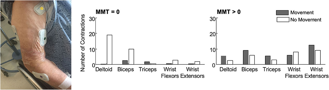
In our work to deploy EMG in this challenging environment, our team prioritized selecting an EMG system that provided wireless sensing in a compact form. The BioStampRC sensors (BioStampRC, MC10, Lexington, MA) included integrated EMG and accelerometer sensors that could concurrently monitor muscle activity and movement. We targeted the muscles most commonly assessed by the clinical team, placing sensors on five muscle groups: the deltoid, biceps, triceps, wrist flexors, and wrist extensors of the affected upper extremity (Figure 1). We followed SENIAM guidelines for placing the sensors, but often had to adjust to accommodate IV's, bandages, or telemetry pads. Loose skin, adipose tissue, and sweat were also common issues that impacted signal quality and sensor adherence.

FIGURE 1
www.frontiersin.org

Figure 1. (Left) BioStamp sensors provided a wireless and low-profile sensor to monitor muscle activity. We monitored muscle activity from five muscle groups on the paretic arm—the deltoid, biceps, triceps, wrist extensors, and wrist flexors. Tegaderm and Coband were placed over the electrodes to ensure they did not fall off or get stuck to bed sheets during 4 h of monitoring. The EMG data were used to evaluate outcome metrics like the median number of contractions (per 30-min of analyzed data) among patients with no observable muscle activity (N = 11, MMT = 0) and patients with some residual muscle activity (N = 10, MMT > 0). Accelerometer data were used to classify each contraction as occurring during periods with or without movement. (Right) Median number of contractions identified for each muscle with and without movement. Importantly, contractions were identified for all five muscles in all patients. For participants with MMT > 0, contractions were identified in all five muscles in a single 30-min monitoring session. Up to 3 h of monitoring was required to detect contractions in all five muscles for the participants with MMT = 0. As expected, participants with MMT > 0 had more contractions with movement. For participants with MMT = 0, contractions during movement likely reflect times when their arm was being moved during care. Participants with MMT = 0 also had more contractions in proximal muscle groups.

We deployed these sensors with stroke survivors who demonstrated impaired arm movement (NIHSS > 1) at a level-one trauma hospital. Patients were excluded if they were on comfort care, but otherwise we had broad inclusion as our main goal was evaluating deployment of the technology and observing muscle activity of all stroke survivors. We recruited patients from the acute stroke unit, where some patients may have received initial care in the intensive care unit. At this hospital, most stroke survivors stay in acute care for <2 weeks, receiving daily evaluations and therapy, before being discharged to inpatient rehabilitation, a skilled nursing facility, or their home. Our primary objective was to evaluate whether muscle activity could be detected during acute stroke care. We were especially interested in determining whether EMG sensors could detect muscle activity for those patients classified as having dense hemiplegia or flaccidity, who could not participate or be evaluated with other clinical measures. For each patient, we collected up to four hours of data. We manually identified contractions for each muscle, marking the start and stop time and coding each contraction as during periods of movement or rest based upon concurrent accelerometer data. Details on the data collection, EMG processing, and analyses can be found in (29, 30) and (REF), while here we aim to share key experiences in deploying this technology.

For the patients we monitored, muscle contractions were detected from all five muscles during a single four hour collection period during standard care (Figure 1). This was true even for the patients who had an MMT score of zero (N = 11), indicating no voluntary movement or muscle activity detected via palpation. For the participants with an MMT >0 (N = 10), only a single 30-min time window was required to identify contractions in all five muscles. For the patients who were initially flaccid, we did find moderate correlations between early contraction characteristics and scores on the MMT at follow-up. These findings indicate that muscle activity is present during the first week after stroke, even among participants characterized as flaccid, and EMG can provide quantitative metrics that may have prognostic value for predicting future function.


Saturday, August 25, 2018

Post-Stroke Rehabilitation Monitoring Using Wireless Surface Electromyography: A Case Study

Well then write up a protocol on its use. Or are you that fucking lazy and incompetent that you won't actually help stroke survivors? This article is the best effort you can do? 

Post-Stroke Rehabilitation Monitoring Using Wireless Surface Electromyography: A Case Study

Abstract:
Post-stroke rehabilitation monitoring provides key insights which can be used for development of customized treatment plans for patients. Rehabilitation monitoring systems available today are limited to observational measurements performed over a short period of time. Long term monitoring of stroke patients is necessary to keep track of stroke recovery and assess the patient’s response to the therapist’s treatment technique. This work is a case study that focuses on investigating the effects on muscle recruitment in bicep and calf muscles with and without orthotic intervention. A wireless surface Electromyography (sEMG) device is developed for monitoring muscle recruitment. Monitoring is done on a hemiplegic subject, before and after the physiotherapy treatment sessions, over duration of four months. An increase in sEMG peak frequency was observed after therapy in the absence of orthotic intervention while there was reduction in the peak frequency post therapy with orthotic intervention. Functional Independence Measurement scale, used to assess a patient’s level of disability as well as change in patient status in response to medical intervention is used as a reference measure to validate the sEMG device. The substantial changes in muscle recruitment due to regular therapy and orthotic intervention found in the study supports the use of the developed sEMG device as a surrogate to existing devices
Date of Conference: 11-13 June 2018
Date Added to IEEE Xplore: 20 August 2018
ISBN Information:
Publisher: IEEE
Conference Location: Rome, Italy, Italy 

I. Introduction

Worldwide, stroke is ranked as the leading cause of disability [1] . Due to stroke, coordination and muscle recruitment are commonly impaired. Up to 88% of people affected by stroke suffer hemiparesis with disorders of gait and balance, which persists even in the chronic phase [2] . Individuals suffering stroke actively overcome sensorimotor issues and asymmetry by using supportive or assistive equipments for increasing their gait ability. Quantifying normative patterns of muscle recruitment and coordination during common clinical tests can provide the neuromuscular demand required for common tasks and provide baselines for evaluating stroke patients. Currently, two main mechanisms contribute to stroke recovery. The first mechanism relates to functional recovery due to compensation based on improved use and refinement of remaining motor functions [3] , [4] . The second postulated mechanism assumes real recovery, i.e. restoration of lost brain functions due to learning-dependent reorganization of the brain [5] . Consequently, significant efforts are focused on gait retraining during rehabilitation following a stroke and efforts to develop and improve locomotor retraining programs are a major focus of rehabilitation research. Electromyographic (EMG) recordings provide a window into the central nervous system to evaluate muscle recruitment and coordination. After stroke, EMG recordings have been used to evaluate synergistic patterns of muscle activity, control assistive devices and guide biofeedback training. Despite improvements in measuring equipment since the discovery of EMG [6] , much of the research in this area is limited to observational measurements performed over short periods of time in laboratory settings. A study with a proper follow-up of patients over long periods could only help capture the temporal changes in muscle activity using EMG. Such long-term follow-up is not easy especially when dealing with subjects affected by stroke or hemiparesis. However, such long-term monitoring of sEMG can help observe sympathetic arousal patterns and previously missed out trends which could help in tracking the progress of patients. Monitoring a stroke patient can also uncover otherwise unperceived coping difficulties.
More at link.