Use the labels in the right column to find what you want. Or you can go thru them one by one, there are only 34,264 posts. Searching is done in the search box in upper left corner. I blog on anything to do with stroke. DO NOT DO ANYTHING SUGGESTED HERE AS I AM NOT MEDICALLY TRAINED, YOUR DOCTOR IS, LISTEN TO THEM. BUT I BET THEY DON'T KNOW HOW TO GET YOU 100% RECOVERED. I DON'T EITHER BUT HAVE PLENTY OF QUESTIONS FOR YOUR DOCTOR TO ANSWER.
Changing stroke rehab and research worldwide now.Time is Brain!trillions and trillions of neuronsthatDIEeach day because there areNOeffective hyperacute therapies besides tPA(only 12% effective). I have 523 posts on hyperacute therapy, enough for researchers to spend decades proving them out. These are my personal ideas and blog on stroke rehabilitation and stroke research. Do not attempt any of these without checking with your medical provider. Unless you join me in agitating, when you need these therapies they won't be there.
What this blog is for:
My blog is not to help survivors recover, it is to have the 10 million yearly stroke survivors light fires underneath their doctors, stroke hospitals and stroke researchers to get stroke solved. 100% recovery. The stroke medical world is completely failing at that goal, they don't even have it as a goal. Shortly after getting out of the hospital and getting NO information on the process or protocols of stroke rehabilitation and recovery I started searching on the internet and found that no other survivor received useful information. This is an attempt to cover all stroke rehabilitation information that should be readily available to survivors so they can talk with informed knowledge to their medical staff. It lays out what needs to be done to get stroke survivors closer to 100% recovery. It's quite disgusting that this information is not available from every stroke association and doctors group.
Did your competent? doctor immediately give you all the appropriate vaccination upon entering the hospital?
Do you
prefer your doctor, hospital and board of director's incompetence NOT
KNOWING? OR NOT DOING? Your choice; let them be incompetent or demand
action!
Respiratory syncytial virus (RSV) vaccination provided substantial protection against RSV-associated thromboembolic complications in older adults during the same season as vaccination, according to a large US retrospective cohort study published in Emerging Infectious Diseases.
Ryan E. Wiegand, Centers for Disease Control and Prevention, Atlanta, Georgia, and colleagues evaluated community-dwelling fee-for-service Medicare beneficiaries aged ≥65 years between October 1, 2023 and March 30, 2024. The analysis included 15,558,386 beneficiaries, of whom 58% were women, 80% lived in urban areas, and 13% had immunocompromising conditions.
RSV-associated thromboembolic events were defined as myocardial infarction, ischaemic stroke, or venous thromboembolism occurring from 7 days before to 30 days after an RSV diagnosis.
After adjusting for confounders, RSV vaccination demonstrated vaccine effectiveness (VE) of 79% (95% confidence interval [CI], 74%-83%) against RSV-associated thromboembolic events among all beneficiaries. Consistent protection was observed across major subgroups, with VE of 82% (95% CI, 77%-86%) in immunocompetent individuals and 69% (95% CI, 56%-78%) in immunocompromised patients. Protection was similar by age -- 75% (95% CI, 63%-83%) in adults aged 65-74 years and 80% (95% CI, 74%-84%) in those aged ≥75 years.
Effectiveness was stable over time, with VE of 80% at 14-59 days, 79% at 60-119 days, and 75% beyond 120 days after vaccination, suggesting minimal waning over the first 4 months. Comparable results were observed for both licensed products, with VE of 76% for Arexvy and 85% for Abrysvo. Sensitivity analyses extending follow-up and restricting to periods of high RSV circulation produced similar estimates.
By contrast, VE against all-cause thromboembolic events, regardless of RSV diagnosis, was lower at 21%.
“This study demonstrates the effectiveness of RSV vaccines against RSV-associated thromboembolic events, including myocardial infarction, ischaemic stroke, and venous thromboembolism,” the authors concluded. “Our findings are consistent with studies demonstrating that influenza and COVID-19 vaccines reduce the likelihood of thromboembolic events in adults.”
By Deborah Lynn Blumberg, American Heart Association News
Stroke survivor Rekha Desai (right) with her granddaughter Kaiya. (Photo courtesy of Dr. Dhaval Desai)
Rekha Desai planned to play Legos and blocks with her 2-year-old grandson, Kaveh, as she watched him in his Atlanta home.
But Rekha never arrived. Instead, the 73-year-old lay in a gurney thousands of feet in the air after having a stroke.
A
helicopter raced Rekha to a hospital that could perform a specialized
procedure. Her son, Dr. Dhaval Desai, drove 20 minutes to meet her. His
wife, Dr. Yogita Tailor, also a doctor, waited nervously at home with
Kaveh and their daughter, Kaiya.
"Will Dida be OK?" Kaiya asked about her grandmother, or dida, her best friend and cooking and slumber party partner.
Yogita hugged Kaiya and hoped the answer would be yes.
Rekha's
medical drama started in July 2022 when her son asked her to watch
Kaveh while he worked so Yogita could take Kaiya to a friend's birthday
party.
That morning, Yogita called Dhaval at the hospital.
"Your mom isn't feeling well," she said. "She doesn't think she should watch Kaveh."
Rekha was dehydrated and vomiting. Yogita speculated that her mother-in-law had a stomach bug.
Dhaval
wasn't overly concerned. With his mother's small stature, vegetarian
diet and "go, go, go" attitude, it wasn't unusual for her to get weak
from not eating or drinking enough. Still, he left work to go check on
her.
He found her in bed. Dhaval took her blood pressure. It was
normal. A COVID-19 test was negative. He gave her water and saltines and
nausea medicine.
Soon after, Rekha vomited again. The room was spinning.
"Something's not right," said Dhaval, who works in internal medicine and pediatrics. "We're going to the ER."
"No," Rekha said. "I'll drink more water and hydrate."
But Dhaval wouldn't take no for an answer. He called a colleague in the ER. "We're on our way," he said.
Rekha
got IV fluids and nausea medicine and had bloodwork done. Still, she
felt nauseous. She got a different nausea medicine. But it didn't agree
with her. She twitched and writhed in bed until it wore off.
A CT
scan didn't show any bleeding or a tumor, though the doctor noticed
calcium deposits in blood vessels in the back of her brain.
Still,
"stroke was the last thing on my mind," Dhaval said. Rekha didn't have
any of the classic signs: face drooping, arm weakness or slurred speech.
Rekha
met with her doctors, his colleagues. One suggested a CTA scan of
Rekha's head and neck. It's a more specialized test that combines a CT
scan with a dye to create pictures of blood vessels and tissues.
Dhaval
agreed. Rekha's doctor said she'd call in a few hours with results.
It's not going to show anything, Dhaval told himself. He went home to
change and check on the kids.
Less than an hour after Dhaval arrived home, his phone rang.
"Your
mom had a cerebellar stroke," the doctor said. This meant Rekha's
stroke affected her cerebellum, the part of the brain that controls
movement. It was caused by a blood clot in her brain. The doctor said
she may need a thrombectomy, a time-sensitive procedure to remove the
clot.
Their hospital wasn't equipped to perform the procedure. Rekha needed to be flown to a hospital that could.
"I don't want a procedure," Rekha told Dhaval on the phone. "I don't want to go in a helicopter."
Dhaval conferenced in his brother, Seemal Desai, who is a doctor in Dallas. Their father got on the phone, too.
"You have to," they told her. "You need to do this."
The main helicopter had a two-hour delay. "I was in near tears," Dhaval said.
With more work, he found a helicopter to transport her right away.
At
the new hospital, doctors determined a thrombectomy was too risky
because of where the clot was located. They gave her medication to thin
her blood in hopes it would dissolve the clot.
Doctors said they
didn't think the plaque in her artery was due to cholesterol, as can be
the case. Dhaval suspects his mother's stroke was related to having had
COVID-19 six months prior. His uncle, Rekha's brother, died from a
post-COVID-19 stroke.
At first, Rekha had trouble with her peripheral vision. She was unstable walking.
Rekha Desai recovering in the hospital. (Photo courtesy of Dr. Dhaval Desai)
Five
days later, Rekha was strong enough to go home. She needed help bathing
and dressing. For three months, she did occupational and vocational
therapy.
Exercises helped Rekha graduate from her walker.
Therapists took her to a grocery store to make sure she could shop on
her own. They assessed her driving ability, too, and cleared her to
drive, though she avoids highways. She takes a daily blood thinner.
"Sometimes
I have little memory lapse, but I'm 99.9% fine now," Rekha said. "And
it's because of the instant reaction on my son's part that I'm here
today."
The reaction was Dhaval in doctor mode. The rest of the time he was in son mode, which proved to be eye-opening.
"It was frightening and stressful being on 'the other side,'" he said.
While her dida was hospitalized, Kaiya worried.
She called to check on her. She made a card Dhaval delivered.
During Rekha's recovery, Kaiya was eager to help. One day, when Rekha had a dizzy spell, Kaiya ran to bring the walker.
"She was always there," Rekha said. "She wanted to make sure I was safe."
"I'm not too little to help," Kaiya said more than once. She bought a word search book to help Rekha with her letters.
Dhaval called Kaiya "a little coach on the sidelines always helping."
Rekha
Desai with her husband, Rohit (far left), and grandchildren, from left:
Kaveh, Kaiya, Armaan and Anya. (Photo courtesy of Dr. Dhaval Desai)
Nine
months after her dida's stroke, Kaiya, who's now 7, decided to write a
book about what happened. She sat down with crayons and markers, drew
pictures, then wrote words.
Kaiya's teacher, Tiffany Churchwell,
helped her piece together and bind the book, "My Life: Dida and the
Stroke." Kaiya read the story to her classmates and filmed a reading.
"I
have a wild story to tell you," she wrote. Then, "I was so scared!!!"
Finally, she wrote about how excited she was that her dida was coming
home.
"What inspired you to write this story?" a classmate asked Kaiya after the reading.
"I
want people to see how I solved my problems," she answered. "Maybe this
can help them or give them ideas on how to solve their own problem."
Stories From the Heart chronicles the inspiring journeys of heart disease and stroke survivors, caregivers and advocates.
In case you are getting bamboozled by vaccine claims. I'm much more worried about brain damage from COVID-19 than the vaccine. I already have enough brain damage, I'm preventing more.
The article references a pre-print study
published in April that explored the long-term neurological effects
observed in those who had been infected with COVID-19. It claims the
study revealed that "spike proteins from mRNA jabs infest the brain
tissue of vaccinated people."
But Dr. Ali Ertürk,
a co-author of the paper and director of the Institute of Tissue
Engineering and Regenerative Medicine in Munich, told USA TODAY the
study did not examine the COVID-19 vaccine of its side effects.
"We have done zero experiments using the vaccine,
and we have shown and claim zero side effects of the vaccine," said
Ertürk in an email. "Our work reports the presence of the spike protein
in the skull of deceased individuals long after their COVID-19
infection, suggesting that the spike's persistence may contribute to
long-term neurological symptoms."
None of the COVID-19 vaccines authorized in the U.S. contain the live virus, according to the Centers for Disease Control and Prevention. Instead, the mRNA in the vaccine
teaches the body's cells to make copies of the COVID-19 spike protein
so they can later recognize and fight off the virus if they become
infected.
"The issue is that during infection,
there is an enormous amount of viral replication and spike protein
production, which impacts many organs including the brain," Ertürk said.
The COVID-19 vaccine is safe and effective, according to the CDC.
USA TODAY reached out to the users who shared the post for comment but did not immediately receive a response.
Hopefully all the vaccines I got will prevent severe COVID-19. I did get COVID-19 this summer during a trip to Mexico but it must have been the Omicron variety since I barely got sick at all.
As
coronavirus disease 2019 (COVID-19) and aging are both accompanied by
cognitive decline, we hypothesized that COVID-19 might lead to molecular
signatures similar to aging. We performed whole-transcriptome analysis
of the frontal cortex, a critical area for cognitive function, in
individuals with COVID-19, age-matched and sex-matched uninfected
controls, and uninfected individuals with intensive care unit/ventilator
treatment. Our findings indicate that COVID-19 is associated with
molecular signatures of brain aging and emphasize the value of
neurological follow-up in recovered individuals.
Main
COVID-19 is an acute respiratory disease often accompanied by neurological sequelae1. Individuals with previous severe COVID-19 exhibit a 10-year average drop in their global cognitive performance2,
mimicking accelerated aging. Complementary studies combining
neuroimaging and cognitive screening implicate COVID-19-induced
impairment of the frontal cortex3,4, a critical area for cognitive function, but molecular evidence of aging-like effects in the brain is lacking.
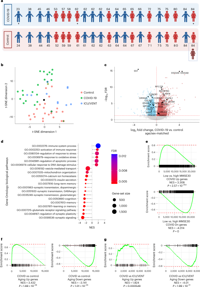
To
address this, we performed RNA-sequencing (RNA-seq) analysis of 54
postmortem frontal cortex samples, including samples from 21 individuals
with severe COVID-19 (previous neurological history was limited to
Alzheimer’s disease in one person and epilepsy in another) and 1
asymptomatic individual aged between 23 and 84 years old, 22 age-matched
(±2 years) and sex-matched uninfected controls with no history of
neurological or psychiatric disorders, an age-matched and sex-matched
uninfected individual with Alzheimer’s disease, and an additional
independent control group of 9 uninfected individuals with history of
intensive care unit (ICU) or ventilator treatment (22–85 years old;
ICU/VENT; Fig. 1a and Supplementary Fig. 1a; see Supplementary Table 1
for clinical information; COVID-19 cohort). All COVID-19-cases were
determined by positive pre-mortem or peri-mortem testing for severe
acute respiratory syndrome coronavirus 2 (SARS-CoV-2) infection via
nasopharyngeal swab qPCR and history of hospitalization, whereas
uninfected control samples were collected before the COVID-19 pandemic
(with three exceptions in the ICU/VENT group that had negative
SARS-CoV-2 qPCR tests at the time of death, and no COVID-19 history
and/or negative serological test).
Fig. 1: Severe COVID-19 is associated with transcriptomic signatures of aging in the human brain.
a, Age and sex of each individual in COVID-19 or uninfected age/sex-matched control (±2 years) groups (n = 22
per group) analyzed in this cohort. An asterisk indicates notable
COVID-19 cases. The 23-year-old male presented with asymptomatic
COVID-19, the 62-year-old female presented with severe COVID-19 history
and comorbid epilepsy and the 84-year-old female who had a history of
severe COVID-19 with comorbid Alzheimer’s disease (AD; an uninfected
individual with AD was also included as an additional control;
Supplementary Table 1). Created with BioRender.com. b, t-distributed stochastic neighbor embedding (t-SNE)
analysis of frontal cortex transcriptomes from COVID-19-infected
individuals, uninfected age-matched and sex-matched controls, and an
independent group of uninfected controls with history of ICU and/or
ventilator treatment (ICU/VENT). Black border, 23-year-old asymptomatic
male with COVID-19. Red border, 62-year-old female with COVID-19 history
and comorbid epilepsy. Blue border, 84-year-old female with COVID-19
history and comorbid AD. Black point, 84-year-old female without
COVID-19 but with AD. Green border, uninfected age-matched and
sex-matched control (non-AD) for the COVID-19-infected individual with
comorbid AD. For age-matched/sex-matched controls and COVID-19 samples n = 22 per group; ICU/VENT-treated uninfected controls n = 9. c,
Volcano plot representing the DEGs of the frontal cortex of individuals
with COVID-19 versus age-matched and sex-matched controls (n = 22
per group). Red points denote significantly upregulated genes among
COVID-19 cases (false discovery rate (FDR) < 0.05). Blue points
denote significantly downregulated genes among COVID-19 cases. Black
points highlight significant genes with corresponding gene symbols
(Supplementary Table 2). d,
Gene Ontology (GO) biological pathway enrichment analysis of COVID-19
versus age-matched/sex-matched control DEGs. Gene ranks were determined
by signed −log10 FDRs of DEGs (Supplementary Table 3). e,
GSEA of cognitive decline-regulated genes using COVID-19 (COVID-19
versus age-matched and sex-matched controls) DEGs. DEG ranks were
assigned by signed −log10 FDR from the frontal cortex
transcriptome of individuals with MMSE scores > 25 (high cognitive
performance) versus the transcriptome of individuals with MMSE
scores < 25 (low cognitive performance/cognitive decline) as measured
in the ROSMAP study. f,g, GSEA of COVID-19 DEGs (COVID-19 versus age-matched/sex-matched control in f and COVID-19 versus ICU/VENT in g),
using significantly upregulated (top) or downregulated genes (bottom)
in our aging cohort as gene sets. DEG ranks were assigned by signed −log10 FDR from COVID-19 versus corresponding control frontal cortex. NES, normalized enrichment score. P, two-tailed GSEA P value (Supplementary Fig. 5).
By
clustering analyses, COVID-19 transcriptomic cases broadly segregated
away from controls, with two of the outliers being from the 23-year-old
asymptomatic individual and the 62-year-old individual with comorbid
epilepsy; the age-matched/sex-matched controls proximal to COVID-19
cases were from older adults (Fig. 1b and Supplementary Figs. 1 and 2).
Uninfected older adults in the ICU/VENT group generally clustered
closer to COVID-19-infected individuals, whereas younger ICU/VENT
individuals clustered relatively close to controls; two cases cluster
separately from all other samples (Supplementary Fig. 1b).
Comparison of COVID-19 cases and their corresponding age-matched and
sex-matched controls revealed 6,993 differentially expressed genes
(DEGs), 3,330 of which were upregulated and 3,663 downregulated (Fig. 1c and Supplementary Table 2). For example, the S100A8 and S100A9 genes, which encode calprotectin and blood circulating levels of which distinguish severe from mild COVID-19 disease5,
were upregulated among individuals with COVID-19. Pathway enrichment
analysis identified numerous significant GO terms associated with aging
in the human brain enriched upon severe COVID-19, including positive
enrichment of immune-related pathways and negative enrichment of
synaptic activity, cognition and memory pathways (Fig. 1d and Supplementary Fig. 3).
We also observed significant associations of cellular response to DNA
damage, mitochondrial function, regulation of response to stress and
oxidative stress, vesicular transport, calcium homeostasis6, and insulin signaling/secretion7 pathways previously associated with aging processes and brain aging6,8.
Altogether, our analyses suggest that many biological pathways that
change with natural aging in the brain also change in severe COVID-19.
As
natural brain aging is associated with cognitive decline, we further
assessed associations of transcriptomic changes in COVID-19 and
cognitive function. We collated frontal cortex transcriptomic data from
633 individuals who underwent the Mini-Mental State Examination (MMSE)
while alive and donated their brains at the time of death as part of the
ROSMAP study9,10.
We split individuals and their corresponding transcriptomic data based
on the median MMSE score: ≥25 as high cognitive performance versus
MMSE < 25 as low cognitive performance. From gene-set enrichment
analysis (GSEA), we found strong associations between low cognitive
performance and COVID-19 (Fig. 1e).
Given
the strong associations between aging-regulated pathways and severe
COVID-19, we sought to directly test whether COVID-19 is associated with
similar gene expression patterns as natural aging in the human brain.
We performed RNA-seq analysis of postmortem frontal cortex samples of 10
young (≤38 years old) and 10 older (≥71 years old) uninfected
individuals (Supplementary Table 1; aging cohort and Supplementary Fig. 4a)
and compared these findings to COVID-19 DEGs. We found striking
similarities between individuals with COVID-19 and aged individuals:
genes upregulated in aging were upregulated in severe COVID-19;
likewise, genes downregulated in aging were also downregulated in severe
COVID-19 (see Fig. 1f
for age-matched/sex-matched controls versus COVID-19). As further
validation, we collated transcriptome-wide datasets from five
independent aging cohorts and confirmed this association (Supplementary
Fig. 5 and Supplementary Table 4).
Intriguingly, we continued to observe a significant association between
aging-associated genes and DEGs from individuals with COVID-19 versus
uninfected individuals treated with ICU/VENT (Fig. 1g).
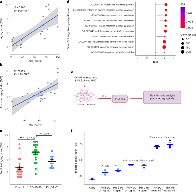
To
delineate the effects of severe COVID-19 on brain aging directly, we
leveraged our aging cohort to derive an aging index (Supplementary Fig. 4),
comprising our aging DEGs and condensed by the first principal
component across these transcriptomic data. As validation, we applied
our predicted aging index to uninfected controls (COVID-19 cohort) and
found similarly strong Pearson correlations between the training and
test sets (Fig. 2a,b).
Applying this model to individuals with COVID-19, we observed a
significant increase in the predicted aging index compared to
corresponding uninfected age-matched/sex-matched control and uninfected
ICU/VENT control groups (Fig. 2c).
Additional analysis revealed that the predicted aging index in
individuals with COVID-19 was not significantly affected by the presence
or absence of cerebrovascular disease (P > 0.05). Thus,
severe COVID-19 appears to shift the molecular age of brains relative to
both uninfected age-matched and sex-matched controls as well as
uninfected ICU/VENT controls. Lastly, using qPCR analysis, we validated
several of the top shared DEGs between our COVID-19 cohort and aging
datasets (n = 22 per group; Supplementary Fig. 6).
Fig. 2: Severe COVID-19 and cytokine treatment of neurons are associated with increases in predicted age.
a,
First, a principal-component analysis using DEGs (FDR < 0.05) of
young versus old uninfected controls estimated principal component 1
(PC1). The graph presents a two-tailed Pearson correlation of
chronological age with aging index (PC1) among young versus old
uninfected controls from the aging cohort (training set). n = 20. Gray shadow indicates the 95% confidence interval from a linear regression fit (R2 (18) = 0.58, P = 9.2 × 10⁻⁵). b,
Two-tailed Pearson correlation of chronological age with predicted
aging index (PC1) among uninfected age-matched and sex-matched controls
from the COVID-19 cohort (test set). n = 22. Gray shadow indicates the 95% confidence interval from a linear regression fit (R2 (20) = 0.438, P = 7.9 × 10⁻⁴). c, Predicted aging index (PC1) of individuals with COVID-19 (n = 22), age-matched/sex-matched uninfected controls (control; n = 22), and an independent group of uninfected cases with ICU/VENT treatment history (n = 9). The line in each group represents the mean ± s.e.m. COVID-19 versus control Welch two-tailed t(42.0) = 5.68, P = 3.4 × 10⁻⁶; COVID-19 versus ICU/VENT Welch two-tailed t(21.0) = 3.14, P = 0.015. A Bonferroni correction was used to adjust for multiple comparisons. d,
Significant interferon and TNF-related pathways identified using GO
biological pathway enrichment analysis of COVID-19 versus
age-matched/sex-matched control frontal cortex DEGs. FDR, GSEA FDR
(Supplementary Table 3). e, Experimental design of in vitro cytokine treatment in human neurons. Created with BioRender.com. f, Effects of
IFN-β, IFN-γ and TNF on predicted aging index, as assessed following in
vitro treatment of primary human neurons. The line in each group
represents the mean ± s.e.m. n = 3 independent wells (1 × 10⁵ cells per well were plated) for each treatment. IFN-β_LO versus control Welch two-tailed t(2.68) = 4.48, P = 0.16; IFN-β_HI versus control Welch two-tailed t(3.51) = 8.26, P = 0.012; IFN-γ_LO versus control Welch two-tailed t(3.58) = 20.8, P = 4.4 × 10⁻⁴; IFN-γ_HI versus control Welch two-tailed t(3.35) = 12.3, P = 4.1 × 10⁻³; TNF_LO versus control Welch two-tailed t(3.63) = 33.6, P = 6.6 × 10⁻⁵; TNF_HI versus control Welch two-tailed t(2.28) = 15.8, P = 0.013. Bonferroni correction was used to adjust for multiple comparisons.
Finally,
we sought to determine pathophysiologic mechanisms that may explain the
association of COVID-19 with aging. We considered that this could be
due to multiple factors, including SARS-CoV-2 viral infection in the
frontal cortex or COVID-19-induced systemic inflammation. In agreement
with previous studies11,12, SARS-CoV-2 viral RNA was not detected in samples from individuals with COVID-19 (at the time of death; Supplementary Fig. 7),
suggesting that the observed gene expression changes are unlikely due
to direct effects of the viral RNA in the frontal cortex. On the other
hand, our transcriptomic pathway analyses identified upregulation of
tumor necrosis factor (TNF) and type I/II interferon response pathways
in the frontal cortex of individuals with COVID-19 (Fig. 2d). Indeed, interferons and TNF have been implicated in brain aging and aging-induced cognitive decline6,13,14,15.
Among individuals with COVID-19 with available peripheral cytokine
data, we indeed observed increased TNF levels (3.5–24.2 pg ml−1 at 0–2 d before death in three individuals; values > 2.8 pg ml−1
are considered elevated; data were not available for the rest of the
individuals). In line with our findings, a mouse model of SARS-CoV-2
infection exhibited elevation of pro-inflammatory cytokines in
cerebrospinal fluid, including interferon gamma (IFN-γ) and TNF, in the
absence of viral neuroinvasion16.
To test whether TNF and type I/II interferons can modulate the
expression of aging-regulated genes, we performed a transcriptomic
analysis (RNA-seq) on human primary neurons treated with different doses
of TNF, interferon beta (IFN-β) or IFN-γ. Interestingly, TNF and to a
lesser extent IFN-γ and IFN-β increased the predicted aging index of
cytokine-treated neurons, suggestive of the induction of an aging-like
effect (Fig. 2e,f).
We also found that cytokines upregulated the expression of
aging-regulated genes that were upregulated in individuals with COVID-19
such as TRIM22, CHI3L1, C1S and IFITM1 and downregulated the expression of aging-regulated genes that were downregulated in individuals with COVID-19 such as CCND2, ACTR3B and EPHA5 (Supplementary Fig. 8).
Taken together, our data suggest that COVID-19-induced TNF and type
I/II interferons may lead to significant deteriorating effects in the
brain in the absence of SARS-CoV-2 neuroinvasion.
Aging is a major
risk factor for the development of cognitive deficits. Our results,
together with previously reported residual cognitive deficits reported
in recovered cases2,
imply that aging-associated and cognitive decline-associated gene
expression changes observed in individuals with COVID-19 may lead to
increased rates of cognitive decline. Furthermore, we provide evidence
that these aging-regulated gene expression changes may be mediated in
part by circulating TNF and type I/II interferons, suggesting that acute
management of severe COVID-19-induced inflammation may be
neuroprotective. We recognize limitations in our study including: the
variability in illness duration, the imperfect quality of several
samples and the specificity of our findings due to COVID-19. Despite
these constraints, our study was sufficiently powered to identify
substantial transcriptome-wide changes in individuals with COVID-19. In
addition to being larger than previously reported COVID-19 brain
transcriptome studies11,12,17,
our COVID-19 cases were matched by age and sex to uninfected controls,
enabling the identification of aging-associated gene expression
signatures in our samples. Furthermore, we included ICU/VENT uninfected
samples as an additional independent control group to distinguish
COVID-19 from other comorbidities requiring ICU monitoring and/or
ventilation. The generalizability of our results to individuals who had
mild COVID-19 or who recovered from COVID-19 remains to be determined.
Given our findings, we advocate for neurological follow-up of
individuals who recovered from COVID-19 and suggest potential clinical
value in modifying risk factors to reduce the risk or delay the
development of aging-related neurological pathologies and cognitive
decline18.
You don't want this risk so get vaccinated and boosted to reduce the severity of any COVID-19 you do get. Which is why at the start of COVID-19 I was going to demand heparin.
Heparin:
Why I'm getting heparin. Heparin
binds to cells at a site adjacent to ACE2, the portal for SARS-CoV-2
infection, and "potently" blocks the virus, which could open up therapy
options.
Individuals with long COVID may have a higher risk for abnormal blood
clotting, especially those who have difficulties with basic exercise
for more than 12 weeks after infection, according to a study published
in Blood Advances.
The findings highlight a potential role for antithrombotic therapy in the management of these patients, researchers noted.
Source: Adobe Stock.
Rationale and methods
Long COVID,
characterized by persistent fatigue, reduced exercise tolerance, chest
pain, shortness of breath and cognitive slowing, is an increasingly
recognized complication of acute COVID-19 infection, according to Nithya Prasannan,
researcher in the department of hematology at University College London
Hospitals NHS Foundation Trust in the U.K., and colleagues.
“Acute COVID-19 is strongly linked with increased risk [for]
thrombosis and a prothrombotic state, quantified by elevated von
Willebrand factor antigen:ADAMTS13 ratio, and is associated with
severity of acute COVID-19 infection,” they wrote.
Investigators sought to examine whether patients with long COVID
referred to a dedicated post-COVID-19 clinic experienced a prothrombotic
state associated with symptom severity. The analysis included 330
patients (median age, 46 years; range, 18-88; 60% female), 97% of whom
had symptoms for 3 months or more after acute COVID-19 infection.
Researchers assessed thrombotic risk and characterized patients as
being in a prothrombotic state if they had significantly more von
Willebrand factor antigen than ADAMTS13 in the bloodstream.
Eighty-four percent of participants (n = 276) completed exercise
tests that included walking on a flat surface and/or repeatedly going
from a sitting to standing position from a chair while wearing oxygen
monitors. Researchers measured oxygen levels and tested participants’
blood before and after exercise to measure lactate levels.
Key findings
Results showed 28% of the entire cohort had a von Willebrand factor
antigen:ADAMTS13 ratio of 1.5 or higher, which researchers considered to
be raised. This was four times more common among patients with impaired
exercise capacity, characterized by desaturation levels 3% or more
and/or an increase in lactate level more than 1 from baseline on
1-minute sit to stand test and/or 6-minute walk test (P < .0001). Among those 56 patients, more than half (55%) had had a raised a von Willebrand factor antigen:ADAMTS13 ratio.
About one-quarter of all patients with long COVID had high factor
VIII levels that ranged from 2.1 IU/mL to 5.1 IU/mL, and 18% had
elevated von Willebrand factor antigen levels of 1.7 IU/mL to 3.3 IU/mL.
Implications
Looking ahead, Prasannan and colleagues plan to assess patients’
bloodwork using different research platforms during the course of their
long COVID to assess how risk for thrombosis may change with progression
of symptoms, according to a press release.
“I hope that people will view this research as a step forward in
understanding what causes long COVID, which will hopefully help us guide
future treatment options,” Prasannan said in the release. “I encourage
people experiencing long COVID to participate in clinical trials when
available because the more data we have, the better we can understand
this condition.”
Findings may point to potential mechanism for brain fog in people with long COVID
by
Judy George, Senior Staff Writer, MedPage Today
February 4, 2022
Brains of COVID-19 patients had some of the same
pathological changes seen in Alzheimer's disease, which may explain the
memory problems people with long COVID experience, a small study
suggested.
The study, based on autopsies of 10 people who died with COVID-19,
linked the inflammatory response found in SARS-CoV-2 infection with
pathways causing tau hyperphosphorylation typically associated with
Alzheimer's disease, reported Andrew Marks, MD, of Columbia University
in New York City, and co-authors.
The
data also indicated a role for leaky ryanodine receptor 2 (RyR2) in the
pathophysiology of SARS-CoV-2 infection, the researchers wrote in Alzheimer's & Dementia.
"The study shows that long COVID-19 brain fog may be a form of
Alzheimer's disease, but much more research needs to be done before we
can make more definitive conclusions," Marks told MedPage Today.
"The major strength of the paper is that they identified
abnormalities in several molecules which help characterize the
neuroglial dysfunction in these patients at a biochemical level," noted
Avindra Nath, MD, of the National Institute of Neurological Disorders
and Stroke in Bethesda, Maryland, who wasn't involved with the study.
"However no histology was performed for Alzheimer's type pathology
and the implications for development of Alzheimer's disease would be
hard to extrapolate from this study," Nath pointed out.
Defective ryanodine receptors have been implicated in diverse processes, including heart and lung disease. Inside neurons, they previously have been linked to an increase in phosphorylated tau.
Persistent
brain fog and cardiac symptoms in people with COVID-19 led Marks and
co-authors to investigate how ryanodine receptors were affected in
COVID-19. "What we found is really, I think, quite unexpected," Marks
said. "Not only did we find defective ryanodine receptors in the hearts
and lungs of deceased COVID patients, we also found them in their
brains."
The researchers analyzed signaling molecules in brain lysates of
COVID-19 patients and controls and found evidence linking SARS-CoV-2
infection to activation of TGF-β signaling and oxidative overload. They
also found high levels of phosphorylated tau in COVID-19 patients'
brains, both in areas where tau is typically located in Alzheimer's and
in other sites. No changes in pathways leading to amyloid beta formation
were seen.
The findings may mean that a COVID-19 immune response causes brain
inflammation which leads to dysfunctional ryanodine receptors and
altered cellular calcium dynamics, then to increases in phosphorylated
tau, Marks and colleagues noted. "We propose a potential mechanism that
may contribute to the neurological complications caused by SARS-CoV-2:
defective intracellular Ca2+ regulation and activation of Alzheimer's disease-like neuropathology," they wrote.
Leaky
RyR2 channels may be a therapeutic target to ameliorate some of the
cognitive defects associated with SARS-CoV-2 infection and long COVID,
the researchers suggested.
Lab studies that treated COVID-19 patient brain samples with a drug
targeting RyR2 channels prevented the calcium leak. The treatment, known
as ARM210, currently is undergoing clinical testing at NIH for RyR1-related myopathy.
"Future experiments will explore calcium channels as a potential
therapeutic target for the neurological complications associated with
COVID-19," Marks and co-authors wrote.
Judy George
covers neurology and neuroscience news for MedPage Today, writing about
brain aging, Alzheimer’s, dementia, MS, rare diseases, epilepsy,
autism, headache, stroke, Parkinson’s, ALS, concussion, CTE, sleep,
pain, and more. Follow
Disclosures
The research was supported by the NIH.
Marks
and Columbia University own stock in Armgo Pharma, which is developing
compounds targeting the ryanodine receptor, and have patents on the
compounds. One co-author has consulted for Armgo Pharma. All other
authors declared no competing interests or conflicts.
You'll want this transformative research, so it it your doctor's and hospital's requirement that they initiate human testing. Anything less is total incompetence.
Your risk of dementia, has your doctor told you of this?
A
promising new approach to potentially treat Alzheimer's disease—and
also vaccinate against it—has been developed by a team of UK and German
scientists.
Both the
antibody-based treatment and the protein-based vaccine developed by the
team reduced Alzheimer's symptoms in mouse models of the disease. The
research is published today in Molecular Psychiatry.
The
work is a collaboration between researchers at the University of
Leicester, the University Medical Center Göttingen and the medical
research charity LifeArc.
Rather than focus on the amyloid
beta protein in plaques in the brain, which are commonly associated
with Alzheimer's disease, the antibody and vaccine both target a
different soluble—form of the protein, that is thought to be highly
toxic. Amyloid beta protein naturally exists as highly flexible,
string-like molecules in solution, which can join together to form
fibers and plaques. In Alzheimer's disease, a high proportion of these
string-like molecules become shortened or 'truncated', and some
scientists now think that these forms are key to the development and
progression of the disease.
Professor
Thomas Bayer, from the University Medical Center Göttingen, said: "In
clinical trials, none of the potential treatments which dissolve amyloid plaques
in the brain have shown much success in terms of reducing Alzheimer's
symptoms. Some have even shown negative side effects. So, we decided on a
different approach. We identified an antibody in mice that would
neutralize the truncated forms of soluble amyloid beta, but would not
bind either to normal forms of the protein or to the plaques."
Dr.
Preeti Bakrania and colleagues from LifeArc adapted this antibody so a
human immune system wouldn't recognize it as foreign and would accept
it. When the Leicester research group looked at how and where this
'humanized' antibody, called TAP01_04, was binding to the truncated form
of amyloid beta, the team had a surprise. They saw the amyloid beta
protein was folded back on itself, in a hairpin-shaped structure.
Professor
Mark Carr, from the Leicester Institute of Structural and Chemical
Biology at the University of Leicester, explained: "This structure had
never been seen before in amyloid beta. However, discovering such a
definite structure allowed the team to engineer this region of the
protein to stabilize the hairpin shape and bind to the antibody in the
same way. Our idea was that this engineered form of amyloid beta could
potentially be used as a vaccine, to trigger someone's immune system to
make TAP01_04 type antibodies."
When the team tested the engineered amyloid beta protein in mice, they found that mice who received this 'vaccine' did produce TAP01 type antibodies.
The
Göttingen group then tested both the 'humanized' antibody and the
engineered amyloid beta vaccine, called TAPAS, in two different mouse
models of Alzheimer's disease. Based on similar imaging techniques to
those used to diagnose Alzheimer's in humans, they found that both the
antibody and the vaccine helped to restore neuron function, increase
glucose metabolism in the brain, restore memory loss and—even though
they weren't directly targeted—reduce amyloid beta plaque formation.
LifeArc's
Dr. Bakrania said: ''The TAP01_04 humanized antibody and the TAPAS
vaccine are very different to previous antibodies or vaccines for
Alzheimer's disease that have been tested in clinical trials, because
they target a different form of the protein.
This makes them really promising as a potential treatment for the
disease either as a therapeutic antibody or a vaccine. The results so
far are very exciting and testament to the scientific expertise of the
team. If the treatment does prove successful, it could transform the
lives of many patients."
Professor
Mark Carr added: "While the science is currently still at an early
stage, if these results were to be replicated in human clinical trials,
then it could be transformative. It opens up the possibility to not only
treat Alzheimer's once symptoms are detected, but also to potentially
vaccinate against the disease before symptoms appear."
The researchers are now looking to find a commercial partner to take the therapeutic antibody and the vaccine through clinical trials.
There’s growing evidence that inoculation confers significant protective benefits.
WSJ Opinion: What's the Coronavirus Priority? Masks or Vaccination?
You may also like
Up Next
WSJ Opinion: What's the Coronavirus Priority? Masks or Vaccination?
Main Street: The CDC should scrap its confusing guidance and
make Covid-19 vaccination the only priority. Images: AFP via Getty
Images Composite: Mark Kelly
By
Print
Text
Covid vaccines enormously reduce the risk of death and
hospitalization in those who have been infected by the novel
coronavirus. But could they also help protect seniors against dementia
and Alzheimer’s disease? There’s reason to hope so.
Growing evidence indicates that seniors who get vaccinated
against illnesses such as tetanus and even the flu are much less likely
to develop Alzheimer’s, the leading cause of dementia, characterized by a
buildup of amyloid plaque and tau tangles in the brain. Scientists
don’t completely understand why, but many hypothesize that vaccines
generate a systemic immune response that can reduce inflammation in the
brain, which results in neuron loss and cognitive decline.