Use the labels in the right column to find what you want. Or you can go thru them one by one, there are only 33,991 posts. Searching is done in the search box in upper left corner. I blog on anything to do with stroke. DO NOT DO ANYTHING SUGGESTED HERE AS I AM NOT MEDICALLY TRAINED, YOUR DOCTOR IS, LISTEN TO THEM. BUT I BET THEY DON'T KNOW HOW TO GET YOU 100% RECOVERED. I DON'T EITHER BUT HAVE PLENTY OF QUESTIONS FOR YOUR DOCTOR TO ANSWER.
Changing stroke rehab and research worldwide now.Time is Brain!trillions and trillions of neuronsthatDIEeach day because there areNOeffective hyperacute therapies besides tPA(only 12% effective). I have 523 posts on hyperacute therapy, enough for researchers to spend decades proving them out. These are my personal ideas and blog on stroke rehabilitation and stroke research. Do not attempt any of these without checking with your medical provider. Unless you join me in agitating, when you need these therapies they won't be there.
What this blog is for:
My blog is not to help survivors recover, it is to have the 10 million yearly stroke survivors light fires underneath their doctors, stroke hospitals and stroke researchers to get stroke solved. 100% recovery. The stroke medical world is completely failing at that goal, they don't even have it as a goal. Shortly after getting out of the hospital and getting NO information on the process or protocols of stroke rehabilitation and recovery I started searching on the internet and found that no other survivor received useful information. This is an attempt to cover all stroke rehabilitation information that should be readily available to survivors so they can talk with informed knowledge to their medical staff. It lays out what needs to be done to get stroke survivors closer to 100% recovery. It's quite disgusting that this information is not available from every stroke association and doctors group.
An organ once thought to become unimportant after childhood could actually be key to longer life, according to new research.
The thymus is a small organ located behind the sternum and is known to play a central role in the immune system. It acts as a training ground for the immune system’s T cells, which help the body recognise and fight infections.
It’s generally believed to become largely inactive after puberty as it shrinks with age and produces fewer new T cells. As a result, scientists say, its role in adult health remains less studied.
A new study at Mass General Brigham challenges the theory that the thymus grows unimportant after childhood.
Clinicians used artificial intelligence to review CT X-ray scans and found that adults with healthier thymus tended to live longer, with lower risks of heart disease and cancer.
Another study shows that the health of the thymus affects how well cancer patients respond to a key treatment.
“The thymus has been overlooked for decades and may be a missing piece in explaining why people age differently and why cancer treatments fail in some patients,” Hugo Aerts, an author of one of the two studies published in the journal Nature, said. “Our findings suggest thymic health deserves much more attention and may open new avenues for understanding how to protect the immune system as we age.”
AI model analyses CT scans to estimate thymic health. Image depicts a comparison between high, average, and low thymic health and corresponding CT images (Mass General Brigham/Nature)
One of the studies analysed data from over 25,000 adults who were part of a national lung cancer screening programme and over 2,500 participants in the Framingham Heart Study, a long-running group of generally healthy individuals.
The thymus of each participant was evaluated by measuring its size, structure and composition, creating a “thymic health” score.
Researchers found those with higher scores had roughly a 50 per cent lower risk of death, a 63 per cent lower risk of dying from heart disease, and a 36 per cent lower risk of developing lung cancer compared to those with lower scores.
These risk reductions held even after accounting for age and other health factors.
When the thymus function declines, researchers suspect the immune system becomes less effective at responding to new threats like cancer. They say chronic inflammation, smoking and higher body weight are linked to poorer thymic health.
The second study corroborates this.
It reviewed CT scans and outcomes from 1,200 patients treated with immunotherapy, a key treatment method for cancer involving the priming of one’s own immune system to fight cancerous cells.
Patients with better thymic health had a 37 per cent lower risk of cancer progression and a 44 per cent lower risk of death, even after adjusting for differences in patients, tumours, and treatments, the study found.
The findings point to a previously under-recognised role for the thymus in determining how patients respond to modern cancer therapies.
More studies are needed to confirm these findings, scientists say, adding that an improved understanding of thymic health can help physicians better assess disease risk and guide treatment decisions.
Ask your competent? doctor how this will be used in your favor to slow down brain aging. NO knowledge; you DON'T have a functioning stroke doctor! RUN AWAY!
Summary: A study reveals how brain cell interactions
influence aging, showing that rare cell types either accelerate or slow
brain aging. Neural stem cells provide a rejuvenating effect on
neighboring cells, while T cells drive aging through inflammation.
Researchers used advanced AI tools and a spatial single-cell atlas to
map cellular interactions across the lifespan in mice.
This work
sheds light on how interventions, such as enhancing neural stem cells,
might combat neurodegeneration. By understanding these cellular
dynamics, scientists can explore tailored therapies to slow aging and
promote brain resilience. The findings also offer insights into
conditions like Alzheimer’s disease, highlighting the importance of
cell-to-cell interactions.
Key Facts:
Rejuvenating Role: Neural stem cells create a supportive environment that rejuvenates nearby cells, even outside their lineage.
Aging Impact: T cells accelerate brain aging through pro-inflammatory signals, especially interferon-γ.
Innovative Tools: Researchers used a spatial transcriptomic atlas and machine learning models to study brain aging at the cellular level.
Source: Stanford
Much
like plants in a thriving forest, certain cells in the brain create a
nurturing environment, enhancing the health and resilience of their
neighbors, while others promote stress and damage, akin to a noxious
weed in an ecosystem.
A new study published in Nature on
December 18, 2024, reveals these interactions playing out across the
lifespan. It suggests local cellular interactions may profoundly
influence brain aging — and offers fresh insights into how we might slow
or even reverse the process.
“What was exciting to us was finding
that some cells have a pro-aging effect on neighboring cells while
others appear to have a rejuvenating effect on their neighbors,”
said Anne Brunet, the Michele and Timothy Barakett Endowed Professor in
Stanford’s Department of Genetics and co–senior investigator of the new
study.
These
findings are important, says Zou, “because they highlight how cellular
interactions — not just the intrinsic properties of individual cells —
shape the aging process.” Credit: Neuroscience News
Specifically,
Brunet said, “We were surprised to discover that neural stem cells,
which we’ve studied for a long, long time, had a rejuvenating effect on
the cells around them. In the future we want to understand the role of
neural stem cells in providing a beneficial environment for resilience
within the brain.”
Brunet collaborated with James Zou, an
associate professor of biomedical data science at Stanford, to conduct
the study, which was spearheaded by graduate student, Eric Sun.
Brunet’s
lab, a leader in brain aging and neural stem cell biology, provided the
biological expertise and experimental framework. Zou’s team brought
cutting-edge AI techniques to analyze the data, while Sun, with a
background in physics and quantitative analysis, acted as the bridge
between these two worlds.
The research was supported by a Catalyst
Award from the Knight Initiative for Brain Resilience at Stanford’s Wu
Tsai Neurosciences Institute.
These findings open new avenues of
research, including examining how rejuvenating interventions like
exercise and reprogramming factors promote brain health, potentially by
enhancing the brain’s natural resilience and repair mechanisms. Such
insights may suggest new strategies to combat neurodegeneration and
cognitive decline.
The findings may also help scientists
understand how diseases such as Alzheimer’s disease change the way cells
interact and drive brain aging.
Cells that age — and rejuvenate — the brain
The
research team set out to tackle a fundamental question: How do cells in
their native environment influence one another during the aging
process?
Previous studies have focused on individual cells in
isolation, overlooking the critical context of their “neighborhoods”
— the cells surrounding them.
By preserving and analyzing these
spatial relationships, the team aimed to uncover whether interactions
between different cell types either drive or mitigate aging in the
brain.
Their investigation revealed a striking finding: Out of the
18 different cell types the researchers identified, two rare cell types
had powerful but opposing effects on nearby cells.
T cells,
immune cells that infiltrate the aging brain, have a distinctly
pro-inflammatory, pro-aging effect on neighboring cells that may be
driven by interferon-γ, a signaling molecule that drives inflammation.
On
the other hand, they found that neural stem cells, though rare,
demonstrate a powerful rejuvenating effect, even on nearby cells outside
the neural lineage.
During brain development, neural stem cells
mature into the major cell types in the brain; in adults, they can also
give rise to new neurons and are important for maintenance and repair of
the nervous system.
Beyond their well-established ability to
generate healthy new neurons, the new study suggests NSCs may help
create a supportive environment for brain cells.
These findings
are important, says Zou, “because they highlight how cellular
interactions — not just the intrinsic properties of individual cells
— shape the aging process.”
Building a map and models
At
the heart of this research are three key innovations by the research
team: a spatial single-cell atlas of gene activity in the mouse brain
across its lifespan and two advanced computational tools, each essential
for piecing together how cells influence one another as they age.
To
map the complex neighborhoods of the brain, the researchers created a
spatial single-cell transcriptomic atlas of the mouse brain, capturing
gene expression data from 2.3 million cells across 20 stages of life,
equivalent to human ages 20 to 95.
Unlike traditional methods that
separate complex tissues, like the brain, into a collection of many
disconnected cells, this experimental approach preserved the spatial
relationships between cells, allowing the team to study how their
spatial proximity shapes aging.
The atlas laid the groundwork for
the first computational tool — a spatial aging clock. The clocks are
machine-learning models designed to predict the biological age of
individual cells based on their gene expression.
“For the first
time, we can use aging clocks as a tool to discover new biology,” says
Sun, instead of just using them to estimate biological age.
The
second tool, built using graph neural networks, provided a powerful way
to model these cell-to-cell interactions. By creating a kind of in
silico brain, the researchers could simulate what happens when specific
cell types are added, removed, or altered. This allowed them to explore
potential interventions that would be nearly impossible to test in a
living brain.
“This computational tool allows us to simulate what
happens when we perturb individuals cell in the brain, which is
something we can’t really test experimentally at scale,” says Zou.
To
ensure the broader scientific community can build on their findings,
Sun has made their tools and code publicly available, providing a
valuable resource for studying cellular interactions across various
tissues and organisms.
Implications and future directions
The
study offers major insights into the drivers of aging, as well as
rejuvenating factors that could help restore resilience and vitality to
the aging brain.
“Different cells respond differently to rejuvenating interventions,” explains Brunet.
“Brain
aging is exceptionally complex, so future therapies will need to be
tailored not only to tissues but also to the specific cell types within
those tissues.”
By demonstrating how spatial context and proximity
influence cellular aging, the research builds on longstanding theories
about the role of immune and senescent cells in the aging process.
Looking ahead, the team hopes to move from observation to causation.
“If
we prevent T cells from releasing their pro-aging factors or enhance
the effects of neural stem cells, how does that change the tissue over
time?” asks Brunet.
While the study focused on mice, the team also
hopes to extend their approach to human tissues. “We’re working to make
these tools broadly applicable to other tissues and biological
processes,” adds Sun.
Funding
The research
was supported by the the Knight Initiative for Brain Resilience at
Stanford’s Wu Tsai Neurosciences Institute, the Stanford Knight-Hennessy
Scholars Program, the National Institutes of Health (P01AG036695,
R01AG071711), a National Science Foundation (Graduate Research
Fellowship, CAREER award 1942926), P.D. Soros Fellowship for New
Americans, Silicon Valley Foundation, Chan Zuckerberg Biohub–San
Francisco Investigator program, Chan Zuckerberg Initiative, the Milky
Way Research Foundation, the Simons Foundation, and a generous gift from
M. and T. Barakett.
Competing Interests
Brunet is a scientific advisory board member of Calico.
About this genetics and neuroscience research news
Author: Nicholas Weiler Source: Stanford Contact: Nicholas Weiler – Stanford Image: The image is credited to Neuroscience News
You do expect your competent? doctor and hospital to get human testing going? OF COURSE NOT! They are way too fucking incompetent to even know about this research, much less do something about it!
The
inflammatory cascade(Call it what is really is: the neuronal cascade of death! Which your doctors are doing nothing about, leaving millions to billions of neurons to die in the first week!) that ensues following an ischemic stroke has been
increasingly recognized(Known since the Rockefeller University report in Jan. 2009! So you're that fucking out-of-date?) as a driving force in the long-term disability
associated with the disease. This synopsis will highlight recent
preclinical studies that aimed to promote a robust Treg (T-regulatory
cell) response via interleukin-2 (IL-2) signaling following an
experimental stroke.
To elucidate the mechanism by which an
expansion in Tregs following experimental ischemic injury promotes white
matter integrity and functional recovery, Yuan et al (Regulatory T cell expansion promotes white matter repair after stroke. Neurobiol Dis. 2023;179:106063. doi: 10.1016/j.nbd.2023.106063)
demonstrated that Treg augmentation via direct intravenous transfer of
Tregs isolated from donor mice 2 hours after transient middle cerebral
artery occlusion (tMCAO) resulted in improved white matter recovery when
compared with splenocyte-treated mice poststroke. To determine whether
promoting an endogenous Treg expansion poststroke would similarly result
in a neuroprotective(Don't ever use the milquetoast word, neuroprotection. It doesn't signal urgency at all! Whereas your doctor telling you they failed to stop the neuronal cascade of death in the first week might engender a few medical malpractice suits. I suggest a payment of $1000 a dead neuron; in my case that would come to 5.571 billion dead neurons; Only 55 trillion dollars!) phenotype, wild-type mice were treated with either
a consecutive intraperitoneal administration of IL-2/IL-2 antibody
complexes (IL-2/IL-2Ab) or equal concentrations of isotype-matched
antibody (IgG) at 6 hours, 1 day, 2 days, 3 days, 10 days, 20 days, and
30 days after stroke. At 14 days after stroke, IL-2/IL-2Ab mice
displayed a significant increase in the CD25+CD4+Foxp3+
Treg cell population in the blood, spleen, and brain parenchyma when
compared with IgG-treated controls. Importantly, IL-2/IL-2Ab treatment
mitigated sensorimotor dysfunction at 35 days, but not at 7 days after
stroke. To evaluate white matter integrity, longitudinal in vivo
diffusion tensor imaging scans 14 and 28 days after tMCAO coupled with
ex vivo diffusion tensor imaging scanning of brains 35 days after stroke
were used to construct fractional anisotropy maps. Fractional
anisotropy mapping showed that IL-2/IL-2Ab–treated mice exhibited
improved white matter integrity at 28 and 35 days after stroke,
indicating that Treg expansion promotes white matter integrity in the
late phase of stroke. Delayed IL-2/IL-2Ab treatment remained protective
even when administered as late as 5 days after stroke, as Luxol fast
blue staining of coronal brain slices of treated mice showed increased
myelin in the external capsule and striatum 21 days after tMCAO in
IL-2/IL-2Ab–treated mice compared with IgG-treated mice.
To determine a precise signaling mechanism through which Tregs promote long-term tissue repair following ischemic stroke, Shi et al (Treg cell-derived osteopontin promotes microglia-mediated white matter repair after ischemic stroke. Immunity. 2021;54:1527–1542.e8. doi: 10.1016/j.immuni.2021.04.022)
utilized single-cell RNA sequencing and flow cytometry to verify that
Tregs are among the immune cell populations that infiltrate the brain
poststroke at 3, 5, 7, 14, and 35 days after stroke. To confirm their
role in improving white matter integrity after stroke, Tregs were
selectively depleted via diphtheria toxin (DT) injections in Foxp3DTR (DTR) transgenic mice that express the DT receptor under the control of the Foxp3
promoter. Treg depletion resulted in diminished functional recovery and
drastically aggravated white matter lesions following an ischemic
stroke. Comparative transcriptomic analysis of sorted CD4+CD25+Foxp3(GFP)+
Treg cells from the ischemic brain and blood of stroke and sham DTR
mice identified differentially expressed genes between
brain-infiltrating Treg cells and peripheral Treg cells; several genes
upregulated in brain-infiltrating Treg cells encoded trophic factors
known to stimulate oligodendrocyte precursor cell differentiation, such
as Igf1, IL-1a, and Osm. Additionally, brain-infiltrating Treg cells displayed higher levels of transcripts encoding cytokines such as Spp1, Il1b, Il1a, and Il10,
suggesting that Treg cell-mediated white matter repair may rely on
immunomodulatory signals and cell-cell interactions. Treg-derived
osteopontin was identified as a potential signaling molecule driving
microglial-mediated white matter repair, as protein-protein interaction
enrichment analysis via STRING (Search Tool for the Retrieval of
Interacting Genes/Proteins) identified interactions between Spp1, which encodes osteopontin, and Itgb1, Itga5, and Itgav,
which encode the integrin subunits of the osteopontin receptor on
microglia. To confirm the role of osteopontin signaling in vivo, Treg
cells derived from wild-type or Spp1−/− mice into DTR+DT mice 6 hours after tMCAO, and myelination was assessed via Luxol fast blue staining. Importantly, Spp1−/−
Treg cell-treated mice displayed a reduction in myelination when
compared with wild-type Treg cell-treated mice. Lastly, this study
showed that IL-2/IL-2Ab treatment boosted the number of osteopontin+
Treg cells in the ischemic brain 3 days after tMCAO, improved
sensorimotor function, spatial learning, and mitigated white matter
injury (fractional anisotropy mapping and dual staining for myelin basic
protein).
Observing the capacity of IL-2 to drive Treg
recruitment and infiltration into the brain following stroke resulting
in improved functional recovery and white matter repair, Yshii et al (Astrocyte-targeted
gene delivery of interleukin 2 specifically increases brain-resident
regulatory T cell numbers and protects against pathological
neuroinflammation. Nat Immunol. 2022;23:878–891. doi: 10.1038/s41590-022-01208-z)
aimed to develop and validate a central nervous system–specific
therapeutic strategy. The adeno-associated virus–based therapeutic
delivery system detailed in this study utilized the GFAP (glial
fibrillary acidic protein) promoter to drive IL-2 expression in
astrocytes specifically while simultaneously avoiding expression in both
the peripheral immune systems and astrocytes. The ability of the
delivery system to drive astrocyte-specific expression of IL-2 and
increase recruit of Tregs to the central nervous system was validated
using ELISA, flow cytometry, immunofluorescent staining, and single-cell
RNA-seq. The efficacy of the gene delivery was evaluated in various
models of central nervous system injury, such as the controlled cortical
impact model for traumatic brain injury, a distal middle cerebral
artery occlusion model, the photothrombotic model stroke, and the
experimental autoimmune encephalomyelitis model of multiple sclerosis.
Cognitive recovery was then assessed 15 days after injury using the
Morris water maze test and the novel object recognition test.
Comparative assessments of tissue injury and lesion size were conducted
using immunofluorescence staining of the cortical tissue and MRI 14 days
after injury. Importantly, PHP.GFAP-IL-2 treatment before injury
mitigated neuroinflammation and improved functional recovery in all
models of central nervous system injury, including traumatic brain
injury, stroke, and multiple sclerosis, and did not impact the
peripheral immune system.
While preclinical studies have shown
promising results in the use of IL-2 to amplify Tregs to mitigate white
matter injury following ischemic stroke, the application of this method
in clinical settings remains sparse. One major hurdle in applying
IL-2–mediated Treg expansion in clinical trials is to do so without
inadvertently activating other immune cells or causing broader side
effects that come with IL-2 treatments.
New Scientist magazine has this to say about this research:
Giving mice antibiotics can protect them from brain damage caused by stroke. Antibiotics change the make-up of the mice's gut bacteria, which in turn alters the immune cells that travel to the brain and would normally cause inflammation. The treatment appears to reduce cell destruction by around 60 percent.
What is your doctor and hospital doing to ensure human testing gets done? Being incompetent like usual and doing nothing?
Commensal
gut bacteria impact the host immune system and can influence disease
processes in several organs, including the brain. However, it remains
unclear whether the microbiota has an impact on the outcome of acute
brain injury. Here we show that antibiotic-induced alterations in the
intestinal flora reduce ischemic brain injury in mice, an effect
transmissible by fecal transplants. Intestinal dysbiosis alters immune
homeostasis in the small intestine, leading to an increase in regulatory
T cells and a reduction in interleukin (IL)-17–positive γδ T cells
through altered dendritic cell activity. Dysbiosis suppresses
trafficking of effector T cells from the gut to the leptomeninges after
stroke. Additionally, IL-10 and IL-17 are required for the
neuroprotection afforded by intestinal dysbiosis. The findings reveal a
previously unrecognized gut-brain axis and an impact of the intestinal
flora and meningeal IL-17+ γδ T cells on ischemic injury.
Xiao Cheng1,2, Jianxin Ye3*, Xiaolei Zhang1 and Kun Meng1
1Department of Neurology, ShanXi Province People's Hospital of Shanxi Medical University, Taiyuan, China
2Shanxi Key Laboratory of Brain Disease Control, Shanxi Provincial People's Hospital, Taiyuan, China
3Department of Neurology, The 900th Hospital
of the Joint Logistics Support Force of the Chinese People's Liberation
Army, Fuzhou, China
Objective: Cell division cycle 42 (CDC42) modulates CD4+
T-cell differentiation, blood lipids, and neuronal apoptosis and is
involved in the pathogenesis of acute ischemic stroke (AIS); however,
the clinical role of CDC42 in AIS remains unanswered. This study aimed
to evaluate the expression of CDC42 in a 3-year follow-up and its
correlation with disease severity, T helper (Th)1/2/17 cells, and the
prognosis in patients with AIS.
Methods: Blood CDC42 was detected in
143 patients with AIS at multiple time points during the 3-year
follow-up period and in 70 controls at admission by reverse
transcription-quantitative polymerase chain reaction (RT-qPCR). In
addition, blood Th1, Th2, and Th17 cells and their secreted cytokines
(interferon-γ (IFN-γ), interleukin-4 (IL-4), and interleukin-17A
(IL-17A)) in patients with AIS were detected by flow cytometry and
enzyme-linked immunosorbent assay (ELISA), respectively.
Results: Compared with controls (p
< 0.001), CDC42 was reduced in patients with AIS. CDC42 was
negatively correlated with the National Institutes of Health Stroke
Scale (NIHSS) score (p < 0.001), whereas, in patients with AIS (all p
< 0.050), it was positively associated with Th2 cells and IL-4 but
negatively correlated with Th17 cells and IL-17A. CDC42 was decreased
from admission to 3 days and gradually increased from 3 days to 3 years
in patients with AIS (P<0.001). In a 3-year follow-up, 24
patients with AIS recurred and 8 patients died. On the 3rd day, 7th day,
1st month, 3rd month, 6th month, 1st year, 2nd year, and 3rd year,
CDC42 was decreased in recurrent patients than that in non-recurrent
patients (all p < 0.050). CDC42 at 7 days (p = 0.033) and 3 months (p = 0.023) was declined in reported deceased patients than in survived patients.
Conclusion: CDC42 is used as a biomarker to constantly monitor disease progression and recurrence risk of patients with AIS.
Introduction
Stroke is a common cerebrovascular disease, which has
affected nearly 104 million people worldwide in the past three decades
and has climbed to the second leading cause of death (second only to
ischemic heart disease), besides, stroke is also known for its high
disability rate (nearly 33.4–71%) (1–4).
Acute ischemic stroke (AIS) is the primary type of stroke (accounting
for ~70% of all stroke cases), which is characterized by immune system
disorder, severe neurological deficits, etc. (5–10).
Gradually, diversified therapeutic strategies (including thrombolysis,
antiplatelet treatment, anticoagulants, and neuroprotective agents) have
been introduced in an attempt to eliminate arterial occlusion, restore
blood flow to the brain, and improve the recovery of neurological
function; however, AIS is disease prone to recurrence, which requires
continuous attention (11–16).
Therefore, it is imperative to develop objective biomarkers to help
identify patients with AIS as soon as possible, predict their outcomes,
and then adjust the treatment regimens accordingly.
Cell division cycle 42 (CDC42), a small hydrolase of
guanosine triphosphate (GTPase), acts as a signal transduction
convergence point that mediates many signaling pathways. Moreover, it is
reported that CDC42 regulates blood lipids, blood vessel development,
CD4+ T-cell differentiation, microglial process, and neuronal
apoptosis in some cardiovascular and cerebrovascular diseases
(including ischemic brain injury, coronary heart disease, and
cerebrovascular malformations) (17–23).
For instance, a study found that CDC42 can act as an upstream activator
of the c-Jun N-terminal kinase (JNK) signaling pathway to govern
neuronal apoptosis in ischemic brain injury (19).
Another study explored the correlation between CDC42 and T helper (Th) 2
cells, Th17 cells, and blood lipids in patients with coronary heart
disease (20).
Interestingly, these CDC42-modulated biological processes (mentioned
above) behave as underlying pathogenesis of AIS, implying that CDC42
might be implicated in the development of AIS (6, 8, 24). Additionally, an in vitro
study reported that the activation of CDC42 promoted the migration of
endogenous neural stem/progenitors cells after ischemic stroke, which
facilitates the recovery of injured brain tissue (25). However, the detailed clinical role of CDC42 in patients with AIS remains unanswered.
In this study, the expression of CDC42 was detected in
patients with AIS during a 3-year follow-up period with the aim of
evaluating the longitudinal changes of CDC42 and its correlation with
disease severity, Th1/2/17 cells, and the prognosis in patients with
AIS.
In mice, so your doctor and stroke hospital have followup to to to ensure this gets tested in humans. With tens of thousands of doctors and stroke hospital presidents calling for this research it would get done. Their responsibility, if they fail at this, call the board of directors and ask when competent people will be employed.
GRPs confocol in brain. Credit: Johns Hopkins Medicine
Transplanted brain stem cells survive without anti-rejection
drugs in mice. By exploiting a feature of the immune system, researchers
open the door for stem cell transplants to repair the brain.
In experiments in mice, Johns Hopkins Medicine researchers say they
have developed a way to successfully transplant certain protective brain
cells without the need for lifelong anti-rejection drugs.
A report on the research, published today (September 16, 2019) in the journal Brain,
details the new approach, which selectively circumvents the immune
response against foreign cells, allowing transplanted cells to survive,
thrive and protect brain tissue long after stopping immune-suppressing
drugs.
The ability to successfully transplant healthy cells into the brain
without the need for conventional anti-rejection drugs could advance the
search for therapies that help children born with a rare but
devastating class of genetic diseases in which myelin, the protective
coating around neurons that helps them send messages, does not form
normally. Approximately 1 of every 100,000 children born in the U.S.
will have one of these diseases, such as Pelizaeus-Merzbacher disease.
This disorder is characterized by infants missing developmental
milestones such as sitting and walking, having involuntary muscle
spasms, and potentially experiencing partial paralysis of the arms and
legs, all caused by a genetic mutation in the genes that form myelin.
“Because these conditions are initiated by a mutation causing
dysfunction in one type of cell, they present a good target for cell
therapies, which involve transplanting healthy cells or cells engineered
to not have a condition to take over for the diseased, damaged or
missing cells,” says Piotr Walczak, M.D., Ph.D., associate professor of
radiology and radiological science at the Johns Hopkins University
School of Medicine.
A major obstacle to our ability to replace these defective cells is
the mammalian immune system. The immune system works by rapidly
identifying ‘self’ or ‘nonself’ tissues, and mounting attacks to destroy
nonself or “foreign” invaders. While beneficial when targeting bacteria
or viruses, it is a major hurdle for transplanted organs, tissue or
cells, which are also flagged for destruction. Traditional
anti-rejection drugs that broadly and unspecifically tamp down the
immune system altogether frequently work to fend off tissue rejection,
but leave patients vulnerable to infection and other side effects.
Patients need to remain on these drugs indefinitely.
In a bid to stop the immune response without the side effects, the
Johns Hopkins Medicine team sought ways to manipulate T cells, the
system’s elite infection-fighting force that attacks foreign invaders.
Specifically, Walczak and his team focused on the series of so-called
“costimulatory signals” that T cells must encounter in order to begin
an attack.
“These signals are in place to help ensure these immune system cells
do not go rogue, attacking the body’s own healthy tissues,” says Gerald
Brandacher, M.D., professor of plastic and reconstructive surgery and
scientific director of the Vascularized Composite Allotransplantation
Research Laboratory at the Johns Hopkins University School of Medicine
and co-author of this study.
The idea, he says, was to exploit the natural tendencies of these
costimulatory signals as a means of training the immune system to
eventually accept transplanted cells as “self” permanently.
To do that, the investigators used two antibodies, CTLA4-Ig and
anti-CD154, which keep T cells from beginning an attack when
encountering foreign particles by binding to the T cell surface,
essentially blocking the ‘go’ signal. This combination has previously
been used successfully to block rejection of solid organ transplants in
animals, but had not yet been tested for cell transplants to repair
myelin in the brain, says Walczak.
In a key set of experiments, Walczak and his team injected mouse
brains with the protective glial cells that produce the myelin sheath
that surrounds neurons. These specific cells were genetically engineered
to glow so the researchers could keep tabs on them.
The researchers then transplanted the glial cells into three types of
mice: mice genetically engineered to not form the glial cells that
create the myelin sheath, normal mice and mice bred to be unable to
mount an immune response.
Then the researchers used the antibodies to block an immune response, stopping treatment after six days.
Each day, the researchers used a specialized camera that could detect
the glowing cells and capture pictures of the mouse brains, looking for
the relative presence or absence of the transplanted glial cells. Cells
transplanted into control mice that did not receive the antibody
treatment immediately began to die off, and their glow was no longer
detected by the camera by day 21.
The mice that received the antibody treatment maintained significant
levels of transplanted glial cells for over 203 days, showing they were
not killed by the mouse’s T cells even in the absence of treatment.
“The fact that any glow remained showed us that cells had survived
transplantation, even long after stopping the treatment,” says Shen Li,
M.D., lead author of the study. “We interpret this result as a success
in selectively blocking the immune system’s T cells from killing the
transplanted cells.”
The next step was to see whether the transplanted glial cells
survived well enough to do what glial cells normally do in the brain —
create the myelin sheath. To do this, the researchers looked for key
structural differences between mouse brains with thriving glial cells
and those without, using MRI images. In the images, the researchers saw
that the cells in the treated animals were indeed populating the
appropriate parts of the brain.
Their results confirmed that the transplanted cells were able to
thrive and assume their normal function of protecting neurons in the
brain.
Walczak cautioned that these results are preliminary. They were able
to deliver these cells and allow them to thrive in a localized portion
of the mouse brain.
In the future, they hope to combine their findings with studies on
cell delivery methods to the brain to help repair the brain more
globally.
###
Other researchers involved in this study include Byoung Chol Oh,
Chengyan Chu, Antje Arnold, Anna Jablonska, Georg Furtmüller, Huamin Qin
and Miroslaw Janowski of The Johns Hopkins University; Shen Li of the
Dalian Municipal Central Hospital and The Johns Hopkins University;
Johannes Boltze of the University of Warwick; and Tim Magnus and Peter
Ludewig of the University of Hamburg.
Reference: “Induction of immunological tolerance to myelinogenic
glial-restricted progenitor allografts” by Shen Li, Byoung Chol Oh,
Chengyan Chu, Antje Arnold, Anna Jablonska, Georg J Furtmüller, Hua-Min
Qin, Johannes Boltze, Tim Magnus, Peter Ludewig, Mirosław Janowski,
Gerald Brandacher and Piotr Walczak, 16 September 2019, Brain.
In addition to maintaining immune tolerance, FOXP3+ regulatory T (Treg) cells perform specialized functions in tissue homeostasis and remodelling1,2. However, the characteristics and functions of brain Treg cells are not well understood because there is a low number of Treg cells in the brain under normal conditions. Here we show that there is massive accumulation of Treg
cells in the mouse brain after ischaemic stroke, and this potentiates
neurological recovery during the chronic phase of ischaemic brain
injury. Although brain Treg cells are similar to Treg cells in other tissues such as visceral adipose tissue and muscle3,4,5, they are apparently distinct and express unique genes related to the nervous system including Htr7, which encodes the serotonin receptor 5-HT7. The amplification of brain Treg
cells is dependent on interleukin (IL)-2, IL-33, serotonin and T cell
receptor recognition, and infiltration into the brain is driven by the
chemokines CCL1 and CCL20. Brain Treg cells suppress
neurotoxic astrogliosis by producing amphiregulin, a low-affinity
epidermal growth factor receptor (EGFR) ligand. Stroke is a leading
cause of neurological disability, and there are currently few effective
recovery methods other than rehabilitation during the chronic phase. Our
findings suggest that Treg cells and their products may
provide therapeutic opportunities for neuronal protection against stroke
and neuroinflammatory diseases.
Previous research investigating the roles of T effector (Teff) and T regulatory (Treg)
cells after injury to the CNS has yielded contradictory conclusions,
with both protective and destructive functions being ascribed to each of
these T cell subpopulations. In this work, we study this dichotomy by
examining how regulation of the immune system affects the response to
CNS trauma. We show that, in response to CNS injury, Teff and Treg
subsets in the CNS-draining deep cervical lymph nodes are activated,
and surgical resection of these lymph nodes results in impaired neuronal
survival. Depletion of Treg, not surprisingly, induces a robust Teff
response in the draining lymph nodes and is associated with impaired
neuronal survival. Interestingly, however, injection of exogenous Treg
cells, which limits the spontaneous beneficial immune response after
CNS injury, also impairs neuronal survival. We found that no Treg accumulate at the site of CNS injury, and that changes in Treg
numbers do not alter the amount of infiltration by other immune cells
into the site of injury. The phenotype of macrophages at the site,
however, is affected: both addition and removal of Treg
negatively impact the numbers of macrophages with alternatively
activated (tissue-building) phenotype. Our data demonstrate that
neuronal survival after CNS injury is impaired when Treg
cells are either removed or added. With this exacerbation of
neurodegeneration seen with both addition and depletion of Treg, we
recommend exercising extreme caution when considering the therapeutic
targeting of Treg cells after CNS injury, and possibly in chronic neurodegenerative conditions.
I'm not sure how this will help us so ask your doctors what they will do with this information. Hopefully not scoff at you. Mine pretty much did when I asked about specific research articles. I doubt he had read anything new since medical school. That lack of acquiring knowledge needs to be brought up to the hospital president because that means the stroke department head is not setting proper goals and expectations.
1Functional Unit of Cerebrovascular Diseases, Hospital Clínic, Barcelona, Spain
2August Pi i Sunyer Biomedical Research Institute (IDIBAPS), Barcelona, Spain
3Department of Brain Ischemia and
Neurodegeneration, Instituto de Investigaciones Biomédicas de Barcelona
(IIBB), Consejo Superior de Investigaciones Científicas (CSIC),
Barcelona, Spain
Brain proteins are detected in the cerebrospinal fluid (CSF) and
blood of stroke patients and their concentration is related to the
extent of brain damage. Antibodies against brain antigens develop after
stroke, suggesting a humoral immune response to the brain injury.
Furthermore, induced immune tolerance is beneficial in animal models of
cerebral ischemia. The presence of circulating T cells sensitized
against brain antigens, and antigen presenting cells (APCs) carrying
brain antigens in draining lymphoid tissue of stroke patients support
the notion that stroke might induce antigen-specific immune responses.
After stroke, brain proteins that are normally hidden from the
periphery, inflammatory mediators, and danger signals can exit the brain
through several efflux routes. They can reach the blood after leaking
out of the damaged blood-brain barrier (BBB) or following the drainage
of interstitial fluid to the dural venous sinus, or reach the cervical
lymph nodes through the nasal lymphatics following CSF drainage along
the arachnoid sheaths of nerves across the nasal submucosa. The route
and mode of access of brain antigens to lymphoid tissue could influence
the type of response. Central and peripheral tolerance prevents
autoimmunity, but the actual mechanisms of tolerance to brain antigens
released into the periphery in the presence of inflammation, danger
signals, and APCs, are not fully characterized. Stroke does not
systematically trigger autoimmunity, but under certain circumstances,
such as pronounced systemic inflammation or infection, autoreactive T
cells could escape the tolerance controls. Further investigation is
needed to elucidate whether antigen-specific immune events could
underlie neurological complications impairing recovery from stroke.
A new study in animals shows that
using a compound to block the body’s immune response greatly reduces
disability after a stroke.
The study by scientists from the University of Wisconsin School of
Medicine and Public Health also showed that particular immune cells—CD4+
T-cells produce a mediator, called interleukin (IL)-21 that can cause
further damage in stroke tissue.
Moreover, normal mice, ordinarily killed or disabled by an ischemic
stroke, were given a shot of a compound that blocks the action of IL-21.
Brain scans and brain sections showed that the treated mice suffered
little or no stroke damage.
“This is very exciting because we haven’t had a new drug for stroke
in decades, and this suggests a target for such a drug,” says lead
author Dr. Zsuzsanna Fabry, professor of pathology and laboratory
medicine.
Stroke is the fourth-leading killer in the world and an important
cause of permanent disability. In an ischemic stroke, a clot blocks the
flow of oxygen-rich blood to the brain. But Fabry explains that much of
the damage to brain cells occurs after the clot is removed or dissolved
by medicine. Blood rushes back into the brain tissue, bringing with it
immune cells called T-cells, which flock to the source of an injury.
The study shows that after a stroke, the injured brain cells provoke
the CD4+ T-cells to produce a substance, IL-21, that kills the neurons
in the blood-deprived tissue of the brain. The study gave new insight
how stroke induces neural injury.
Similar Findings in Humans
Fabry’s co-author Dr. Matyas Sandor, professor of pathology and
laboratory medicine, says that the final part of the study looked at
brain tissue from people who had died following ischemic strokes. It
found that CD4+ T-cells and their protein, IL-21 are in high
concentration in areas of the brain damaged by the stroke.
Sandor says the similarity suggests that the protein that blocks
IL-21 could become a treatment for stroke, and would likely be
administered at the same time as the current blood-clot dissolving
drugs.
“We don’t have proof that it will work in humans,” he says, “but
similar accumulation of IL-21 producing cells suggests that it might.”
The paper was published in the Journal of Experimental Medicine.
Source: University of Wisconsin School of Medicine and Public Health
The immune response can
contribute to the risk of stroke as well as to brain injury following
stroke. Directed modulation of the immune response to attenuate risk or
injury is thus a potential therapeutic strategy for treating stroke.
Inducing a population of antigen-specific regulatory T cells is one such
strategy and can be used to locally modulate the immune response to the
organs in which the antigen is present, thereby limiting potential side
effects. This chapter addresses the use of mucosal tolerance to both
prevent and treat ischemic stroke.


 Xiao Cheng1,2,
  Jianxin Ye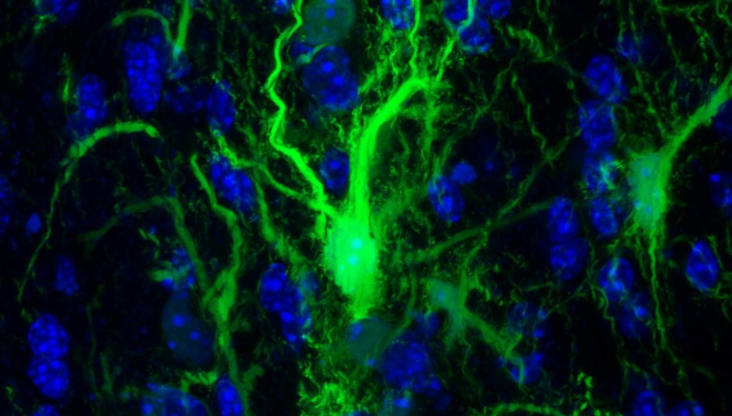
 Francesc Miró
 Anna M. Planas