Introduction
Excitotoxicity is a form of neuronal death triggered by
excessive and/or sustained exposure to the amino acid glutamate, the
primary excitatory neurotransmitter in the brain. Evidence accumulated
in the past four decades indicates that excitotoxicity is a critical
contributor to the neuronal demise occurring upon acute and chronic
neurological conditions, like stroke, Alzheimer’s disease (AD),
Huntington’s disease (HD), Amyotrophic Lateral Sclerosis (ALS), and
Parkinson’s disease (PD) (Mehta et al., 2013).
Although, 50 years have passed since the first description of glutamate’s neurotoxic activity (Olney, 1969),
therapeutic strategies set at counteracting these processes have been
only partially exploited. In that regard, the targeting of upstream
mechanisms of glutamate-driven neurotoxicity has produced, in the late
80s, an early wave of enthusiasm and fueled a level of optimism that has
not been corroborated in the following years. These approaches have
been found promising in preclinical models (Lee et al., 1999) but failed in clinical trials (Lee et al., 1999; Ikonomidou and Turski, 2002; Chamorro et al., 2016; Choi, 2020).
Only riluzole and memantine, two drugs that target glutamate-driven
neuronal death, have been approved for the treatment of ALS and AD,
respectively.
Although, most of the preclinical findings failed “the
bench to bed” translation, this experimental evidence has significantly
helped dissect the molecular underpinnings of excitotoxicity. These
studies have also helped provide support for the excitotoxic cascade
hypothesis (Zivin and Choi, 1991; Choi, 2020).
The construct posits that excitotoxic neuronal death is primarily
mediated by the glutamate-driven activation of N-methyl-D-aspartate
receptors (NMDARs) and the subsequent toxic intraneuronal accumulation
of calcium (Ca2+). The NMDAR-driven Ca2+ overload
is, in fact, a mandatory step in the process as most of the downstream
mechanisms of the cascade, like the generation of reactive oxygen
species (ROS; of mitochondrial and non-mitochondrial origin), or
reactive nitrogen species (RNS), the concurrent mitochondrial
dysfunction, metabolic impairment, as well as the activation of
necrotic/apoptotic pathways, are all Ca2+-dependent processes (Lee et al., 1999; Lai et al., 2014; Bano and Ankarcrona, 2018; Choi, 2020; Swanson and Wang, 2020). However, Ca2+ is not alone, and other cations find a way to participate in the death banquet. Zinc (Zn2+) is, for instance, a VIP guest.
In the review, we provide a brief overview of the role of Zn2+
in the brain and discuss its neurotoxic properties and how they
intertwine with the excitotoxic cascade. Finally, we focus on the
distinct features of the NADPH-diaphorase neurons, a subpopulation
spared from excitotoxic insults offering an intriguing model to further
our understanding of neuroprotective mechanisms.
Zinc Homeostasis and Its Role in Brain Functions
After iron, Zn2+ is the most abundant metal ion in the brain. The cation can be found in either structural or labile forms (Sensi et al., 2009). Structural Zn2+
is tightly bound to proteins/peptides and acts as a critical component
for proper protein folding or as the catalytic/co-catalytic element
required for several enzymes (McCall et al., 2000).
Labile, free Zn2+ is either stored in the
lumen of intracellular organelles, like synaptic “zinkergic” vesicles,
mitochondria, lysosomes, the endoplasmic reticulum (ER), and the Golgi
apparatus, or bound to metallothioneins (MTs), a class of metal-binding
redox-sensitive proteins (Maret, 1994). Under physiological conditions, cytosolic Zn2+ concentrations are kept in a picomolar to a low nanomolar range (Outten and O’Halloran, 2001) through the carefully orchestrated activity of Zn2+ transporters (ZnTs), Zrt-, Irt-related proteins (ZIPs), Zn2+-stores and binding proteins (Sekler et al., 2007; Sensi et al., 2009).
Zn2+ within synaptic vesicles is released, along with glutamate, during excitatory neurotransmission (Sensi et al., 2009).
Once released in the synaptic cleft, the cation shapes the
post-synaptic glutamate responses by modulating the activity of
glutamatergic receptors, like NMDARs and the
α-amino-3-hydroxy-5-methyl-4-isoxazolepropionic acid receptors (AMPARs) (Paoletti et al., 1997; Kalappa et al., 2015). Zn2+
exerts an inhibitory effect on NMDARs by acting on a high (nanomolar)
and a low (micromolar) affinity site that is located on the GluN2A and
GluN2B subunits, respectively (Rachline et al., 2005).
As for AMPARs, the cation has been proposed to exert an inhibitory
activity by acting on the histidine residues of the receptor
ligand-binding domain (Kalappa et al., 2015).
However, direct structural evidence for this interaction is still
missing. Of note, recent findings indicate that ZnT1-dependent cation
extrusion in the proximity of synaptic NMDARs is required for the Zn2+-dependent inhibition of the receptor (Mellone et al., 2015; Krall et al., 2020).
A novel post-synaptic mechanism that may put under a new light the
cation’s modulating activities as the metal has been so far thought to
only act via its presynaptic release. Extracellular Zn2+ is
also implicated in the modulation of neurotrophic signaling as the
cation is critical for the activity of the matrix metalloproteinases
(MMPs), a class of enzymes involved in matrix remodeling and the
maturation of the brain-derived neurotrophic factor (BDNF) from its
precursor form proBDNF (Hwang et al., 2005), a process activated by Zn2+ supplementation and impaired by metal chelation (Corona et al., 2010; Frazzini et al., 2018).
The proBDNF/BDNF balance is critical for neuronal functioning as the
two peptides exert opposite effects. BDNF affects long-term memory
processes and neuronal survival. On the contrary, proBDNF inhibits
GABAergic neurotransmission (Riffault et al., 2014), facilitates long-term depression (Woo et al., 2005), and activates neuronal death-related pathways (Teng et al., 2005; Mizui et al., 2016). Zn2+
has also been shown to activate the BDNF receptor TrkB directly. This
process, called transactivation, is triggered by synaptically-released
Zn2+ and/or ROS-driven intracellular Zn2+ elevations (Huang et al., 2008; Huang and McNamara, 2012). However, the mechanisms described “in vitro” settings do not entirely translate in “in vivo” conditions as, surprisingly, TrkB activation has been found to be unaffected in transgenic mice devoid of vesicular Zn2+ (Helgager et al., 2014).
Unlike what is known about vesicular Zn2+,
the intracellular labile pools’ physiological significance has been only
partially unraveled. Along with its role as a metal reservoir,
compelling evidence indicates that releasable Zn2+ can affect mitochondria and lysosomal functioning and, in close analogy with Ca2+, act as a signaling molecule (Yamasaki et al., 2007).
Most importantly, like Ca2+, when dysregulated, Zn2+ can turn into a potent neurotoxin (Sensi et al., 2009).
Zinc as Neurotoxin
The contribution of Zn2+ in neurodegenerative
processes has been extensively investigated. In conditions
characterized by the overactivation of excitatory signaling,
synaptically released Zn2+ can flux into post-synaptic neurons through Zn2+ permeable channels (Sensi et al., 2009). Zn2+ entry occurs primarily through voltage-gated Ca2+ channels (VGCCs) and Ca2+ permeable AMPARs (CP-AMPARs) (Sensi et al., 1997, 1999b; McDonald et al., 1998; Colvin et al., 2000; Sheline et al., 2002). AMPAR permeability to Ca2+ and Zn2+
ions is restricted to certain neuronal populations or occurs upon
disease associated challenges. The distinct expression pattern and the
high permeability to Ca2+ and Zn2+ contribute to the unique role of CP-AMPAR in selective neurodegeneration (see Weiss, 2011 for an extensive review on the topic). NMDARs are poorly permeable to Zn2+ ions (Sensi et al., 1997).
Additional routes of entry are the Na+-Zn2+
exchanger and transient receptor potential channels (TRP); however,
their contribution to the cation’s toxic accumulation is still mostly
unexplored (Bouron and Oberwinkler, 2014). The exact amount of Zn2+
released from presynaptic terminals and the degree of its contribution
to cation accumulation in the post-synaptic neurons are also not
completely clear and have been matters of lively debates (Kay and Toth, 2008; Vergnano et al., 2014).
Zn2+ released from intracellular pools also participates in the cation’s cytosolic build-up (McCord and Aizenman, 2014). In this regard, MTs are a significant source of intracellular Zn2+ (Maret, 1994; Aizenman et al., 2000). MTs mobilize a large amount of Zn2+ (ranging 10–100 nM) in response to Ca2+-driven
generation of ROS/RNS as well as in conditions of mild acidosis, a
combination found in excitotoxic settings and several neurodegenerative
conditions. The critical role played by Zn2+ released from
MTs in the production of neuronal death is supported by the ability of
oxidative agents [i.e., 2,2′-dithiodipyridine (DTDP) or N-ethylmaleimide
(NEM)] to trigger widespread apoptotic neuronal death (Aizenman et al., 2000; Gibon et al., 2010). This process is mostly reduced by Zn2+chelators and independent of Ca2+ load. Intraneuronal Zn2+
rises are not the final step of the toxic cascade but are critical to
trigger mitochondrial and lysosomal dysfunction, as well as the
activation of neurotoxic pathways in the cytosol (Sensi et al., 2009; Ji et al., 2019; Koh et al., 2019).
Mitochondria are a primary target of intracellular Zn2+ as the cation accumulates in the organelles thanks to their steep electrochemical gradient (Δp). Once sequestered, Zn2+, along with Ca2+, contributes to Δp loss and promotes ROS generation (Sensi et al., 1999a; Ji and Weiss, 2018). Zn2+
mobilization is an essential prerequisite to trigger irreversible
mitochondrial dysfunction as the cation, by acting in close synergy with
Ca2+ damaging effects, promote the full demise of the organelles and, eventually, cell death (Jiang et al., 2001; Granzotto and Sensi, 2015). Within mitochondria, Zn2+
acts by inhibiting complexes of the electron transport chain (ETC) and
α-ketoglutarate dehydrogenase (αKGDH) of the Krebs cycle, thereby
promoting aberrant ROS production and metabolic failure (Sensi et al., 2009; Ji et al., 2019). Zn2+
interactions with αKGDH and the matrix-facing complexes of the ETC
support the presence of the cation in the mitochondrial matrix.
Moreover, recent findings indicate that mitochondrial Zn2+ uptake through the activation of the mitochondrial Ca2+ uniporter (MCU) participates in producing the neuronal death found in preclinical models of brain ischemia (Ji et al., 2019, 2020). Zn2+
also triggers the permeabilization of the mitochondrial membrane
through the activation of the mitochondrial permeability transition pore
(MPTP; a key promoter of cell death; Bernardi et al., 2015), thereby generating the release/production of pro-apoptotic factors [like cytochrome c, apoptosis-inducing factor (AIF), and ΔN-Bcl-XL] (Jiang et al., 2001; Bossy-Wetzel et al., 2004; Bonanni et al., 2006; Ji et al., 2019).
In addition, Zn2+ elevations target lysosomes (Koh et al., 2019). Lysosomal Zn2+
rises, coupled with the accumulation of lipid peroxidation byproducts
(4-hydroxinonenal), are instrumental for organelle membrane
permeabilization (LMP). LMP results in cation release in the cytosol,
along with the activation of lysosomal degrading enzymes. These events
are critical for neuronal and astrocyte death when exposed to oxidative
challenges (Lee and Koh, 2010; Koh et al., 2019).
Zn2+ also affects many cytosolic pathways to
promote demise in the CNS cells, including activation of
apoptotic/necrotic pathways, modulation of plasma membrane channels,
depletion of metabolic substrates, and the induction of cytosolic
oxidative enzymes. In neurons and astrocytes, the metal contributes to
NADPH oxidase activation, resulting in aberrant O2– generation (Noh and Koh, 2000; Brennan et al., 2009; Swanson and Wang, 2020). Similarly, Zn2+
activates the neuronal isoform of the nitric oxide synthase (nNOS),
thereby promoting increased production of nitric oxide (NO) (Kim and Koh, 2002). These two pathways converge in a process in which O2– + NO generate ONOO– (peroxynitrite), a potently neurotoxic RNS (Bossy-Wetzel et al., 2004). Of note, the Zn2+-driven
ROS/RNS production promotes further metal release from intracellular
redox-sensitive stores (like MTs), thereby exacerbating a vicious
feed-forward loop of cation dyshomeostasis (Corona et al., 2011; Slepchenko et al., 2017). At the cytosolic level, Zn2+ promotes NAD+
depletion, thereby resulting in glyceraldehyde-3-phosphate
dehydrogenase (GAPDH, a critical enzyme in the glycolytic pathway)
inhibition, ATP breakdown, and eventually energetic neuronal failures (Sheline et al., 2000). This NAD+ depletion may critically impinge on mechanisms that are relevant to aging (Lautrup et al., 2019). Zn2+ also targets and promotes activation of PARP (Kim and Koh, 2002), cyclin-dependent kinase 5 (Cdk5; Tuo et al., 2018), and AMPK (Kim et al., 2020), three molecules involved in cell death pathways.
Finally, Zn2+ mobilization, by contributing to the activation of the CamKII/p38/syntaxin/calcineurin axis, promotes outward potassium (K+) currents, a critical step in the production of neuronal apoptosis (Yu et al., 1997; McCord and Aizenman, 2013; Shah and Aizenman, 2014; Aizenman et al., 2020).
The Resilience of nNOS (+) Neurons: a Model to Investigate Excitotoxic Mechanisms
Intriguingly, some neuronal subpopulations are mostly
insensitive to excitotoxicity. The phenomenon is present in oculomotor
neurons, Onuf’s nucleus neurons, and NADPH-diaphorase neurons (Koh et al., 1986; Brockington et al., 2013).
NADPH-diaphorase neurons are a subset of medium-sized aspiny
interneurons that are largely spared following excitotoxic hits (Koh et al., 1986; Koh and Choi, 1988; Uemura et al., 1990; Weiss et al., 1994; Granzotto and Sensi, 2015). The subpopulation is characterized by the overexpression of nNOS [also known as NOS1; hereafter termed nNOS (+) neurons (Dawson et al., 1991; Hope et al., 1991)].
nNOS (+) neurons are present with a relatively more significant
percentage in the striatum but are also expressed in good numbers in the
hippocampus and the cerebral cortex. The subpopulation encompasses
various cellular subtypes characterized by distinct morphological,
transcriptomic, and functional features (Tricoire and Vitalis, 2012).
Early studies have shown that these neurons survive instead of the
widespread neuronal loss documented by brain autopsy of AD, HD, and PD
patients, three conditions characterized by a robust glutamatergic
overdrive (Ferrante et al., 1985; Graveland et al., 1985; Mufson and Brandabur, 1994).
Our group has recently exploited this neuronal
subpopulation’s unique features to evaluate the mechanisms that promote
resilience to excitotoxicity. Employing an array of single-cell imaging
and biochemical approaches, we have demonstrated that nNOS (+) neurons
fail to generate ROS in response to excitotoxic stimuli (Canzoniero et al., 2013; Granzotto and Sensi, 2015), a critical step that contributes to their resilience and enhanced survival upon glutamate-driven neurodegeneration.
The investigation of these processes has indicated an intriguing scenario in which the ROS-dependent release of intracellular Zn2+ acts as a critical intermediate step of the excitotoxic process (Granzotto and Sensi, 2015). Thus, experimental data support the notion that Zn2+ participates, with glutamate and Ca2+, in a neurotoxic ménage-à-trois.
Overactivation of NMDARs is the first mandatory step in
the excitotoxic cascade; compelling evidence indicates that the receptor
triggers the activation of early signaling pathways involving PSD95 and
nNOS recruitment as well as aberrant Ca2+-driven induction of nNOS (Szydlowska and Tymianski, 2010; Fricker et al., 2018; Wu and Tymianski, 2018). Disruption of the NMDAR/PSD95/nNOS axis prevents excitotoxic damage in in vitro and in vivo preclinical models of cerebral ischemia (Aarts et al., 2002).
Functional, transcriptomic, and biochemical analysis, however, indicate
that nNOS (+) neurons express fully operational NMDARs that do not
differ from the ones present in the general population of nNOS (−)
neurons (Price et al., 1993; Landwehrmeyer et al., 1995; Canzoniero et al., 2013; Granzotto and Sensi, 2015; Granzotto and Sensi, 2015,
observations). Interestingly, additional studies have also indicated
that nNOS (+) neurons are positive to cobalt staining, a maneuver
employed to identify CP-AMPARs, thereby suggesting that these cells
possess a significant number of these glutamate receptor subtypes (Weiss et al., 1994). This set of findings supports the notion that NMDAR-driven Ca2+
overload and nNOS activation are necessary but not sufficient steps for
the initiation and development of the excitotoxic cascade. Additional
downstream processes are required, and Zn2+ participates in these mechanisms with a leading role.
Mitochondria, the Final Common Pathway
Early studies indicated that mitochondria are critical hubs for the development of the excitotoxic cascade (Ankarcrona et al., 1995). The organelles participate in the clearance of NMDAR-driven cytosolic Ca2+ raises and are instrumental for the activation of apoptotic and necrotic processes (Ankarcrona et al., 1995; Schinder et al., 1996). Mitochondrial Ca2+ overload results in organelle dysfunction, aberrant ROS generation, and, ultimately, neuronal loss (Dugan et al., 1995; Stout et al., 1998; Duchen, 2012; Rizzuto et al., 2012).
Mitochondria of nNOS (+) cells are insensitive to
excitotoxicity and have emerged as a critical switch to turn off the
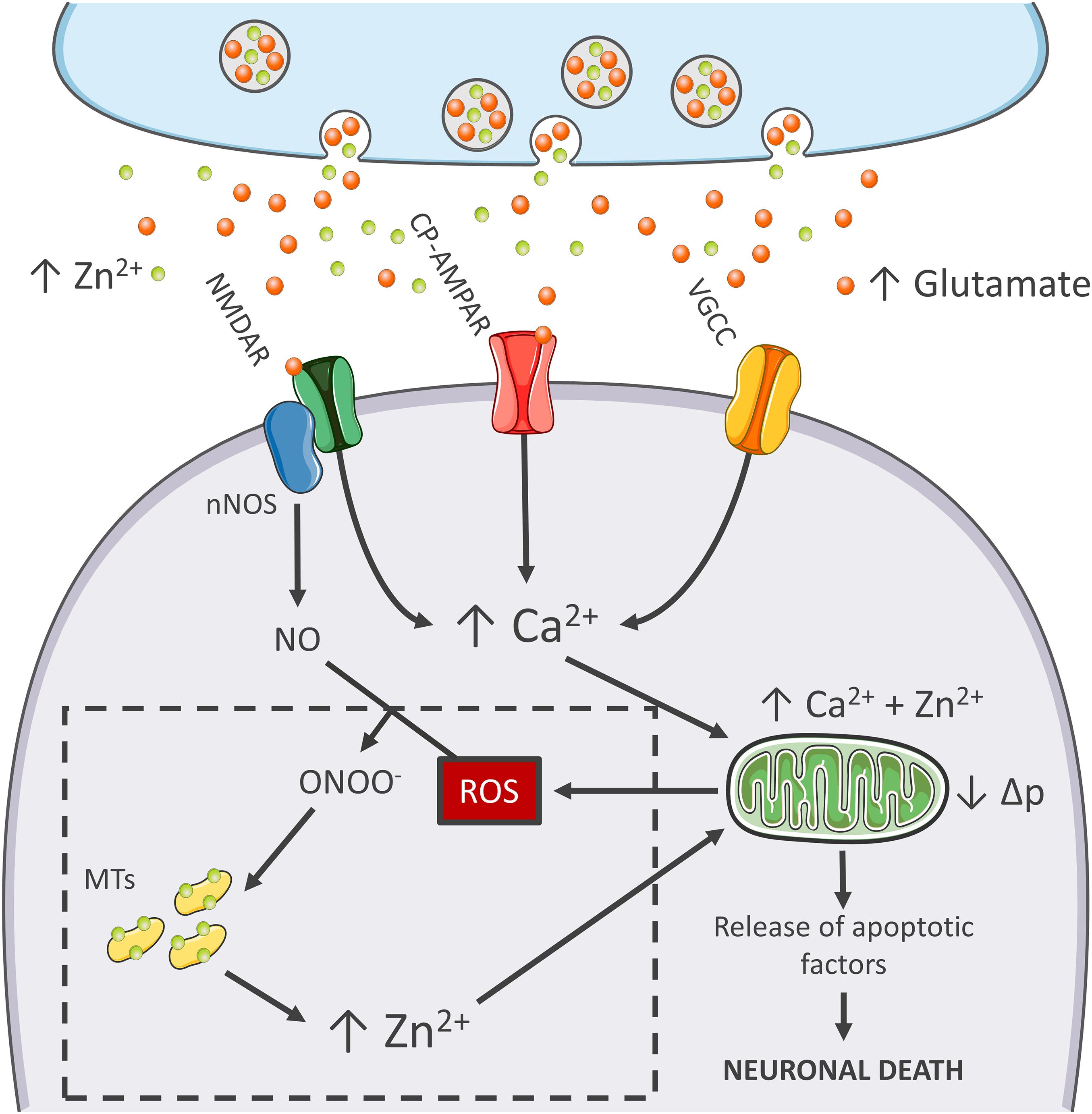
injurious process (Canzoniero et al., 2013; Granzotto and Sensi, 2015; Figure 1). Although, mitochondria of these neurons take up large amounts of Ca2+, the organelles respond with minimal Δp losses and negligible generation of ROS (Canzoniero et al., 2013; Granzotto and Sensi, 2015).
Early studies have shown that, to counteract the detrimental effects
linked to peroxynitrite generation, nNOS (+) neurons express high levels
of SOD2, the ROS quenching enzyme that is strategically localized
inside of mitochondria (Gonzalez-Zulueta et al., 1998).
Therefore, it is conceivable that this constitutive overexpression of
SOD2 makes the subpopulation better equipped to cope with the oxidative
surge produced by the excitotoxic challenges.
The idea that mitochondrial
dysfunction and oxidative stress are prerequisites for NMDA-driven
neuronal loss is in line with the “source-specific” hypothesis of
excitotoxicity. The construct posits that the neurotoxic cascade depends
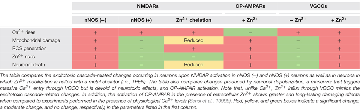
on the route of Ca2+ entry, mainly NMDARs, rather than the magnitude of cation load (Wu and Tymianski, 2018). In agreement with this view, abundant Ca2+ entry through VGCCs, a maneuver devoid of neurotoxic effects, fails to trigger ROS and Δp changes (Table 1). This phenomenon shows great analogies with the effects of Ca2+ rises observed in nNOS (+) neurons following NMDAR activation (Granzotto and Sensi, 2015). Although, NMDAR and VGCC activation produces large Ca2+ rises, differences can be found when dissecting the temporal progression of the two stimuli. Unlike VGCC-driven Ca2+ entry, NMDAR overactivation promotes a prolonged and sustained build-up of Ca2+,
a phenomenon likely due to impaired cation handling. Conceivably, the
NMDAR-driven generation of RNS and ROS can severely affect the
mitochondrial Ca2+ buffering as well as the defective extrusion of the cation.
RNS/ROS can per se contribute to mitochondrial damage (Murphy, 2009). However, an alternative angle is offered by the mitotoxic properties of Zn2+. The cation represents a critical point of convergence between Ca2+, ROS, and mitochondrial failure. By missing ROS generation, nNOS (+) neurons fail to mobilize intracellular Zn2+ upon NMDAR overactivation (Granzotto and Sensi, 2015). Chelation experiments support the hypothesis that NMDAR-triggered Zn2+ rises are required for the full development of the excitotoxic cascade. In that respect, chelation prevents Zn2+ rises without affecting the upstream mechanisms of the cascade (i.e., Ca2+ entry or the Ca2+-driven generation of RNS/ROS). In nNOS (−) neurons, blockade of excitotoxic-driven Zn2+ elevations results in reduced mitochondrial dysfunction and improved intracellular Ca2+ cycling; two functional changes that closely match the ones observed in the nNOS (+) subpopulation (Granzotto and Sensi, 2015) and provide neuroprotection in excitotoxic settings (Wang and Thayer, 2002). On the contrary, nNOS (+) neurons are extremely vulnerable to pharmacological maneuvers that promote Zn2+
elevations, thereby suggesting that the damaging effects of the cation
can override the protective machinery of nNOS (+) neurons (Granzotto and Sensi, 2015, and unpublished observations).
These results are in line with several studies showing that Zn2+ chelation is highly neuroprotective as the maneuver prevents mitochondrial failure, irreversible dysregulation of Ca2+ homeostasis, and eventually neuronal demise (Jiang et al., 2001; Bossy-Wetzel et al., 2004; Medvedeva et al., 2009; Vander Jagt et al., 2009; Clausen et al., 2013; Medvedeva and Weiss, 2014; Ji and Weiss, 2018; Ji et al., 2020; Table 1).
The peculiar milieu offered by nNOS (+) neurons
replicates these mechanisms in a naïve, patho-physiologically relevant
setting and allows inference on the central role played by Zn2+ in the excitotoxic process (Figure 1). Zn2+ is, therefore, not an accomplice or an amplifier of Ca2+-driven toxicity but rather the downstream executioner. Zn2+ actively promotes mitochondrial dysfunction, Ca2+ dysregulation and, eventually, neuronal death.
More at link.
 Study: Dietary zinc intake associated with stroke in American adults - Image credit: Evan Lorne/Shutterstock.com

 Alberto Granzotto
 Lorella M. T. Canzoniero
 Stefano L. Sensi