Changing stroke rehab and research worldwide now.Time is Brain! trillions and trillions of neurons that DIE each day because there are NO effective hyperacute therapies besides tPA(only 12% effective). I have 523 posts on hyperacute therapy, enough for researchers to spend decades proving them out. These are my personal ideas and blog on stroke rehabilitation and stroke research. Do not attempt any of these without checking with your medical provider. Unless you join me in agitating, when you need these therapies they won't be there.

What this blog is for:

My blog is not to help survivors recover, it is to have the 10 million yearly stroke survivors light fires underneath their doctors, stroke hospitals and stroke researchers to get stroke solved. 100% recovery. The stroke medical world is completely failing at that goal, they don't even have it as a goal. Shortly after getting out of the hospital and getting NO information on the process or protocols of stroke rehabilitation and recovery I started searching on the internet and found that no other survivor received useful information. This is an attempt to cover all stroke rehabilitation information that should be readily available to survivors so they can talk with informed knowledge to their medical staff. It lays out what needs to be done to get stroke survivors closer to 100% recovery. It's quite disgusting that this information is not available from every stroke association and doctors group.

Showing posts with label somebody else's problem. Show all posts

Thursday, April 11, 2024

Devices used for photobiomodulation of the brain—a comprehensive and systematic review

 Hasn't your competent? doctor been using this for years? Oh, you don't have a competent doctor or hospital, do you?


Well fuck, we really need someone to write and distribute a protocol on this. All this previous research; OR ARE WE WAITING FOR SOMEONE ELSE TO SOLVE THE PROBLEM?     


A new treatment protocol using photobiomodulation and muscle/bone/joint recovery techniques having a dramatic effect on a stroke patient's recovery: a new weapon for clinicians Sept. 2012      


Interplay between up-regulation of cytochrome-c-oxidase and hemoglobin oxygenation induced by near-infrared laser June 2017      


Photobiomodulation therapy promotes neurogenesis by improving post-stroke local microenvironment and stimulating neuroprogenitor cells Oct. 2017    


Increased Functional Connectivity Within Intrinsic Neural Networks in Chronic Stroke Following Treatment With Red/Near-Infrared Transcranial Photobiomodulation: Case Series With Improved Naming in Aphasia November 2019

The latest here which of course NOTHING will happen with.  Your stroke hospital is completely fucking incompetent.


Devices used for photobiomodulation of the brain—a comprehensive and systematic review

Abstract

A systematic review was conducted to determine the trends in devices and parameters used for brain photobiomodulation (PBM). The revised studies included clinical and cadaveric approaches, in which light stimuli were applied to the head and/or neck. PubMed, Scopus, Web of Science and Google Scholar databases were used for the systematic search. A total of 2133 records were screened, from which 97 were included in this review. The parameters that were extracted and analysed in each article were the device design, actuation area, actuation site, wavelength, mode of operation, power density, energy density, power output, energy per session and treatment time. To organize device information, 11 categories of devices were defined, according to their characteristics. The most used category of devices was laser handpieces, which relate to 21% of all devices, while 28% of the devices were not described. Studies for cognitive function and physiological characterisation are the most well defined ones and with more tangible results. There is a lack of consistency when reporting PBM studies, with several articles under defining the stimulation protocol, and a wide variety of parameters used for the same health conditions (e.g., Alzheimer’s or Parkinson’s disease) resulting in positive outcomes. Standardization for the report of these studies is warranted, as well as sham-controlled comparative studies to determine which parameters have the greatest effect on PBM treatments for different neurological conditions.

Background

Photobiomodulation (PBM) is a non-invasive therapy that entails the use of red to infrared (IR) light (wavelengths of 600 to 1100 nm) to stimulate cellular processes that promote tissue healing and regeneration [1,2,3]. Additionally to being studied and used as a therapy for pain relief [4, 5], wound healing and skin rejuvenation [6, 7], the neuromodulation effects of PBM, when applied to the scalp, have been increasingly more studied, with several clinical studies showing that it could be a safe, non-invasive, and non-destructive alternative to conventional treatments for various neurological disorders.

Functional near-infrared spectroscopy (NIRS) studies have shown that PBM can effectively increase cerebral oxygenation, which has a great impact on cognitive tasks, such as memory and attention, among others [8,9,10,11,12,13]. For traumatic brain injury (TBI), there are several case studies in which patients exhibited improvement in symptoms, measured through standard neurological tests and self-assessments [14,15,16,17,18]. PBM has also reduced depression symptoms in patients diagnosed with depression disorder [19,20,21,22]. For neurodegenerative conditions, such as dementia, Alzheimer’s disease (AD), and Parkinson’s disease (PD), several studies showed improvements in cognition, quality of life, and clinical signs of these conditions [23,24,25,26]. Specifically, a randomized controlled trial on the effects of transcranial PBM in patients diagnosed with PD showed improvements in gait, further establishing the relevance of this therapy for neurodegenerative conditions [27].

The effect of PBM on the brain has been studied to understand the mechanisms behind these positive results and to determine which parameters are more beneficial in these treatments. Although brain PBM has been studied for more than two decades, there is great variability in studies using distinct PBM parameters for the same neurological pathologies, such as wavelength, mode of operation (i.e., continuous or pulsed), area of actuation and energy delivered to the head. Often, authors point out the need for further research to confirm methods to establish PBM as an effective treatment for neurological conditions [17, 22, 28,29,30].

This review aims to draw conclusions from the devices and parameters used for PBM, to determine, if possible, optimal procedures for different pathologies, to promote and accelerate scientific research in this area. Furthermore, since there is some inconsistency in the reporting of these studies, it is also intended to provide further insights into which parameters are more relevant for a full characterisation of the brain PBM.

More at link.

Saturday, June 17, 2023

Exposure to near-infrared light before bed linked to better sleep, daytime function

Well fuck, we really need someone to write and distribute a protocol on this. All this previous research; OR ARE WE WAITING FOR SOMEONE ELSE TO SOLVE THE PROBLEM?     


A new treatment protocol using photobiomodulation and muscle/bone/joint recovery techniques having a dramatic effect on a stroke patient's recovery: a new weapon for clinicians Sept. 2012      


Interplay between up-regulation of cytochrome-c-oxidase and hemoglobin oxygenation induced by near-infrared laser June 2017      


Photobiomodulation therapy promotes neurogenesis by improving post-stroke local microenvironment and stimulating neuroprogenitor cells Oct. 2017    


Increased Functional Connectivity Within Intrinsic Neural Networks in Chronic Stroke Following Treatment With Red/Near-Infrared Transcranial Photobiomodulation: Case Series With Improved Naming in Aphasia November 2019

The latest here which of course NOTHING will happen with.  Your stroke hospital is completely fucking incompetent. 

The latest here:


Exposure to near-infrared light before bed linked to better sleep, daytime function

Key takeaways:

  • Thirty adults were randomized to wear near-infrared light-emitting cervical collar or sham device every other night for 5 weeks.
  • Patients with NIR collar self-reported improvement in sleep-related symptoms.

INDIANAPOLIS — Transdermal exposure to near-infrared light before bed via a wearable device was linked to improved sleep, relaxation and next-day functionality compared with sham, according to research presented at SLEEP 2023.

“Given the emerging field of photobiomodulation and its potential neuroprotective and vasodilating effects, this red-light and near-infrared emitting device may be useful if milliwatt power level, dosage and frequency of use are refined,” Kathryn E.R. Kennedy, BS, lead study author and PhD candidate in the department of psychiatry at the University of Arizona, said in a related release.

Sleep duration, sleep timing, social jetlag and shift work were not associated with fecundability or live birth. Source: Shutterstock
Results of a randomized, sham-controlled clinical trial featuring a cervical near-infrared light device found that exposure was linked to better sleep, daytime function. Image: Adobe Stock

Noting therapeutic effects, such as relaxation, with delivery of transdermal near-infrared (NIR) light, Kennedy and colleagues examined the effects of NIR exposure on sleep and daytime functioning when given before bed.

Their randomized, sham-controlled, 5-week study included 30 individuals, aged 30 to 60 years, who self-reported complaints about sleep but were not diagnosed with a sleep disorder. Following a 2-week baseline period, participants wore a NIR-emitting cervical collar (combined 660 nm, 740 nm, 810 nm and 870 nm) or sham device every other night before bed for 3 weeks.

Researchers assessed physical symptoms and sleep-related experiences using the Systematic Assessment of Treatment Emergent Effects (SAFTEE) questionnaire. They also examined differences between groups with the Insomnia Severity Index (ISI), daily sleep diaries, Oura rings and daily participant ratings of sleep quality and feeling refreshed, daytime function, energy levels, relaxation and performance.

According to results, significant differences between active and sham groups were reported in SAFTEE total score (–9.4 vs. +13.8) and sleep-related symptoms (–2.9 vs. +4.1), while no differences were reported in ISI scores, Oura recordings or sleep diary data.

While most changes were consistent between groups, the active group had decreased sleep latency (–6 mins) and REM% (–0.9%), increased average sleep quality (+0.6 pts) and feeling refreshed (+0.7 pts), and perceived increase in relaxation (17.5 pts) and daytime function (17.5 pts). Conversely, the sham group had decreases in perceived overall performance (18.7 pts) and energy level (26 pts).

“This novel phototherapy device — while still being explored and in need of further research — appeared to be generally well-tolerated by a small group of participants,” Kennedy said in the release. “Those with active, light-emitting devices — as opposed to the inert sham devices — self-reported an increase in relaxation and better sleep with use.”

Reference:

Sunday, March 5, 2023

Healio launches easy-to-use guide for nutrition


Your doctor incompetently WAITED LONG ENOUGH FOR SOMEONE ELSE TO SOLVE THE PROBLEM? 

Still missing all these needs:

For stroke prevention; for dementia prevention; for cognitive improvement; for cholesterol reduction; for plaque removal; for Parkinsons prevention; for inflammation reduction; for blood pressure reduction. 

YOUR DOCTOR'S RESPONSIBILITY TO GET THESE OTHER DIET PROTOCOLS CREATED!


Healio launches easy-to-use guide for nutrition

Healio has announced that it is now offering a special report on nutrition that physicians can use as a resource to guide conversations with patients about healthy diet options.

The new resource — Food is Medicine: A Healio Guide to Diets — serves as an easily accessible, one-stop-shop for details about diets and evidence on their potential health benefits.

PC0323HealioDiet_Graphic_01_WEB

“This special report was created with the busy clinician in mind, to make it easier to discuss nutrition with their patients and share in the decisions that are right for them,” John Schoen, an editorial director at Healio, said in a press release. “We're really excited to share this resource with our readers.”

Justin F. Tondt, MD, an assistant professor of family and community medicine at Penn State College of Medicine, and Kurt R. Wharton, MD, FACOG, chief of the Women’s and Children’s Clinical Care Programs for Beaumont Health in Michigan and a professor of obstetrics and gynecology at Oakland University’s William Beaumont School of Medicine, contributed to the report. It includes recommendations from the Dietary Guidelines for Americans, 2020-2025, and offers information on specific diets, what they entail and their health benefits based on current scientific evidence.

Click here to downloaded the report, or visit Healio.com/Diet.

Monday, October 10, 2022

Flavonoids from Stems and Leaves of Scutellaria baicalensis Georgi Improve Composited Aβ-induced Alzheimer's Disease Model Rats' Memory and Neuroplasticity Disorders


WHOM will be doing the human testing? And then get the protocol rolled out to all stroke hospitals? With NO stroke leadership I'm afraid nothing will be done, other promising animal research never seems to get to human testing and protocols.  

With your elevated risk of Alzheimers/dementia you'll want your doctor following this carefully.  Is your doctor ensuring further studies occur?

Or is your doctor incompetently WAITING FOR SOMEONE ELSE TO SOLVE THE PROBLEM? 

Your risk of dementia, has your doctor told you of this?

1. A documented 33% dementia chance post-stroke from an Australian study?   May 2012.

2. Then this study came out and seems to have a range from 17-66%. December 2013.`    

3. A 20% chance in this research.   July 2013.

4. Dementia Risk Doubled in Patients Following Stroke September 2018 

What is your doctor's EXACT PROTOCOL TO PREVENT DEMENTIA?

The latest here:

Flavonoids from Stems and Leaves of Scutellaria baicalensis Georgi Improve Composited Aβ-induced Alzheimer's Disease Model Rats' Memory and Neuroplasticity Disorders 

Affiliations

Abstract

Aim: To investigate the effects and mechanism of flavonoids from stems and leaves of Scutellaria baicalensis Georgi (SSF) on the disorders in learning and memory and neuroplasticity induced by beta amyloid 25-35 (Aβ25-35) combined with aluminum trichloride (AlCl3) and human recombinant transfer factor-β1 (RHTGF-β1) (composited Aβ) in rats.

Methods: A rat Alzheimer's disease (AD) model was established by intracerebroventricular injection of Aβ25-35 combined with AlCl3 and RHTGF-β1. The successful AD model of rats was screened with Morris water maze. The successful model rats were randomly divided into a model group and three doses of SSF treated group. The Morris water maze was used to detect the rats' learning and memory abilities. The real-time fluorescence quantitative (qPCR) was applied to assay the mRNA expressions of CaM, CamkIV and Ferritin, as well as the neuroplasticity factors of HuB, HuC and HuD. The Western blotting was used to measure the protein expressions of CaM, CamkIV, HuB/D, HuC+HuD and Ferritin in CaM-CamkIV-CREB signal pathway.

Results: Compared with sham group, the abilities of learning and memory in the model group were significantly impaired (P<0.01), and the mRNA or protein expressions of CaM, CamkIV, HuB, HuC, HuD, HuB/D, HuC+HuD and Ferritin in CaM-CamkIV-CREB signal pathway were abnormal changed in model group. However, the three doses of SSF can differently ameliorated the impaired learning and memory and regulate the abnormal expressions of mRNA or protein in rats' CaM, CamkIV, HuB, HuC, HuD, HuB/D, HuC+HuD and Ferritin induced by composited Aβ.

Conclusion: The improvement of SSF on the learning and memory disorder induced by composited Aβ is primarily derived from the positive regulation in CaM-CamkIV-CREB signal pathway and activation in neuroplasticity.

Keywords: Alzheimer's disease; CaM-CamkIV-CREB signal pathway; Flavonoids from stems and leaves of Scutellaria baicalensis Georgi; learning and memory; neuroplasticity.

Friday, September 17, 2021

Functional Plasticity Induced by Mirror Training: The Mirror as the Element Connecting Both Hands to One Hemisphere

 When this research came out 9 years ago did your doctor do ONE DAMN THING with it? Or is your doctor incompetently WAITING FOR SOMEONE ELSE TO SOLVE THE PROBLEM? 

Well shit, the problem has been solved, all your doctor had to do was implement it, buy a few mirrors and do a little training for the therapists.  But your doctor did absolutely nothing? Right?

Functional Plasticity Induced by Mirror Training: The Mirror as the Element Connecting Both Hands to One Hemisphere

First Published January 13, 2012 Research Article Find in PubMed 

Background

Mirror therapy (MT) is a promising therapeutic approach in stroke patients with severe hand paresis.  

Objective

The ipsilateral (contralesional) primary sensorimotor cortex (SMC) and the mirror neuron system have been suggested to play decisive roles in the MT network. The present study investigated its underlying neural plasticity. Methods. Two groups of healthy participants (n = 13 in each group) performed standardized fine motor tasks moving pegs and marbles (20 min/d for 4 days) with their right hand with either a mirror (mirror training group, MG) or a nonreflective board (control training group, CG) positioned orthogonally in front of them. The number of items moved by each hand was tested after each training session. Functional MRI (fMRI) was acquired before and after the training procedure to investigate the mirror training (MTr)-specific network by the analysis of the factors Time and Group.  

Results

The hand performance test of the trained right hand did not differ between the 2 groups. The untrained left hand improved significantly more in the MG compared with the CG. fMRI analysis of action observation and imitation of grasping tasks demonstrated MTr-specific activation changes within the right dorsal and left ventral premotor cortex as well as in the left SMC (SMCleft). Analysis of functional and effective connectivity showed a MTr-specific increase of functional coupling between each premotor region and the left supplementary motor area, which in turn showed an increased functional interaction with the ipsilateral SMCleft.  

Conclusions

MTr remodels the motor system by functionally connecting hand movement to the ipsilateral SMC. On a system level, it leads to interference of the neural circuit related to motor programming and observation of the trained hand with the illusionary movement of the untrained hand.

Effective stroke management is important as the treatment of stroke consumes a significant amount of resources.1 Functional recovery can occur even in the chronic stage of stroke with plastic changes induced in the affected primary motor cortex (M1) by intense motor training of the paretic hand.2,3 Additional commitment of noninvasive stimulation over the affected M1 by repetitive transcranial magnetic stimulation (TMS)4 or by transcranial direct current stimulation5 promotes functional recovery in association with plastic changes within the affected M1. However, the effectiveness of such strategies is limited to patients with mild to moderate hand paresis. Thus, an effective therapy regime for patients with severe hand paresis or loss of hand function is still lacking.

Mirror therapy (MT) is a promising therapeutic approach in stroke patients, even in those with complete loss of hand function. By using a mirror that is positioned orthogonally in front of the patient, the reflection of the right arm in the mirror provides an illusion that the left arm is being moved. MT was first described by Ramachandran and colleagues6 in patients with phantom pain after limb amputation and has been suggested to improve hand functional deficit in stroke patients.7 Ramachandran and Altschuler8 suggested that the ipsilateral (contralesional) corticospinal tracts from the M1 play a role while training with a mirror,8 but a recent functional MRI (fMRI) study reported an activation shift toward the ipsilesional sensorimotor cortex (SMC) after MT in chronic stroke patients in contrast with a control group.9 It has also been suggested that the mirror neuron system (MNS) plays an important role while observing one’s hand in the mirror and training via action observation (AO) and imitation (IMI).8,10

The underlying neural plasticity of MT is currently unknown. Therefore, in the present study, fMRI was used to address this issue. We focused on training-induced plasticity by training with a mirror rather than the direct effect of observing one’s hand in the mirror, as was investigated with the use of functional imaging11,12 or TMS.13-15


Tuesday, August 31, 2021

Cognitive Impairment After Intracerebral Hemorrhage: A Systematic Review of Current Evidence and Knowledge Gaps

 So you've described a problem. What research are you initiating to solve this problem?

Or once again are you sitting on your asses WAITING FOR SOMEONE ELSE TO SOLVE THE PROBLEM?  If you're doing nothing to actually solve stroke, why are you here?

Cognitive Impairment After Intracerebral Hemorrhage: A Systematic Review of Current Evidence and Knowledge Gaps

  • 1Center for Outcomes Research, Houston Methodist Research Institute, Houston Methodist, Houston, TX, United States
  • 2Department of Neurology, Beth Israel Deaconess Medical Center, Boston, MA, United States
  • 3Department of Nuclear Engineering, Texas A&M University, College Station, TX, United States
  • 4Department of Neurology and Rehabilitation Medicine, University of Cincinnati College of Medicine, Cincinnati, OH, United States
  • 5Glenn Biggs Institute for Alzheimer's and Neurodegenerative Diseases, University of Texas Health Science Center at San Antonio, San Antonio, TX, United States
  • 6Neurological Institute, Houston Methodist, Houston, TX, United States

Background: Cognitive impairment (CI) is commonly observed after intracerebral hemorrhage (ICH). While a growing number of studies have explored this association, several evidence gaps persist. This review seeks to investigate the relationship between CI and ICH.

Methods: A two-stage systematic review of research articles, clinical trials, and case series was performed. Initial search used the keywords [“Intracerebral hemorrhage” OR “ICH”] AND [“Cognitive Impairment” OR “Dementia OR “Cognitive Decline”] within the PubMed (last accessed November 3rd, 2020) and ScienceDirect (last accessed October 27th, 2020) databases, without publication date limits. Articles that addressed CI and spontaneous ICH were accepted if CI was assessed after ICH. Articles were rejected if they did not independently address an adult human population or spontaneous ICH, didn't link CI to ICH, were an unrelated document type, or were not written in English. A secondary snowball literature search was performed using reviews identified by the initial search. The Agency for Healthcare research and Quality's assessment tool was used to evaluate bias within studies. Rates of CI and contributory factors were investigated.

Results: Search yielded 32 articles that collectively included 22,631 patients. Present evidence indicates a high rate of post-ICH CI (65–84%) in the acute phase (<4 weeks) which is relatively lower at 3 (17.3–40.2%) and 6 months (19–63.3%). Longer term follow-up (≥1 year) demonstrates a gradual increase in CI. Advanced age, female sex, and prior stroke were associated with higher rates of CI. Associations between post-ICH CI and cerebral microbleeds, superficial siderosis, and ICH volume also exist. Pre-ICH cognitive assessment was missing in 28% of included studies. The Mini Mental State Evaluation (44%) and Montreal Cognitive Assessment (16%) were the most common cognitive assessments, albeit with variable thresholds and definitions. Studies rarely (<10%) addressed racial and ethnic disparities.

Discussion: Current findings suggest a dynamic course of post-ICH cognitive impairment that may depend on genetic, sociodemographic and clinical factors. Methodological heterogeneity prevented meta-analysis, limiting results. There is a need for the methodologies and time points of post-ICH cognitive assessments to be harmonized across diverse clinical and demographic populations.

Introduction

Intracerebral hemorrhage (ICH) is the most common type of hemorrhagic stroke, accounting for 10–20% of all strokes (1), with a global incidence of 24.6 per 100,000 person-years (2). Spontaneous ICH primarily results from either hypertensive microangiopathy or cerebral amyloid angiopathy (CAA) (3), which are likely to produce varied phenotypes. Hypertensive ICH likely occurs in deep brain structures while CAA-related ICH generally occurs in lobar locations (2). Regardless of the cause, ICH is associated with poor outcomes that include early mortality (2, 4) and the loss of functional independence (2).

Cognitive Impairment (CI) commonly coexists with ICH. The majority of ICH patients exhibit acute phase CI, with impairments reported in up to 84% of patients (5). While the immediate post-ICH cognitive effects and the potential for long-term CI (6) are broadly recognized, several evidence gaps persist. The trajectory of post-ICH CI is poorly characterized and demonstrates considerable variability. Some ICH patients experience favorable recovery after an acute cognitive decline while others exhibit persistent or worsening CI (7). The significant contribution of cognitive function toward quality of life among ICH survivors has driven an increased research focus on post-ICH CI and dementia. With the growing body of literature focused on post-ICH CI, it is important to integrate the available evidence and characterize cognitive function among ICH patients. This systematic review aims to collect and summarize current evidence regarding the risk factors and trajectory of CI after spontaneous ICH, report the strength and validity of study methodologies, and highlight current knowledge gaps in the study of post-ICH CI.


Tuesday, July 6, 2021

Submitted to Neurochem int potential therapeutic agents for ischemic white matter damage

 This seems to make the assumption that myelin sheaths are damaged in ischemic injury(stroke?). What specific person is going to do the followup research to prevent and repair this damage from a stroke?  Or once again are your doctors and stroke hospital sitting on their asses WAITING FOR SOMEONE ELSE TO SOLVE THE PROBLEM? 

Submitted to Neurochem int potential therapeutic agents for ischemic white matter damage

ZhongChenacWei-weiHua

Highlights

Ischemic white matter damage is a leading cause of multiple neurological disorders in both adults and infants.

Oligodendrocytes and myelin sheaths are particularly vulnerable to ischemic injury, via different mechanisms, compared to other cells in the CNS.

Multifaceted agents with both actions on demyelination and remyelination would be preferable for the treatment of white matter damage.

Abstract

Ischemic white matter damage (WMD) is increasingly being considered as one of the major causes of neurological disorders in older adults and preterm infants. The functional consequences of WMD triggers a progressive cognitive decline and dementia particularly in patients with ischemic cerebrovascular diseases. Despite the major stride made in the pathogenesis mechanisms of ischemic WMD in the last century, effective medications are still not available. So, there is an urgent need to explore a promising approach to slow the progression or modify its pathological course. In this review, we discussed the animal models, the pathological mechanisms and the potential therapeutic agents for ischemic WMD. The development in the studies of anti-oxidants, free radical scavengers, anti-inflammatory or anti-apoptotic agents and neurotrophic factors in ischemic WMD were summarized. The agents which either alleviate oligodendrocyte damage or promote its proliferation or differentiation may have potential value for the treatment of ischemic WMD. Moreover, drugs with multifaceted protective activities or a wide therapeutic window may be optimal for clinical translation.


Sunday, May 9, 2021

Impact of Early Blood Pressure Lowering in Patients Presenting with Acute Ischemic Stroke

So still no protocol for blood pressure management. What we need is an EFFECTIVE STROKE LEADER THAT WILL SOLVE THIS PROBLEM. Alas we have none.  Hope you like you doctor guessing what to do.

Are we ever going to get a blood pressure protocol? Or will we wait until stroke survivors are in charge? This question has been out there forever. Is your doctor and stroke hospital sitting on their asses WAITING FOR SOMEONE ELSE TO SOLVE THE PROBLEM? 

Systolic Blood Pressure Control and Mortality After Stroke in Hypertensive Patients July 2015


BP Lowering in Acute Stroke Flops for Improving Outcomes February 2015


After stroke, compared with Systolic Blood Pressure in the high range, low to normal SBP is associated with poorer mortality outcomes. May 2015


Systolic Blood Pressure and Mortality After Stroke May 2015


Blood pressure-lowering treatment with candesartan had no beneficial effect on activities of daily living and level of care at 6 months June 2015


Blood pressure reduction in acute ischemic stroke according to time to treatment: a subgroup analysis of the China Antihypertensive Trial in Acute Ischemic Stroke trial May 2017


Intensive blood pressure lowering in patients with acute intracerebral haemorrhage: Clinical outcomes and haemorrhage expansion. Systematic review and meta-analysis of randomised trials February 2017 

But this to think about:

Don't go too low with blood pressure in hypertensive CAD patients


The latest here:

 Impact of Early Blood Pressure Lowering in Patients Presenting with Acute Ischemic Stroke

Abstract

Purpose of Review

In this review article we will discuss the acute hypertensive response in the context of acute ischemic stroke and present the latest evidence-based concepts of the significance and management of the hemodynamic response in acute ischemic stroke.

Recent Findings

Acute hypertensive response is considered a common hemodynamic physiologic response in the early setting of an acute ischemic stroke. The significance of the acute hypertensive response is not entirely well understood. However, in certain types of acute ischemic strokes, the systemic elevation of the blood pressure helps to maintain the collateral blood flow in the penumbral ischemic tissue. The magnitude of the elevation of the systemic blood pressure that contributes to the maintenance of the collateral flow is not well established. The overcorrection of this physiologic hemodynamic response before an effective vessel recanalization takes place can carry a negative impact in the final clinical outcome. The significance of the persistence of the acute hypertensive response after an effective vessel recanalization is poorly understood, and it may negatively affect the final outcome due to reperfusion injury.

Summary

Acute hypertensive response is considered a common hemodynamic reaction of the cardiovascular system in the context of an acute ischemic stroke. The reaction is particularly common in acute brain embolic occlusion of large intracranial vessels. Its early management before, during, and immediately after arterial reperfusion has a repercussion in the final fate of the ischemic tissue and the clinical outcome.

Introduction

Blood pressure is usually elevated in the acute phase of all types of hemorrhagic and ischemic strokes. This acute hypertensive (presumably physiologic) response is common in the early phase of acute ischemic stroke. Approximately two-third of the ischemic strokes present with elevated systolic and diastolic blood pressure [1]. The significance of this type of hemodynamic response is unclear. In large vessel occlusion, the acute hypertensive response is believed to be responsible for the maintenance of the retrograde collateral leptomeningeal circulation. The optimal management of the blood pressure in this scenario includes balancing the risk of inappropriate lowering of the blood pressure that can jeopardize the ischemic penumbra and the excessive arterial hypertension that can promote hemorrhagic transformation of the ischemic tissue. Unfortunately, the best strategy for the management of blood pressure in patient with acute ischemic stroke is not yet established. In this review article we will focus on the latest evidence pertaining blood pressure reduction in the early phase of an acute ischemic stroke and the management of the pre- and post-operative hemodynamic response in patient with acute ischemic stroke due to large vessel occlusion undergoing to mechanical thrombectomy.

Natural History of the Acute Hypertensive Response in Acute Ischemic Stroke

After an acute occlusion of a major intracranial artery, the cerebral tissue is able to sense a decrease in the interstitial oxygen tension [2]. The resulting ischemia around the cerebral tissue is able to trigger a glio-neurovascular signal that produces an increased sympathetic outflow from the central nervous system into the cardiovascular system that induces an elevation of the vascular tone [3]. This explains in part why subjects without premorbid arterial hypertension can experience severe elevation of the systolic and diastolic blood pressure minutes after suffering an embolic occlusion of a proximal intracranial artery. The magnitude of the hypertensive response tends to correlate with the size of tissue at risk. Large areas of ischemic penumbral cerebral tissue can provoke more dramatic acute hypertensive response. This neuroendocrine response can also be augmented by undiagnosed or untreated premorbid systemic arterial hypertension [4].

The acute hypertensive response observed in the acute phase of ischemic stroke is self-limiting, and it tends to decline over the course of the next several days and return to the premorbid baseline levels. In embolic strokes, this tends to happen at the same time when spontaneous recanalization occurs. Mechanical thrombectomy for large vessel occlusion can shorten this period, and it is not uncommon to observe a dramatic reduction of the systolic blood pressure after the vessel is recanalized. On the contrary, in cases of ineffective vessel recanalization, the acute hypertensive response may persist for several days.

Impact of Early Lowering of the Blood Pressure Across All Acute Ischemic Strokes

Sudden and aggressive reduction of the blood pressure is deleterious across all types of ischemic strokes. This is also true in intracerebral hemorrhage. In the case of hypertensive intracerebral hemorrhage, two different randomized clinical trials showed a tendency for reduction in the hematoma expansion with early and moderate reduction of the hypertensive response [5, 6]. However, more aggressive reduction in the blood pressure not only did not result in further reduction of the hematoma expansion but also resulted in adverse complication related to cerebral, , and cardiac hypoperfusion. Unfortunately, the timing, intensity, and the duration of the correction of the acute hypertensive response in both ischemic and hemorrhagic forms of stroke is not yet determined.

Less than one-third of acute ischemic stroke can present without elevation of the blood pressure, and some of the can present with low blood pressure level. Subjects presenting with acute ischemic stroke and lack of acute hypertensive response can harbor other cardiovascular comorbidities including concomitant congestive heart failure and severe aortic and mitral valvular disease. These patients are well known to face worse outcomes in spite of the successful acute interventions. In a recent secondary analysis of the Head Positioning in acute Stroke Trial (HeadPoST), patients with acute ischemic stroke presenting initially with low blood pressure, defined as a systolic blood pressure less than 120 mmHg and a diastolic blood pressure less than 70 mmHg, had an increased risk of death or dependency (adjusted OR 1.27, 95% CI 1.02–1.58) compared with patients presenting with acute hypertensive physiologic response [7]. The association with poor clinical outcome in patients with acute ischemic stroke and failure to activate a hypertensive response persisted after adjusting for several confounding factors including baseline heart failure and cardiac disease. Importantly, these patients were at increased risk for severe adverse cardiac effects as well as worse functional outcome from their stroke.

Acute ischemic stroke patients presenting with an acute physiologic hypertensive response represent the majority of the cases. For the subset of patients arriving with the first 3–4½ h after the symptoms onset guidelines recommend to maintain the systolic blood pressure below 185 mmHg and the diastolic blood pressure below 105 mmHg. The original studies that tested the effectiveness of intravenous r-tPA for acute ischemic stroke used these blood pressure thresholds to reduce the risk of symptomatic hemorrhagic transformation that could potently offset the benefit of the thrombolytic treatment. However, this trials did not assess a specific blood pressure target for the lower limit of the goal. A U-shaped association between the initial blood pressure and the final unfavorable outcome in acute ischemic stroke was demonstrated in several observational studies. The extremes (low blood pressure and excessively elevated blood pressure) range are associated with worse outcome, and the best outcomes appear to be present with a modest initial hypertension. However, none of these observational studies were able to prove causality. Two large registries of intravenous thrombolysis in acute ischemic stroke reported the association between hypertension and the risk of the symptomatic hemorrhagic transformation [8, 9]. In both registries, the subjects presenting with systolic blood pressure higher than 170 mmHg have four more chances of symptomatic intracerebral hemorrhage as compared with subjects presenting with systolic blood pressure between 140 and 150 mmHg. In a meta-analysis of the effect of blood pressure lowering in early ischemic stroke, a total of 12,703 individuals were included, with 6392 participants randomly assigned to the active treatment group (lowering of blood pressure) and 6311 to the control group [10]. The active group underwent to modest reduction of systolic and diastolic blood pressure in the first 24 h. However, the active group did not showed any significant reduction in the risk of death or dependency at 3 months. The international, randomized, open-label, blinded-endpoint, phase 3 trial specifically assessed the potential benefit of lowering the blood pressure to reduce the risk hemorrhagic transformation during administration of intravenous Alteplase (ENCHANTED trial) [11••]. The study randomized subjects with acute ischemic stroke presenting with a systolic blood pressure equal or higher than 150 mmHg if they fulfilled the standard criteria for intravenous r-r-tPA into two groups: intensive systolic blood pressure control (130–140 mmHg) versus guideline-directed systolic blood pressure control (< 185 mmHg). The primary outcome was the percentage of death or disability at 3 months and unfortunately it did not differ between the groups. Several lessons can be learned from the ENCHANTED trial. Majority of the ischemic stroke were mild to moderate (average NIHSS score of 7 points) and even more important the difference in the systolic blood pressure between the two groups was only 7 mmHg (146 versus 153 mmHg mean systolic arterial blood pressure in the active versus the control arm). This was perhaps the main reason why the trial was negative. However, there was a clear tendency toward a lower incidence of hemorrhagic transformation particularly major intracerebral hemorrhage in the intensive blood pressure arm but unfortunately it did not improve the final primary clinical outcome. Until further evidence are available, the management of the initial arterial blood pressure in the acute setting of acute ischemic stroke triage particularly in the first three to four and a half hours after symptoms onset in subjects considered candidates for intravenous thrombolysis should follow the latest guidelines recommendations of a systolic blood pressure equal or less than 185 mmHg and a diastolic blood pressure equal or less than 105 mmHg. In the future, randomize clinical trials assessing the impact of arterial blood pressure manipulation to improve the outcome should focus in the subset of ischemic stroke in which the elevation of the systemic blood pressure may play a more prominent role in the maintenance of the collateral blood flow like in subjects with proximal large vessel occlusion amenable to endovascular recanalization.

Management of the Blood Pressure in Acute Ischemic Stroke Before and During Endovascular Recanalization

Large vessel occlusion is perhaps the subtype of acute ischemic stroke in which the relevance of the acute hypertensive response and its adequate management are of great significance in the final outcome. The acute hypertensive response is noticeable immediately after the embolic occlusion of a proximal intracranial artery. This instantaneous systemic hemodynamic physiologic response is key for the maintenance of the retrograde leptomeningeal collateral circulation. It is also responsible of the initial minimal neurological deficit present in patient with acute large vessel occlusions presenting and low NIHSS score (equal or lower than 5 points) or even with a complete resolution of the clinical symptoms (transient ischemic attack). However, this could be a precarious situation, and it is calculated that approximately 20 to 40% of this subjects will deteriorate their neurologic condition during the subsequent 24–72 h. In this context, arterial hypotension (spontaneous or induced) well known to be obviously deleterious for the final fate of the penumbral tissue and finally the clinical outcome. Spontaneous hypotension can be seen in subjects with large vessel occlusion and concomitant cardiac disease that impairs the stroke volume. Iatrogenic systemic hypotension can be seen in patients with large vessel occlusion that undergo to mechanical thrombectomy under general anesthesia. During the induction of general anesthesia, the inhaled volatile anesthetics can cause inappropriate lowering of the systolic and mean arterial blood pressure that can overcorrect the acute hypertensive physiologic response. In a retrospective study of 371 patients that underwent to mechanical thrombectomy under general anesthesia, a linear association between arterial hypotension and worse outcome was demonstrated [12]. Even a single (less than 10%) drop from baseline resulted in worse neurologic outcome. Even single mean arterial blood pressure drop during mechanical thrombectomy under general anesthesia could result in poor neurologic outcome. In retrospective cohort study of 115 subjects that underwent to mechanical thrombectomy under general anesthesia, it was found that subjects that suffered from drops in the mean arterial blood pressure below 60 mmHg faced a worse outcome [13]. If mechanical thrombectomy will be performed under general anesthesia, the anesthesia team has to be aware that even short episodes of the hypotension during the general anesthesia induction can result in impairment of the collateral circulation. A dedicated cerebrovascular anesthesia team that can provide prompt intubation and general anesthesia in a fast and effective manner without delaying the recanalization times and without causing inappropriate lowering of the systemic blood pressure should be a critical component of comprehensive stroke centers [14]. As a general rule, patients with large vessel occlusion undergoing to mechanical thrombectomy and concomitant intravenous thrombolysis should follow the same recommendation the systolic and diastolic blood pressure parameters for intravenous rt-PA candidates (systolic blood pressure equal or less than 185 mmHg and diastolic blood pressure equal or less than 105 mmHg). In patients with large vessel occlusion undergoing to mechanical thrombectomy without concomitant intravenous thrombolysis, the systolic and diastolic blood pressure parameters should follow the blood pressure recommendation of acute ischemic stroke patients not candidate to intravenous thrombolysis (systolic blood pressure equal or less than 220 mmHg and diastolic blood pressure equal or less than 120 mmHg).

Management of the Blood Pressure in Acute Ischemic Stroke Immediately After Successful Endovascular Recanalization

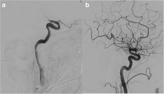
Successful endovascular recanalization in large vessel occlusion is the most important predictor of subsequent successful clinical outcome at 90 days(Is your definition of successful clinical outcome 100% recovery? If not you are in the wrong business. I suggest screaming at your hospital if they don't get you100% recovered, that is their job ). A successful recanalization implies an almost complete or complete reperfusion of the occluded vessel, defined as TICI (Thrombolysis In Cerebral Ischemia) 2b or higher (Fig. 1) score. The endovascular recanalization has to happen in a timely fashion before a large infarcted tissue is established. The survival of the penumbral tissue (cerebral tissue in the process to die but not death yet) largely depend in the effectiveness of the collateral system that as we pointed before it is highly dependent of changes in the systemic blood pressure.

Fig. 1
figure1

Panel a Lateral view from digital subtraction angiography of the brain showing an acute embolic occlusion of the right internal carotid artery terminus corresponding to a thrombolysis in cerebral ischemia score of zero. Panel b Lateral view from digital subtraction angiography of the brain showing complete recanalization after mechanical thrombectomy corresponding to a thrombolysis in cerebral ischemia score of three

A successful recanalization is commonly associated with spontaneous regression and disappearance of the acute hypertensive physiologic response. The lack of spontaneous resolution of the acute hypertensive response after a successful recanalization has been correlated with worse neurologic outcome in several observational studies. The reason behind the persistence of arterial hypertension in spite of successful recanalization is probably multifactorial. Uncontrolled premorbid hypertension might explain some cases but also the fast progression to ischemia in spite of early and effective vessel recanalization is also suspected. In this last circumstance, the ischemic gliovascular tissue might continue sending a signal that keeps the sympathetic outflow from the central nervous system into the cardiovascular system. Post-operative arterial hypertension in patients that underwent to a successful recanalization has been correlated with higher chances of hemorrhagic transformation.

The impact of blood pressure levels within the first 24 h after mechanical thrombectomy on the clinical outcome was reported in a recent retrospective study that included 700 patients with large vessel occlusion that underwent to mechanical thrombectomy [15]. The study found that subjects with lower levels of blood pressure (less than 140 mmHg) 24 h after the mechanical thrombectomy tend to have better outcome and lower mortality at 3 months. It is unknown if the early correction of the persistent hypertensive response would result in better outcomes including less incidence of hemorrhagic complications. In a multicenter retrospective study of the relationship between systemic blood pressure reduction and outcome after successful reperfusion included a total of 1454 patients were included [16]. More than half of the sample underwent to a complete angiographic reperfusion (TICI: 3 score) with a mean time of onset to groin puncture of 216 min. The systolic blood pressure reduction was associated with lower odds of poor outcome. This was particularly consistent in subjects with complete recanalization and no history of premorbid hypertension. Another retrospective study from Taiwan showed that the uncontrolled elevation of the blood pressure in the immediate first 6 h after a successful mechanical thrombectomy is an important factor in the final clinical outcome [17••]. The study found that 50% of the subjects that underwent to a successful recanalization were able to be functionally independent at 90 days. In this particular group, the blood pressure levels were analyzed in the first 24 h after mechanical thrombectomy. The groups were divided into four intervals (0–6, 7–12, 22–19, and 19–24 h). There was a linear association between post-operative hypertension and worse outcome at 90 days. The association was stronger if the average highest blood pressure recording was registered in the first 6 h.

Areas of hyperatenuation in the brain parenchyma (on computerized tomography of the head obtained without intravenous injection of iodinated contrast material) represent regions of the brain tissue with broken brain blood barrier due to established ischemia after mechanical thrombectomy. This area of hyperatenuation is more prone to hemorrhagic transformation after effective recanalization. In a retrospective study of a prospectively collected cohort of the consecutive acute ischemic stroke patients due to large vessel occlusion that underwent to a successful mechanical thrombectomy, 50% of the subjects exhibited areas of the hyperatenuation in the post-procedure non contrast CT [18]. In this particular group, the probability of symptomatic hemorrhagic transformation (parenchymal hematoma) due to reperfusion injury increased with each increment of the post-operative maximum systolic blood pressure in the first 24 h.

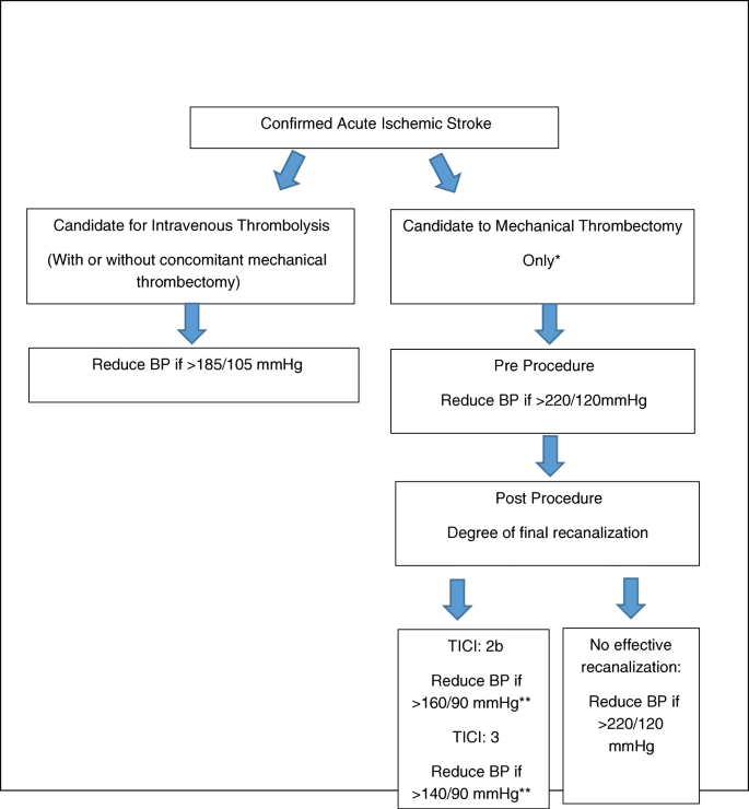
Until further evidence from randomized clinical trials are available, it appears prudent to try to correct the persistence of an acute hypertensive response after a successful mechanical thrombectomy to avoid hemorrhagic reperfusion injury. This is particularly important in subjects with early signs of established ischemia as evidenced by areas of hyperatenuation in the immediate post-operative non contrast CT of the head. Based on the best current evidence, the systolic blood pressure should range between 140 and 160 mmHg and diastolic blood pressure below 90 mmHg after successful thrombectomy. Caution should be applied with subjects with history of premorbid of arterial hypertension. The control of the blood pressure should start in the Cath lab immediately after the clot is removed. We suggest the placement of an invasive arterial line to facilitate a continuous and accurate blood pressure monitoring for at least 24–72 h as well as to facilitate the accurate titration of the doses of the intravenous short-acting vasodilators. If the Cath lab has the capability to perform flat panel detector-computerized tomography of the head, we do suggest to acquire the information of hypertanuated areas of the brain parenchyma to better identify subjects with higher risk of the reperfusion injury and implement therapeutic measurements (including blood pressure control) to improve the final clinical and neurologic outcome (Fig. 2).

Fig. 2
figure2

Algorithm for intervention in the acute hypertensive response in patients presenting with acute ischemic stroke considered for acute reperfusion therapies. Cautious and stepwise blood pressure reduction (no more than 20% if the baseline MAP) is advised. Placement of arterial line for continuous real time blood pressure monitoring is suggested. Intravenous bolus and intravenous infusion of short-acting vasodilators is recommended. *If mechanical thrombectomy is performed concomitant to IV rt-PA infusion then the blood pressure parameters for intravenous thrombolysis should be followed. **Consider history of premorbid arterial hypertension and baseline systolic and diastolic blood pressure


Acute hypertensive response is considered a physiologic hemodynamic reaction of the cardiovascular system that is commonly seen in patient with acute ischemic stroke. The lack of presence of this physiologic hemodynamic response or its iatrogenic over correction has a negative impact in the final neurologic outcome, particularly in subject with large vessel occlusion before recanalization. The persistence of the hypertensive response after effective recanalization appears to negatively affect the final outcome, and the cautious lowering of the blood pressure to a safer range appears to be reasonable.