Changing stroke rehab and research worldwide now.Time is Brain! trillions and trillions of neurons that DIE each day because there are NO effective hyperacute therapies besides tPA(only 12% effective). I have 523 posts on hyperacute therapy, enough for researchers to spend decades proving them out. These are my personal ideas and blog on stroke rehabilitation and stroke research. Do not attempt any of these without checking with your medical provider. Unless you join me in agitating, when you need these therapies they won't be there.

What this blog is for:

My blog is not to help survivors recover, it is to have the 10 million yearly stroke survivors light fires underneath their doctors, stroke hospitals and stroke researchers to get stroke solved. 100% recovery. The stroke medical world is completely failing at that goal, they don't even have it as a goal. Shortly after getting out of the hospital and getting NO information on the process or protocols of stroke rehabilitation and recovery I started searching on the internet and found that no other survivor received useful information. This is an attempt to cover all stroke rehabilitation information that should be readily available to survivors so they can talk with informed knowledge to their medical staff. It lays out what needs to be done to get stroke survivors closer to 100% recovery. It's quite disgusting that this information is not available from every stroke association and doctors group.

Showing posts with label testosterone. Show all posts

Monday, September 1, 2025

Application of hair HPG axis hormone levels before onset in predicting the risk of stroke

With no explanation of what should be done to prevent a stroke; THIS RESEARCH WAS TOTALLY FUCKING USELESS! You're fired!

Does anyone in the stroke medical world actually think?

 Application of hair HPG axis hormone levels before onset in predicting the risk of stroke


  • 1Rehabilitation Center, The First Affiliated Hospital with Nanjing Medical University, Nanjing, China
  • 2School of Biological Science and Medical Engineering, Southeast University, Nanjing, China
  • 3Department of Rehabilitation Medicine, The Eighth Affiliated Hospital of Southern Medical University (The First People’s Hospital of Shunde Foshan), Foshan, China

Background: There have been many studies on the relationship between sex hormones and stress, mood, blood pressure, etc., but its impact on the incidence of stroke remains unknown.

Objective: To investigate the expression levels of hypothalamic–pituitary-gonadal (HPG) axis related hormones such as testosterone (T) and progesterone (P) in hair before stroke and their effects on the risk of stroke.

Methods: 48 patients with stroke were recruited from November 2022 to May 2023 as the observation group and 35 healthy subjects were recruited as the control group. There was no obvious difference in age, gender and BMI between the two groups (p > 0.05). T and P levels in hair were tested by LC-MS/MS, and the correlation with the risk of stroke was analyzed.

Results: The T and P levels of hair before the onset of stroke in the observation group were significantly lower than those in the control group (p < 0.01). The T level of men’s hair before the onset of stroke in the observation group was significantly lower than those in the control group (p < 0.05). The T and P levels of women’s hair before the onset of stroke in the observation group were significantly lower than those in the control group (p < 0.01). The results of ROC curve showed that the cut-off value of T level in men’s hair before the onset of stroke was 4.35 pg/mg, the AUC was 0.690 (0.545, 0.835), the sensitivity was 62.50%, and the specificity was 82.61%. The cut-off value of T level in women’s hair before the onset of stroke was 5.00 pg/mg, the AUC was 0.818 (0.658, 0.978), the sensitivity was 75.00%, and the specificity was 83.33%. The cut-off value of P level in women’s hair before the onset of stroke was 8.00 pg/mg, the AUC was 0.891 (0.754, 1.000), the sensitivity was 81.25%, and the specificity was 100.00%.

Conclusion: This preliminary report is the first to suggest that HPG axis hormones such as T and P in hair could have predictive value in screening for stroke risk.

1 Introduction

Cerebrovascular diseases have now ascended to the foremost cause of mortality in China, with stroke constituting the single most prevalent etiology of disability, exerting a substantial burden on both individuals and society (1). Stroke is a preventable and manageable disease, and early screening combined with active intervention can markedly enhance patient prognosis (2).

Emerging evidence suggests associations between hypothalamic–pituitary-gonadal (HPG) axis hormones including testosterone (T), estradiol (E2), and progesterone (P) and stroke incidence (34). Haya et al. (5) conducted a 29-year follow-up study involving 4,615 adult males and 4,724 adult females to investigate endogenous hormone profiles, revealing that extremely low serum T concentrations were significantly associated with elevated ischemic stroke risk in males. However, this study faced methodological limitations, including the absence of precise temporal data on stroke onset in community-based populations, which has led to the need for studies in larger samples. Furthermore, the requirement for continuous serological monitoring and extended follow-up until stroke occurrence resulted in prolonged study duration, substantial costs, and potential confounding from unadjusted variables such as aging.

Hormone levels in human hair serve as endogenous biomarkers for retrospective assessment of hypothalamic–pituitary-adrenocortical (HPA) and HPG axis activity (6). The measurement of hormone levels in hair is retrospective, with 1 cm of hair corresponding to approximately 1 month of hormonal accumulation, and a reliable retrospective window extending up to 6 months (7). Compared to traditional biological matrices such as blood, serum, and urine, hair hormone analysis offers distinct advantages, including cumulative representation of long-term exposure, non-invasive sampling, and the ability to reflect baseline levels over extended periods (similar to how hemoglobin A1c is used to assess serum glucose levels) (79). While clinical studies on hair hormones have proliferated (1011), current research predominantly focuses on associations between hair hormone levels and psychiatric, emotional, and sleep-related disorders. For instance, Wright et al. (12) demonstrated in a systematic review that cortisol quantification in hair can evaluate chronic stress exposure in elderly populations. Deng et al. (11) investigated functional characteristics of the HPA and HPG axes, as well as their interplay, in patients with schizophrenia by analyzing expression levels of eight biomarkers in hair samples. Similarly, Wang et al. (13) identified a significant correlation between pre-onset HPA axis hyperactivity and post-stroke emotional disorders.

Current research lacks substantial evidence on the correlation between pre-stroke HPG axis hormone levels in hair and stroke risk. Considering the influence of age and sex on HPG axis activity (14), this study investigates the relationship between the expression levels of HPG axis hormones in the hair and the risk of stroke onset among individuals aged over 50, across different genders. The findings aim to provide novel insights and methodologies for early stroke screening and intervention strategies.

2 Materials and methods

This study was reviewed and approved by the Ethics Committee of Jiangsu Provincial People’s Hospital (2022-SR-553), and was prospectively registered with the Chinese Clinical Trial Registry (ChiCTR2200065803). All participants provided written informed consent to participate in the study.

2.1 Participants

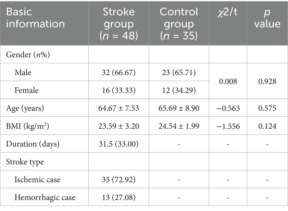
Study participants included 48 stroke patients (32 males, 16 females) hospitalized at Jiangsu Provincial People’s Hospital and its Qixia Rehabilitation Branch between November 2022 and May 2023. Subjects in the stroke group had a mean age of 64.67 ± 7.53 years, with a mean disease duration of 31.50 days (IQR 33.00). Stroke group included 35 ischemic and 13 hemorrhagic cases. A control group of 35 healthy individuals (23 males, 12 females; mean age 65.69 ± 8.90 years) was recruited. No statistically significant differences were observed between groups in age, sex, or body mass index (BMI) (p > 0.05) (Table 1).

Table 1
www.frontiersin.org

Table 1. Comparison of general data between the two groups [M (IQR)] or M ± SD.

Inclusion Criteria for Stroke Group were as follows: ① Age 50–80 years; ➁ Stroke diagnosis confirmed by neuroimaging and compliant with diagnostic guidelines established by the Chinese Stroke Society; ➂ First-ever unilateral stroke; ➃ Disease duration ≤ 3 months; ➄ Clinically stable with vital signs within normal ranges; ➅ Postmenopausal status (confirmed by amenorrhea ≥ 12 months) for female participants.

Inclusion Criteria for Control Group were as follows: ① Age 50–80 years; ➁ No history of stroke or major neurological disorders; ➂ Postmenopausal status for female participants.

Exclusion Criteria (Both Groups) were as follows: ① Auditory comprehension deficits or inability to follow verbal commands; ➁ Concurrent with other central nervous system lesions (e.g., traumatic brain injury, brain neoplasms); ➂ History of reproductive system disorders or surgeries; ➃ Substance abuse, hormone therapy within the past year, or chronic alcoholism; ➄ Occipital hair length < 3 cm; ➅ Recent hair treatments affecting analysis.

2.2 Sample collection

Hair samples were collected from all subjects on their enrollment day. Hair samples were collected by cutting 1 cm of hair from the occipital region, located at the back of the subject’s head near the scalp. Specimens were wrapped in aluminum foil, labeled, and stored at room temperature protected from light.

2.3 Hormonal analysis

Hormonal concentrations in hair were analyzed via high-performance liquid chromatography–tandem mass spectrometry (LC-MS/MS), where an Agilent 1,200 liquid chromatograph (Agilent Technologies, Inc., United States) was combined with an API 3200 Q-TRAP mass spectrometer (Applied Biosystems, Inc., United States), following a previously developed method (15). Hair samples corresponding to the period before the onset of lesions were selected based on the disease course (If the duration of the patient’s disease is 30 days at the time of admission, the 1 cm of hair closest to the scalp is the total amount of hormones accumulated in the hair in the month when the stroke occurred, and the 2 cm close to the scalp is the total amount of hormones accumulated in the hair in the month before the stroke occurred). Hair samples were rinsed with 5 mL of methanol for 2 min, and then heated to 50°C for drying. In a 2 mL centrifuge tube, hair samples were weighed after measuring 1–2 mm in length and cut into powder. After incubation in methanol for 24 h at 25°C, the mixture was centrifuged at 1.2 × 104 r/min for 5 min. Transfer 800 μL of supernatant into another clean centrifuge tube and evaporate nitrogen at 40°C. The residue was then redissolved in 50 mL of mobile phase for LC-MS/MS analysis.

There was good performance, with limits of detection and quantification of 0.3 and 1.0 pg/mg for T, and 0.2 and 0.5 pg/mg for P, respectively. The recovery, intra-day, and inter-day coefficients of variation met the requirements. Analyses were conducted at the Jiangsu Provincial Key Laboratory of Biomaterials and Devices, Southeast University.

2.4 Statistical analysis

SPSS 25.0 software was used for data analysis. Normality of continuous variables was assessed via the Shapiro–Wilk test. Normally distributed data are presented as mean ± standard deviation, while non-normally distributed data are expressed as median (interquartile range, IQR). Group comparisons for normally distributed continuous variables utilized independent samples t-tests, whereas non-parametric Mann–Whitney U tests were applied to non-normally distributed data. Receiver operating characteristic (ROC) curve analysis was conducted to evaluate the predictive efficacy and the cutoff values of T and P for stroke risk. Categorical variables are reported as frequencies and percentages, with between-group comparisons performed using chi-square tests. A two-tailed p < 0.05 was considered statistically significant.

3 Results

3.1 Comparison of T and P expression levels between the two groups

The T and P levels of hair before the onset of stroke were significantly lower in the stroke group compared to the healthy control group (T: 3.60 vs. 6.90 pg/mg, Z = −3.52, p = 0.001; P: 5.85 vs. 9.80 pg/mg, Z = −3.08, p = 0.002). The median reductions in T and P levels were 48% and 40%, respectively (Table 2).

Wednesday, August 31, 2022

Aging, testosterone, and neuroplasticity: friend or foe?

 Ask your doctor what the full article says about using testosterone for neuroplasticity.

All this other stuff for your doctor to know about;

Well your doctor has a lot of studying to do. 

Testosterone Improves Woman’s Brain Functions

FDA Concludes Testosterone Use May Increase Risk of Cardiovascular Events


FDA warns about blood clot risk with testosterone products


Testosterone increases neurotoxicity of glutamate in vitro and ischemia-reperfusion injury in an animal model


Thinking with your gonads: testosterone and cognition


Effect of testosterone on functional recovery in a castrate male rat stroke model


Lower Testosterone Levels Predict Incident Stroke and Transient Ischemic Attack in Older Men


Could androgens maintain specific domains of mental health in aging men by preserving hippocampal neurogenesis?


Single-Dose Testosterone Administration Impairs Cognitive Reflection in Men

The latest here:

Aging, testosterone, and neuroplasticity: friend or foe?

Kiarash Saleki ORCID logo, Mohammad Banazadeh ORCID logo, Amene Saghazadeh and Nima Rezaei

Abstract

Neuroplasticity or neural plasticity implicates the adaptive potential of the brain in response to extrinsic and intrinsic stimuli. The concept has been utilized in different contexts such as injury and neurological disease. Neuroplasticity mechanisms have been classified into neuroregenerative and function-restoring processes. In the context of injury, neuroplasticity has been defined in three post-injury epochs. Testosterone plays a key yet double-edged role in the regulation of several neuroplasticity alterations. Research has shown that testosterone levels are affected by numerous factors such as age, stress, surgical procedures on gonads, and pharmacological treatments. There is an ongoing debate for testosterone replacement therapy (TRT) in aging men; however, TRT is more useful in young individuals with testosterone deficit and more specific subgroups with cognitive dysfunction. Therefore, it is important to pay early attention to testosterone profile and precisely uncover its harms and benefits. In the present review, we discuss the influence of environmental factors, aging, and gender on testosterone-associated alterations in neuroplasticity, as well as the two-sided actions of testosterone in the nervous system. Finally, we provide practical insights for further study of pharmacological treatments for hormonal disorders focusing on restoring neuroplasticity.


Corresponding author: Nima Rezaei, Research Center for Immunodeficiencies, Children’s Medical Center, Tehran University of Medical Sciences, 14197 33151 Tehran, Iran; Department of Immunology, School of Medicine, Tehran University of Medical Sciences, 14176 13151 Tehran, Iran; and Network of Immunity in Infection, Malignancy and Autoimmunity (NIIMA), Universal Scientific Education and Research Network (USERN), 14197 33151 Tehran, Iran, E-mail:
Kiarash Saleki and Mohammad Banazadeh contributed equally to this work.

Thursday, December 13, 2018

Neuroprotective role of hypothermia in hypoxic-ischemic brain injury: combined therapies using estrogen

But is your doctor already treating you with testosterone? Would the combination of testosterone and estrogen cause problems?

Testosterone gel shown for first time to benefit men over 65


Testosterone Improves Woman’s Brain Functions


Low testosterone and the risk of dementia in elderly men


Neuroprotective role of hypothermia in hypoxic-ischemic brain injury: combined therapies using estrogen


Hypoxic-ischemic brain injury is a complex network of factors, which is mainly characterized by a decrease in levels of oxygen concentration and blood flow, which lead to an inefficient supply of nutrients to the brain. Hypoxic-ischemic brain injury can be found in perinatal asphyxia and ischemic-stroke, which represent one of the main causes of mortality and morbidity in children and adults worldwide. Therefore, knowledge on underlying mechanisms triggering these insults may help establish neuroprotective treatments. Selective Estrogen Receptor Modulators and Selective Tissue Estrogenic Activity Regulators exert several neuroprotective effects, including decrease of reactive oxygen species, maintenance of cell viability, mitochondrial survival, among others. However, these strategies represent a traditional approach of targeting a single factor of pathology without satisfactory results. Hence, combined therapies, such as the administration of therapeutic hypothermia with a complementary neuroprotective agent, constitute a promising alternative. In this sense, the present review summarizes the underlying mechanisms of hypoxic-ischemic brain injury and compiles several neuroprotective strategies, including Selective Estrogen Receptor Modulators and Selective Tissue Estrogenic Activity Regulators, which represent putative agents for combined therapies with therapeutic hypothermia.

Thursday, January 4, 2018

Associations of Endogenous Estradiol and Testosterone Levels With Plaque Composition and Risk of Stroke in Subjects With Carotid Atherosclerosis

You'll have to ask your doctor what the hell this means.

Associations of Endogenous Estradiol and Testosterone Levels With Plaque Composition and Risk of Stroke in Subjects With Carotid Atherosclerosis


Marija Glisic, Blerim Mujaj, Oscar L. Rueda-Ochoa, Eralda Asllanaj, Joop S.E. Laven, Maryam Kavousi, M. Kamran Ikram, Meike W. Vernooij, M. Arfan Ikram, Oscar H. Franco, Daniel Bos, Taulant Muka

Abstract

Rationale: Sex steroids may play a role in plaque composition and in stroke incidence.
Objectives: To study the associations of endogenous estradiol and testosterone with carotid plaque composition in elderly men and postmenopausal women with carotid atherosclerosis, as well as with risk of stroke in this population.
Methods and Results: Data of 1023 postmenopausal women and 1124 men (≥45 years) with carotid atherosclerosis, from prospective population-based RS (Rotterdam Study), were available. At baseline, total estradiol (TE) and total testosterone (TT) were measured. Carotid atherosclerosis was assessed by ultrasound, whereas plaque composition (presence of calcification, lipid core, and intraplaque hemorrhage) was assessed by magnetic resonance imaging. TE and TT were not associated with calcified carotid plaques in either sex. TE was associated with presence of lipid core in both sexes (in women odds ratio, 1.48 [95% confidence interval [CI], 1.02–2.15]; in men odds ratio, 1.23 [95% CI, 1.03–1.46]), whereas no association was found between TT and lipid core in either sex. Higher TE (odds ratio, 1.58 [95% CI, 1.03–2.40]) and lower TT (odds ratio, 0.82 [95% CI, 0.68–0.98]) were associated with intraplaque hemorrhage in women but not in men. In women, TE was associated with increased risk of stroke (hazard ratio, 1.98 [95% CI, 1.01–3.88]), whereas no association was found in men. TT was not associated with risk of stroke in either sex.
Conclusions: TE was associated with presence of vulnerable carotid plaque as well as increased risk of stroke in women, whereas no consistent associations were found for TT in either sex.

Friday, November 3, 2017

Single-Dose Testosterone Administration Impairs Cognitive Reflection in Men

Well your doctor has a lot of studying to do. 

Testosterone Improves Woman’s Brain Functions

FDA Concludes Testosterone Use May Increase Risk of Cardiovascular Events


FDA warns about blood clot risk with testosterone products


Testosterone increases neurotoxicity of glutamate in vitro and ischemia-reperfusion injury in an animal model


Thinking with your gonads: testosterone and cognition


Effect of testosterone on functional recovery in a castrate male rat stroke model


Lower Testosterone Levels Predict Incident Stroke and Transient Ischemic Attack in Older Men


Could androgens maintain specific domains of mental health in aging men by preserving hippocampal neurogenesis?


Single-Dose Testosterone Administration Impairs Cognitive Reflection in Men

First Published August 3, 2017 Research Article


In nonhumans, the sex steroid testosterone regulates reproductive behaviors such as fighting between males and mating. In humans, correlational studies have linked testosterone with aggression and disorders associated with poor impulse control, but the neuropsychological processes at work are poorly understood. Building on a dual-process framework, we propose a mechanism underlying testosterone’s behavioral effects in humans: reduction in cognitive reflection. In the largest study of behavioral effects of testosterone administration to date, 243 men received either testosterone or placebo and took the Cognitive Reflection Test (CRT), which estimates the capacity to override incorrect intuitive judgments with deliberate correct responses. Testosterone administration reduced CRT scores. The effect remained after we controlled for age, mood, math skills, whether participants believed they had received the placebo or testosterone, and the effects of 14 additional hormones, and it held for each of the CRT questions in isolation. Our findings suggest a mechanism underlying testosterone’s diverse effects on humans’ judgments and decision making and provide novel, clear, and testable predictions.

Monday, February 27, 2017

Increase in noncalcified plaque associated with testosterone treatment in older men

Well your doctor has a lot of studying to do. 

Testosterone Improves Woman’s Brain Functions

FDA Concludes Testosterone Use May Increase Risk of Cardiovascular Events


FDA warns about blood clot risk with testosterone products


Testosterone increases neurotoxicity of glutamate in vitro and ischemia-reperfusion injury in an animal model


Thinking with your gonads: testosterone and cognition


Effect of testosterone on functional recovery in a castrate male rat stroke model


Lower Testosterone Levels Predict Incident Stroke and Transient Ischemic Attack in Older Men


Could androgens maintain specific domains of mental health in aging men by preserving hippocampal neurogenesis?


Single-Dose Testosterone Administration Impairs Cognitive Reflection in Men

The latest here:

Increase in noncalcified plaque associated with testosterone treatment in older men

Testosterone treatment in older men was linked to a significantly greater increase in coronary artery noncalcified plaque volume, according to a new study in JAMA.
“Although testosterone replacement is increasingly being used clinically, the [CV] benefits and risk of testosterone administration to older men with age-related decline in testosterone levels remain uncertain,” Matthew J. Budoff, MD, professor of medicine at David Geffen School of Medicine at UCLA and program director and director of cardiac CT at Harbor-UCLA Medical Center, and colleagues wrote. “Several observational studies show an inverse association between serum testosterone concentration and adverse [CV] outcomes, the metabolic syndrome, diabetes, and mortality, independent of traditional [CV] risk factors.”
In a double blind, placebo-controlled trial, Budoff and colleagues analyzed 170 men aged 65 years or older with an average of two serum testosterone levels lower than 275 ng/dL and symptoms suggestive of hypogonadism. Participants were enrolled between June 2010 and June 2014 and were randomly assigned testosterone gel (n = 82) or placebo gel (n = 88) for 12 months.
Matt Budoff
Matthew J. Budoff
Of those enrolled, data were available for 138 participants (73 receiving intervention, 65 receiving placebo; mean age, 71 years; 81% white). At baseline, 50.7% participants (n = 70) had a coronary artery calcification (CAC) score higher than 300 Agatston units.
The primary outcome was noncalcified plaque volume and was determined by coronary CTA.
From baseline to 12 months, participants in the testosterone arm had a significantly greater increase in noncalcified plaque volume (from median value 204 mm3 to 232 mm3) compared with placebo (from median value 317 mm3 to 325 mm3) with an estimated difference of 41 mm3 (95% CI, 14-67; P = .003).
The median total plaque volume increased in the testosterone group from 272 mm3 to 318 mm3, compared with 499 mm3 to 541 mm3 for placebo (estimated difference, 47 mm3; 95% CI, 13-80; P = .006). The median CAC score decreased in in the testosterone group (from 255 to 244 Agatston units) and increased in the placebo group (from 494 to 503 Agatston units) from baseline to 12 months (estimated difference, –27 Agatston units; 95% CI, –80 to 26 Agatston units), according to the researchers.
No one in either group had a major adverse CV event.
“The increase in coronary artery noncalcified and total plaque volumes in men treated with testosterone is concerning because any limitation of the vascular lumen could be considered deleterious,” Budoff and colleagues wrote. “The clinical significance of these increases could depend on the differential effects of testosterone on the individual components of noncalcified plaque.”
The researchers concluded that larger studies are needed to understand the clinical implications. – by Cassie Homer

Sunday, February 26, 2017

Increased testosterone decreases medial cortical volume and neurogenesis in territorial side-blotched lizards (Uta stansburiana)

Well your doctor has a lot of studying to do.

Well your doctor has a lot of studying to do. 

Testosterone Improves Woman’s Brain Functions

FDA Concludes Testosterone Use May Increase Risk of Cardiovascular Events


FDA warns about blood clot risk with testosterone products


Testosterone increases neurotoxicity of glutamate in vitro and ischemia-reperfusion injury in an animal model


Thinking with your gonads: testosterone and cognition


Effect of testosterone on functional recovery in a castrate male rat stroke model


Lower Testosterone Levels Predict Incident Stroke and Transient Ischemic Attack in Older Men


Could androgens maintain specific domains of mental health in aging men by preserving hippocampal neurogenesis?


Single-Dose Testosterone Administration Impairs Cognitive Reflection in Men

The latest here:


Increased testosterone decreases medial cortical volume and neurogenesis in territorial side-blotched lizards (Uta stansburiana)

Lara Ladage1*, Timothy Roth2, Cynthia Downs3, Barry Sinervo4 and Vladimir Pravosudov5
  • 1Penn State University- Altoona campus, USA
  • 2Franklin & Marshall College, USA
  • 3Hamilton College, USA
  • 4University of California- Santa Cruz, USA
  • 5University of Nevada, Reno, USA
Variation in an animal’s spatial environment can induce variation in the hippocampus, an area of the brain involved in spatial cognitive processing. Specifically, increased spatial area use is correlated with increased hippocampal attributes such as volume and neurogenesis. In the side-blotched lizard (Uta stansburiana), males demonstrate alternative reproductive tactics and are either territorial - defending large, clearly defined spatial boundaries - or non-territorial - traversing home ranges that are smaller than the territorial males’ territories. Our previous work demonstrated cortical volume (reptilian hippocampal homologue) correlates with these spatial niches. We found that territorial holders have larger medial cortices than non-territory holders, yet these differences in the neural architecture demonstrated some degree of plasticity as well. Although we have demonstrated a link among territoriality, spatial use, and brain plasticity, the mechanisms that underlie this relationship are unclear. Previous studies found that higher testosterone levels can induce increased use of the spatial area and can cause an upregulation in hippocampal attributes. Thus, testosterone may be the mechanistic link between spatial area use and the brain. What remains unclear, however, is if testosterone can affect the cortices independent of spatial experiences and whether testosterone differentially interacts with territorial status to produce the resultant cortical phenotype. In this study, we compared neurogenesis as measured by the total number of doublecortin-positive cells and cortical volume between territorial and non-territorial males supplemented with testosterone. We found no significant differences in the number of doublecortin-positive cells or cortical volume among control territorial, control non-territorial, and testosterone-supplemented non-territorial males, while testosterone-supplemented territorial males had smaller medial cortices containing fewer doublecortin-positive cells. These results demonstrate that testosterone can modulate medial cortical attributes outside of differential spatial processing experiences but that territorial males appear to be more sensitive to alterations in testosterone levels compared with non-territorial males.

Saturday, February 20, 2016

Testosterone gel shown for first time to benefit men over 65

Hmmm, better walking ability and less depression. That might be useful post stroke. Don't start taking this on your own.

Testosterone gel shown for first time to benefit men over 65

The University of Pittsburgh Graduate School of Public Health was among a dozen sites nationwide to participate in the first clinical trial to show that testosterone treatment for men aged 65 and older improves sexual function, walking ability and mood.
 
Results of The Testosterone Trials (TTrials), led by the Perelman School of Medicine at the University of Pennsylvania and funded by the National Institutes of Health (NIH), will be published in tomorrow’s New England Journal of Medicine.
 
“Previous testosterone trials in older men yielded equivocal and inconsistent results,” said coauthor and chair of the TTrials recruitment committee, Jane A. Cauley, Dr.P.H., professor in Pitt Public Health’s Department of Epidemiology and principal investigator at the study’s Pittsburgh site. “We showed that testosterone improved men’s impression that their sexual function and walking ability had improved, suggesting that these effects are clinically important.”
 
The TTrials are a coordinated group of seven trials testing the effect of a testosterone gel compared with a placebo gel, and researchers have analyzed the results of the three primary trials – sexual function, physical function and vitality. They found that testosterone treatment increased the blood testosterone level in the men 65 and older to levels comparable to mid-normal for young men. Testosterone also improved all aspects of sexual function, including sexual activity, sexual desire and the ability to get an erection. Testosterone treatment did not significantly improve distance walked in six minutes when only men enrolled in the physical function trial were considered, but did increase the distance walked when all men in the TTrials were considered. The treatment did not improve energy, but did improve mood and depressive symptoms.
 
“The results of the TTrials show for the first time that testosterone treatment of older men who have unequivocally low testosterone levels does have some benefit,” said lead author and principal investigator of the TTrials Peter J. Snyder, M.D., a professor in the Division of Endocrinology, Diabetes and Metabolism at Penn. “However, decisions about testosterone treatment for these men also will depend on the results of the other four trials – cognitive function, bone, cardiovascular and anemia – and the risks of testosterone treatment.”
 
In 2003, the Institute of Medicine reported that there was insufficient evidence to support any beneficial effect of testosterone in such men. This report was the impetus for TTrials, which are now the largest trials to examine the efficacy of testosterone treatment in men 65 and older whose testosterone levels are low due seemingly to age alone. TTrials researchers screened 51,085 men to find 790 who qualified with a sufficiently low testosterone level and who met other criteria; 78 of the men were enrolled from the Pittsburgh area.
 
The men enrolled were randomized into two groups: one to apply the daily testosterone gel and the other a daily placebo gel, for one year. Efficacy was then evaluated at months three, six, nine and 12. Sexual function was assessed by questionnaires; physical function was measured by questionnaires and the distance walked in six minutes; and vitality, mood and depressive symptoms also were evaluated using questionnaires.
 
Across the three trials, adverse events – including heart attack, stroke, other cardiovascular events and prostate conditions – were similar in men who received testosterone and those who received placebo. However, the number of men in the TTrials was too small to draw conclusions about the risk of testosterone treatment, which the researchers say would require a larger and longer trial.
 
The TTrials were conducted at 11 additional medical centers across the country: Albert Einstein College of Medicine, Baylor College of Medicine, Brigham and Women’s Hospital, Harbor-UCL Medical Center, University of Alabama at Birmingham, Northwestern University Feinberg School of Medicine, Puget Sound Health Care System, University of California at San Diego School of Medicine, University of Florida School of Medicine, University of Minnesota School of Medicine and Yale School of Medicine.
 
The TTrials were supported by NIH National Institute on Aging grant U01 AG030644, and supplemented by funds from the National Heart, Lung, and Blood Institute, National Institute of Neurological Diseases and Stroke, and National Institute of Child Health and Human Development. AbbVie (formerly Solvay and Abbot Laboratories) also provided funding, AndroGel and placebo gel, but did not participate in the design or conduct of the trials, nor analysis or reporting of the data.