Changing stroke rehab and research worldwide now.Time is Brain! trillions and trillions of neurons that DIE each day because there are NO effective hyperacute therapies besides tPA(only 12% effective). I have 523 posts on hyperacute therapy, enough for researchers to spend decades proving them out. These are my personal ideas and blog on stroke rehabilitation and stroke research. Do not attempt any of these without checking with your medical provider. Unless you join me in agitating, when you need these therapies they won't be there. Did your doctor somehow miss the recovery protocol that Pedro Bach-y-Rita used to recover fully back in 1958 with only a partial brain?

What this blog is for:

My blog is not to help survivors recover, it is to have the 10 million yearly stroke survivors light fires underneath their doctors, stroke hospitals and stroke researchers to get stroke solved. 100% recovery. The stroke medical world is completely failing at that goal, they don't even have it as a goal. Shortly after getting out of the hospital and getting NO information on the process or protocols of stroke rehabilitation and recovery I started searching on the internet and found that no other survivor received useful information. This is an attempt to cover all stroke rehabilitation information that should be readily available to survivors so they can talk with informed knowledge to their medical staff. It lays out what needs to be done to get stroke survivors closer to 100% recovery. It's quite disgusting that this information is not available from every stroke association and doctors group.

Showing posts with label weasel out. Show all posts

Monday, December 2, 2024

Visceral Fat May Indicate Alzheimer’s Risk Decades Before Symptoms

 Your competent? doctor needs to first prevent this visceral fat and then if it occurs, remove it! You need excellent brain blood flow for recovery, so don't let your doctor weasel out of this requirement!

In case your doctor is incompetent in this matter, use this to train them:


With your chances of getting dementia post stroke, you need prevention solutions. YOUR DOCTOR IS RESPONSIBLE FOR PREVENTING THIS!

1. A documented 33% dementia chance post-stroke from an Australian study?   May 2012.

2. Then this study came out and seems to have a range from 17-66%. December 2013.`    

3. A 20% chance in this research.   July 2013.

4. Dementia Risk Doubled in Patients Following Stroke September 2018 

The latest here:

Visceral Fat May Indicate Alzheimer’s Risk Decades Before Symptoms

Summary: A new study links visceral fat, a deep layer of abdominal fat, to increased brain amyloid and tau proteins—key markers of Alzheimer’s disease—decades before dementia symptoms arise. Researchers found that visceral fat accounted for 77% of the relationship between high BMI and amyloid accumulation, while other fat types showed no similar impact.

The study highlights how managing visceral fat through lifestyle changes or medication could reduce Alzheimer’s risk. Midlife interventions are especially critical, as participants in their 40s and 50s exhibited these pathologies early on. The findings further reveal that metabolic factors like insulin resistance and cholesterol levels amplify obesity-related brain damage. This research emphasizes the urgent need for targeted prevention strategies against obesity-related Alzheimer’s risk.

Key Facts:

  • Visceral fat is strongly associated with amyloid and tau protein buildup in the brain.
  • Insulin resistance and low HDL cholesterol worsen obesity-related Alzheimer’s pathology.
  • Managing visceral fat through weight loss or metabolic treatments may reduce dementia risk.

Source: RSNA

Researchers have linked a specific type of body fat to the abnormal proteins in the brain that are hallmarks of Alzheimer’s disease up to 20 years before the earliest symptoms of dementia  appear, according to a study being presented today at the annual meeting of the Radiological Society of North America (RSNA).

The researchers emphasized that lifestyle modifications targeted at reducing this fat could influence the development of Alzheimer’s disease.

This shows a brain.
The effects of visceral fat on amyloid pathology were partially reduced in people with higher HDL. Credit: Neuroscience News

“This crucial result was discovered because we investigated Alzheimer’s disease pathology as early as midlife—in the 40s and 50s—when the disease pathology is at its earliest stages, and potential modifications like weight loss and reducing visceral fat are more effective as a means of preventing or delaying the onset of the disease,” said lead study author Mahsa Dolatshahi, M.D., M.P.H., post-doctoral research associate at Mallinckrodt Institute of Radiology (MIR) at Washington University School of Medicine in St. Louis, Missouri.

An estimated 6.9 million Americans, aged 65 and older, are living with Alzheimer’s disease, according to the Alzheimer’s Association. The association estimates this number could grow to 13 million by 2050, barring the development of medical breakthroughs to prevent or cure the disease.

For the study, the researchers focused on the link between modifiable lifestyle-related factors, such as obesity, body fat distribution and metabolic aspects, and Alzheimer’s disease pathology.

A total of 80 cognitively normal midlife individuals (average age: 49.4 years, female: 62.5%,) were included in the study. Approximately 57.5% of participants were obese, and the average body mass index (BMI) of the participants was 32.31.

The participants underwent brain positron emission tomography (PET), body MRI and metabolic assessment (glucose and insulin measurements), as well as a lipid (cholesterol) panel.

MRI scans of the abdomen were performed to measure the volume of the subcutaneous fat (the fat under skin) and visceral fat (deep hidden fat surrounding the organs).

“We investigated the association of BMI, visceral fat, subcutaneous fat, liver fat fraction, thigh fat and muscle, as well as insulin resistance and HDL (good cholesterol), with amyloid and tau deposition in Alzheimer’s disease,” said Dr. Dolatshahi, a member of the Raji Lab at MIR’s Neuroimaging Labs Research Center.

Thigh muscle scans were used to measure volume of muscle and fat. Alzheimer’s disease pathology was measured using PET scans with tracers that bind to amyloid plaques and tau tangles that accumulate in the brains of people with Alzheimer’s disease.

The findings revealed that higher levels of visceral fat were related to increased amyloid, accounting for 77% of the effect of high BMI on amyloid accumulation. Other types of fat did not explain obesity-related increased Alzheimer’s pathology.

“Our study showed that higher visceral fat was associated with higher PET levels of the two hallmark pathologic proteins of Alzheimer’s disease—amyloid and tau,” Dr. Dolatshahi said.

“To our knowledge, our study is the only one to demonstrate these findings at midlife where our participants are decades out from developing the earliest symptoms of the dementia that results from Alzheimer’s disease.”

The study also showed that higher insulin resistance and lower HDL were associated with high amyloid in the brain. The effects of visceral fat on amyloid pathology were partially reduced in people with higher HDL.

“A key implication of our work is that managing Alzheimer’s risk in obesity will need to involve targeting the related metabolic and lipid issues that often arise with higher body fat,” said senior study author Cyrus A. Raji, M.D., Ph.D., associate professor of radiology at MIR.

Although previous studies have shown the role of high BMI in damaging the cells of the brain, no similar study has investigated the differential role of visceral and subcutaneous fat or metabolic profile, especially in terms of Alzheimer’s amyloid pathology as early as midlife, Dr. Dolatshahi pointed out.

“This study goes beyond using BMI to characterize body fat more accurately with MRI and, in so doing, reveals key insights about why obesity can increase risk for Alzheimer’s disease,” Dr. Dolatshahi said.

Drs. Raji, Dolatshahi and colleagues are also presenting a study at RSNA 2024 that shows how obesity and visceral fat reduce blood flow in the brain.

In that study, the researchers performed brain and abdominal MRI on cognitively normal midlife individuals with a wide range of BMI and compared whole-brain and regional cerebral blood flow on brain MRI in individuals with high vs. low visceral and subcutaneous fat. 

The high visceral fat group showed lower whole-brain blood flow. No significant difference was observed in cerebral blood flow in the groups with high vs. low subcutaneous fat.

“This work will have a considerable impact on public health because nearly three out of four Americans are overweight or obese,” Dr. Raji said.

“Knowing that visceral obesity negatively affects the brain opens up the possibility that treatment with lifestyle modifications or appropriate weight-loss drugs could improve cerebral blood flow and potentially lower the burden of and reduce the risk for Alzheimer’s disease.”

Other co-authors are Paul K. Commean, B.E.E., Mahshid Naghashzadeh, M.S., Sara Hosseinzadeh Kassani, Ph.D., Jake Weeks, B.S., Caitlyn Nguyen, B.S., Abby McBee-Kemper, B.S., Nancy Hantler, B.S., LaKisha Lloyd, M.Sc., Shaney Flores, M.S., Yifei Xu, M.S., Jingxia Liu, Ph.D., Claude B. Sirlin, M.D., Bettina Mittendorfer, Ph.D., Joseph E. Ippolito, M.D., Ph.D., John C. Morris, M.D., and Tammie L.S. Benzinger, M.D., Ph.D.

This study was awarded the RSNA Trainee Research Prize.

About this Alzheimer’s disease research news

Author: Linda Brooks
Source: RSNA
Contact: Linda Brooks – RSNA
Image: The image is credited to Neuroscience News

Original Research: The findings will be presented at the 110th Scientific Assembly and Annual Meeting of the Radiological Society of North America

Monday, November 25, 2024

Inside your body, aging unfolds at remarkably different rates

 Your COMPETENT? DOCTOR is responsible for recovering your lost 5 cognitive years from your stroke. Don't let them weasel their way out of that responsibility! Ask EXACTLY how they will accomplish that, screaming may be required.  In my opinion, a competent doctor would know how to do that from medical school!

Of course, your COMPETENT? DOCTOR started working on this 3.5 years ago! NO? So, you DON'T have a functioning stroke doctor, do you?

Inside your body, aging unfolds at remarkably different rates

Gretchen Reynolds
Updated
11 min read
By using advanced molecular biology, genetics, and big data to analyze blood samples, scientists can tell that some people are “heart agers,” meaning their hearts appear much older than the rest of their bodies.

Recently, scientists at Stanford University began to wonder why identical lab mice, bred with the same DNA and brought up in identical conditions, wound up so different in their old age.

Some mice could ace cognitive tests and race around on their running wheels. Others would forget simple tasks and hobble from place to place. Genetically, they remained indistinguishable, but their twilight years could hardly have been more distinct.

Subscribe to The Post Most newsletter for the most important and interesting stories from The Washington Post.

The scientists’ attempts to untangle what was going on inside these mice is redefining how we think about aging. It has opened up a new area of research into what scientists are calling “organ aging,” which looks at how different parts of our bodies seem to start aging earlier than others, affecting what diseases we develop and how long we live.

The research suggests aging isn’t strictly temporal, not solely about minutes and years passing. Once considered a steady, predictable decline, affecting everything in our bodies, everywhere, all at once, aging is much more haphazard than we once thought, starting in different parts of our bodies at different times, possibly long before we’re even thinking about aging.

It’s also personal, occurring at a unique molecular level inside each of us, and the process may be partially within our control. Once we know how our own organs are aging, we may be able to brake or speed that process by how we live.

By using advanced molecular biology, genetics and big data to analyze blood samples from people, scientists can tell that some of us are “heart agers,” meaning our hearts appear much older than the rest of our bodies, or we’re “brain agers,” with a relatively old brain in our skulls, or if we’re fortunate, we might be “brain youthers,” with a brain relatively younger than any other organ we possess. Or we could be “muscle agers” or “liver youthers.” Almost any organ can be the first to show signs of extreme aging.

The consequences for our health are considerable. In one of the largest human studies to date of organ aging, Stanford scientists found that heart agers are far more likely to develop heart failure than other people, while brain youthers are about 80 percent less likely to develop dementia in later years than people with average or old brains.

The results underscore “just how complicated aging is,” said Hamilton Se-Hwee Oh, a postdoctoral researcher who led the study while at Stanford.

They also represent one of the first and potentially most practical takeaways from the broader and often wildly hyped science of human aging.

- - -

The newest way to think about biological age

Aging is far more erratic than many of us might expect.

“You can take animals that are genetically identical, that are raised in the same cages with the same food and same handling, everything about them is exactly the same, but they show different molecular changes with age and different functional decline and diseases at different times,” said Tony Wyss-Coray, the director of the Phil and Penny Knight Initiative for Brain Resilience at Stanford University and senior author of the recent Stanford study of organ aging.

What separates the animals seems to be their biological age, a concept at the heart of the organ-aging research. We all have, of course, a chronological age, according to our birth date. And scientists for years have been refining the definition of a separate biological age, which marks how well our bodies are functioning. Our biological age can be older or younger than our birth age.

“We all know 50-year-olds who don’t look that age, for better or worse,” said Thomas Rando, a molecular biologist and the director of the Broad Stem Cell Research Center at the University of California at Los Angeles, who studies aging and longevity. Those people’s biological age are likely to differ from their calendar years.

In the past decade, scientists have begun harnessing the power of machine learning and complex new biotechnology to create “clocks” designed to estimate biological age. These clocks rely on the analysis of blood and other tissue samples from thousands of people and animals of all ages.

By analyzing these samples, scientists have zeroed in on similarities by age, focusing on specific patterns of gene activity or molecular levels commonly seen at certain ages. These patterns can then be used as biomarkers of biological age.

If a 40-year-old’s gene activity matches that seen in most other 40-year-olds, for instance, that person would be biologically 40. But if his gene patterns more closely resemble those of a typical 30- or 50-year-old, he’d be considered biologically out of alignment with his birth age.

Dozens of at-home tests now promise to estimate biological age from a cheek swab or other sample. None have been approved by the Food and Drug Administration. Prices are as high as $500. And many scientists worry they rely on vastly oversimplified and unreliable interpretations of the aging-clock algorithms scientists originally developed.

“I’ve taken quite a few” of the over-the-counter tests, said Nir Barzilai, director of the Institute for Aging Research at the Albert Einstein College of Medicine in New York. “They showed me being anywhere from 20 years younger to three years older” than his actual age of 68.

But a deeper, more-intractable concern is that the DIY biological age tests offer a single estimate of how an entire body is aging. It’s increasingly clear aging doesn’t work that way.

- - -

Aging fast or slow

One of the first major studies of organ aging appeared in 2020 in the journal Nature, with Wyss-Coray and other researchers tracking gene activity and other cellular changes in 17 organs from lab mice of all ages. The results showed that some of the rodents’ organs were growing old faster or slower, from mouse to mouse, organ to organ, and even from one cell to another within the same organ.

That finding reinforced the growing sense among scientists that “aging isn’t linear,” Wyss-Coray said.

That mouse study and others also hinted that the sequence of organ aging could affect lifelong health, with fast-aging organs influencing later disease risk, and also that aging might, in some fashion, be “contagious,” with whichever organs aged earliest releasing biochemicals that jump-started aging elsewhere in the body.

But, of course, mice aren’t us, so the next obvious step was to repeat the mouse study in people and see if the results would be similar. There were, however, hurdles.

It’s not possible to obtain tissue samples from certain organs, such as the brain, in living people. So Wyss-Coray and his colleagues needed to come up with a different approach than they’d used with mice.

For a study featured on the cover of Nature in 2023, they settled on proteins, “the building blocks of life,” Wyss-Coray said.

Created in cells during gene expression and released constantly into the bloodstream, tens of thousands of proteins course through our bodies all the time. But some are unique to specific organs. Certain genes in liver cells, for instance, produce proteins that can only have come from the liver. Ditto for genes in neurons and lung cells and so on. Prior research had categorized these organ-specific proteins, some of which already are used to check people’s health during standard blood tests.

With that data, the scientists gathered records from existing health databases that included blood samples from about 5,700 men and women of various ages. Using the data, elaborate machine-learning models created molecular, age-related signatures for each of 11 organs: the heart, lungs, arteries, brain, fat, immune system, intestines, kidneys, liver, muscles and pancreas.

At that point, the scientists could identify a prototypical 40-year-old’s liver from a 50-year-old’s by the patterns of proteins in their bloodstreams. They could also identify gaps between the age signatures of people’s organs and their actual, chronological ages.

And many people’s organs, it turned out, were relatively elderly. About 20 percent of the almost 5,700 men and women harbored at least one organ that was significantly older than their birth age, according to their plasma proteins. These affected organs differed from person to person, creating what the scientists called an “ageotype.” People whose hearts were extremely old, compared to their other organs, were heart agers, while those with elderly fat tissue were fat agers.

Perhaps most significant, each aged organ was associated with substantially elevated risks for related diseases.

Heart agers, whether middle aged or older, were as much as 250 percent more likely to develop heart failure in subsequent years as other people, with muscle agers at heightened risk for gait problems, and, well, you get the idea.

- - -

The benefits of a youthful brain

But a pool of about 5,700 study participants, while larger than in many biological studies, was too small for the researchers to delve into the existence or effects of youthful organs.

So, for the newest study, published in June, most of the same scientists turned to the immense trove of data in the UK Biobank, gathering blood and health records from about 44,530 men and women ages 40 to 70 when they joined the biobank. The researchers analyzed blood for organ-aging proteins, compared the same 11 organs’ biological ages to people’s actual ages and also checked for diseases or deaths among people during the next decade or so. (The study is in the preprint stage, meaning it hasn’t been fully peer-reviewed, but the authors expect peer review and publication to be completed soon.)

The associations proved even stronger in this larger group. About 33 percent of the men and women harbored at least one organ that was “extremely” aged, compared with their actual ages. Another 26 percent had two or more extremely aged organs and some as many as eight.

The researchers also again found links between organs’ ages and disease and also lifespan. Heart agers risked heart failure and atrial fibrillation; lung agers developed chronic obstructive pulmonary disease; and for liver agers, it was chronic liver disease.

But the effects of brain aging were the most striking. People with extremely old brains were 3.4 times more likely to develop Alzheimer’s disease than everyone else, while, on the flip side, those with relatively youthful brains had 81 percent less risk of Alzheimer’s than people with brains the same age as them.

The benefits of a youthful brain also extended to longevity, with brain youthers generally outliving brain agers. In fact, an old brain was, of all organs, “most strongly predictive of mortality,” the study authors wrote, “suggesting that the brain may be a central regulator of lifespan in people.”

- - -

Changing our organ age

Perhaps the most important finding in the new study, though, was that organ age “appears to be malleable,” Oh said.

When he and the other scientists compared people’s organ ages to their lifestyles in the new study, they found that those who often smoked, drank or ate processed meats were prone to accelerated organ aging, while anyone who regularly exercised or ate oily fish was far more likely to have youthful organs.

Interestingly, taking estrogen also noticeably affected organ aging in menopausal women, the researchers found. Women who’d used supplemental estrogen wound up with relatively youthful immune systems, livers and arteries, compared to those who hadn’t.

Why and how diet, exercise, hormones or other lifestyle and medical options affect organ aging isn’t clear, Oh said. It’s still impossible, in fact, to pinpoint precisely why our organs age at different rates at all. It might be genetics, lifestyle, luck or all or none of those.

Still, knowing our ageotype could help guide some of our health decisions, Oh said. Someone who knows he or she is a heart ager, for instance, even with no current symptoms, should probably consider more-frequent cardiac testing and diet and exercise changes, such as cutting back on ultra-processed foods and walking for at least 30 minutes a day, while a muscle ager could be advised to take up weight training, which is known to build muscular health at a cellular level.

The organ-aging research “is really interesting, because it does suggest it might be possible to give more granularity into how someone is aging across different systems,” said Morgan Levine, a principal investigator at Altos Lab in San Diego who is widely considered one of the world’s experts on clocks for biological aging. She had no part in the new research.

Blood tests to diagnose ageotype could be on the horizon, although it may still be years before they’re validated by the FDA and available as part of standard health testing, Oh said. “We always need more studies.” (Unvalidated consumer tests could arrive much earlier, he said, but might not be reliable.)

The hope is that these tests will show which of our body parts - our heart, brain, kidneys, skin, fat, intestines or something else - is aging fastest and what, if anything, we can do about it.

The idea that we might be able to change how we age “is what’s really exciting” about this research, Oh said.

Friday, November 15, 2024

A New and Early Predictor of Dementia? Frailty

 EXACTLY HOW is your competent? doctor ensuring that you leave the hospital without frailty? This is your doctor's responsibility. Don't let them weasel out of it! Screaming may be required.

A New and Early Predictor of Dementia?

Signs of frailty may signal future dementia more than a decade before cognitive symptoms occur, in new findings that may provide a potential opportunity to identify high-risk populations for targeted enrollment in clinical trials of dementia prevention and treatment.

Results of an international study assessing frailty trajectories showed frailty levels notably increased in the 4-9 years before dementia diagnosis. Even among study participants whose baseline frailty measurement was taken prior to that acceleration period, frailty was still positively associated with dementia risk, the investigators noted.

"We found that with every four to five additional health problems, there is on average a 40% higher risk of developing dementia, while the risk is lower for people who are more physically fit," study investigator David Ward, PhD, of the Centre for Health Services Research, The University of Queensland, Brisbane, Australia, told Medscape Medical News.

photo of David Ward PhD
David Ward, PhD

The findings were published online on November 11 in JAMA Neurology.

A Promising Biomarker

An accessible biomarker for both biologic age and dementia risk is essential for advancing dementia prevention and treatment strategies, the investigators noted, adding that growing evidence suggests frailty may be a promising candidate for this role.

To learn more about the association between frailty and dementia, Ward and his team analyzed data on 29,849 participants aged 60 years or above (mean age, 71.6 years; 62% women) who participated in four cohort studies: the English Longitudinal Study of Ageing (ELSA; n = 6771), the Health and Retirement Study (HRS; n = 9045), the Rush Memory and Aging Project (MAP; n = 1451), and the National Alzheimer’s Coordinating Center (NACC; n = 12,582).

The primary outcome was all-cause dementia. Depending on the cohort, dementia diagnoses were determined through cognitive testing, self- or family report of physician diagnosis, or a diagnosis by the study physician. Participants were excluded if they had cognitive impairment at baseline.

Investigators retrospectively determined frailty index scores by gathering information on health and functional outcomes for participants from each cohort. Only participants with frailty data on at least 30 deficits were included.

Commonly included deficits included high blood pressure, cancer, and chronic pain, as well as functional problems such as hearing impairment, difficulty with mobility, and challenges managing finances.

Investigators conducted follow-up visits with participants until they developed dementia or until the study ended, with follow-up periods varying across cohorts.

After adjusting for potential confounders, frailty scores were modeled using backward time scales.

Among participants who developed incident dementia (n = 3154), covariate-adjusted expected frailty index scores were, on average, higher in women than in men by 18.5% in ELSA, 20.9% in HRS, and 16.2% in MAP. There were no differences in frailty scores between sexes in the NACC cohort.

When measured on a timeline, as compared with those who didn't develop dementia, frailty scores were significantly and consistently higher in the dementia groups 8-20 before dementia onset (20 years in HRS; 13 in MAP; 12 in ELSA; 8 in NACC).

Increases in the rates of frailty index scores began accelerating 4-9 years before dementia onset for the various cohorts, investigators noted.

In all four cohorts, each 0.1 increase in frailty scores was positively associated with increased dementia risk.

Adjusted hazard ratios [aHRs] ranged from 1.18 in the HRS cohort to 1.73 in the NACC cohort, which showed the strongest association.

In participants whose baseline frailty measurement was conducted before the predementia acceleration period began, the association of frailty scores and dementia risk was positive. These aHRs ranged from 1.18 in the HRS cohort to 1.43 in the NACC cohort.

The 'Four Pillars' of Prevention

The good news, investigators said, is that the long trajectory of frailty symptoms preceding dementia onset provides plenty of opportunity for intervention.

To slow the development of frailty, Ward suggested adhering to the "four pillars of frailty prevention and management," which include good nutrition with plenty of protein, exercise, optimizing medications for chronic conditions, and maintaining a strong social network.(I'm good at all of these.)

Ward suggested neurologists track frailty in their patients and pointed to a recent article focused on helping neurologists use frailty measures to influence care planning.

Study limitations include the possibility of reverse causality and the fact that investigators could not adjust for genetic risk for dementia.

Unclear Pathway

Commenting on the findings for Medscape Medical News, Lycia Neumann, PhD, senior director of Health Services Research at the Alzheimer's Association, noted that many studies over the years have shown a link between frailty and dementia. However, she cautioned that a link does not imply causation.

photo of Lycia Neumann PhD
Lycia Neumann, PhD

The pathway from frailty to dementia is not 100% clear, and both are complex conditions, said Neumann, who was not part of the study.

"Adopting healthy lifestyle behaviors early and consistently can help decrease the risk of — or postpone the onset of — both frailty and cognitive decline," she said. Neumann added that physical activity, a healthy diet, social engagement, and controlling diabetes and blood pressure can also reduce the risk for dementia as well as cardiovascular disease.

The study was funded in part by the Deep Dementia Phenotyping Network through the Frailty and Dementia Special Interest Group. Ward and Neumann reported no relevant financial relationships.

Friday, September 23, 2022

Cerebral embolic protection during TAVR reduces disabling stroke but not overall stroke

 So how are your doctors guaranteeing your 100% recovery from that stroke if they can't prevent it? Don't let them weasel out of truthfully answering.

Cerebral embolic protection during TAVR reduces disabling stroke but not overall stroke

BOSTON — In PROTECTED TAVR, the largest-ever randomized trial of transcatheter aortic valve replacement, cerebral embolic protection did not decrease incidence of periprocedural stroke.

However, cerebral embolic protection was associated with reduced incidence of disabling stroke, so the results do not necessarily rule out the benefit of cerebral embolic protection in patients undergoing TAVR, Samir R. Kapadia, MD, chairman of the Robert and Suzanne Tomsich Department of Cardiovascular Medicine, Sydell and Arnold Miller Family Heart, Vascular & Thoracic Institute at Cleveland Clinic and professor of medicine at the Cleveland Clinic Lerner College of Medicine of Case Western Reserve University, said during a press conference at TCT 2022.

Heart Brain 2019 Adobe
Source: Adobe Stock
Samir R. Kapadia

“As a scientific trial, you would say that the primary endpoint was negative,” Kapadia said at the press conference. “However, the disabling strokes are reduced, so it would be the wrong message to say there was no benefit. [We had to state] the conclusion without overstating it or misguiding the readership.”

The researchers randomly assigned 3,000 patients with aortic stenosis to undergo transfemoral TAVR with or without a cerebral embolic protection device (Sentinel, Boston Scientific).

The primary endpoint was stroke within 72 hours of TAVR or before discharge, whichever came first, in the intention-to-treat population. Secondary endpoints included disabling stroke, death, transient ischemic attack, delirium, major or minor vascular complications at the cerebral embolic protection access site and acute kidney injury. All patients were assessed by a neurologist before and after the procedure.

The results were simultaneously published in The New England Journal of Medicine.

Among the patients from the cerebral embolic protection arm in whom an attempt was made, 94.4% had the device successfully deployed, according to the researchers.

The primary endpoint occurred in 2.3% of the cerebral embolic protection group and 2.9% in the control group (difference, –0.6 percentage points; 95% CI, –1.7 to 0.5; P = .3), Kapadia and colleagues found.

Disabling stroke occurred in 0.5% of the cerebral embolic protection group and 1.3% of the control group (difference, –0.8 percentage points; 95% CI, –1.5 to –0.1; P = .02), according to the researchers. The number needed to treat with cerebral embolic protection to prevent one disabling stroke was 125, Kapadia said at the press conference.

The difference in disabling stroke was driven by disabling ischemic stroke, with six occurring in the cerebral embolic protection group and 17 occurring in the control group, Kapadia said.

There were no differences between the groups in death (cerebral embolic protection, 0.5%; controls, 0.3%), stroke/TIA/delirium (cerebral embolic protection, 3.1%; controls, 3.7%) or acute kidney injury (0.5% in both groups), Kapadia and colleagues found.

One patient in the cerebral embolic protection group had a vascular complication at the device access site, according to the researchers.

A cerebral embolic protection device could not be deployed in 5.5% of patients assigned to that group, but a per-protocol analysis of patients who received the assigned treatment did not differ from the intention-to-treat analysis, the researchers reported.

There were no differences in the primary outcome by subgroup except that it significantly favored the cerebral embolic protection group in patients from the U.S. but not in patients from outside the U.S., and the researchers do not have an explanation for why that happened, Kapadia said.

“Stroke remains unpredictable,” Kapadia said at the press conference. “Despite a big effort, we did not identify who is likely to have them.”

Reference:

Wednesday, September 21, 2022

Odds of cognitive impairment increase after stroke in certain populations

It is your doctor's responsibility to ensure you don't have cognitive impairment post stroke. Don't let your doctor weasel out of that responsibility by quoting the craptastic saying: 'All strokes are different, all stroke recoveries are different'.  That statement means your doctor has given up on solving stroke

Odds of cognitive impairment increase after stroke in certain populations

History of stroke was associated with increased odds of cognitive impairment in both non-Hispanic white and Mexican American individuals, researchers reported.

The magnitude of the impact of stroke on cognition was less in Mexican American participants than in non-Hispanic white participants, according to the researchers.

Dementia
Source: Adobe Stock

The association between stroke and dementia is well established, but less is known about the prevalence in underrepresented ethnic groups, according to the study background.

Christopher J. Becker, MD, neurologist in the University of Michigan Health System, and colleagues conducted a population-based, cohort study in Nueces County, Texas; residents younger than 65 years were recruited via telephone or door-to-door from May 2018 to December 2021. The county is a bi-ethnic community with a primarily Mexican American population. Participants lived in the community for at least 6 months of the year, were aged 65 years or older, spoke English or Spanish and were living in the community or a nursing-care facility.

Of the 1,801 participants, 50% were Mexican American, 44% were non-Hispanic white and 6% were another race/ethnicity. Crude prevalence of stroke history was 12% for Mexican Americans, 12.3% for non-Hispanic white participants and 17.2% for participants of other races/ethnicities (P = .34).

Becker and colleagues found stroke was associated with cognitive impairment in each race/ethnicity, but the magnitude of effect was greater in non-Hispanic white participants (cumulative OR = 3.81; 95% CI, 2.37-6.12) than in Mexican American participants (cumulative OR = 1.58; 95% CI, 1.04-2.41).

Older age and lower educational attainment were also associated with cognitive impairment in all races/ethnicities, the researchers wrote.

“Overall, our findings underscore the importance of aggressive stroke prevention measures not only to minimize the morbidity of poststroke cognitive impairment for patients and caregivers, but also to reduce the burden of dementia at a population level,” Becker and colleagues wrote.

Tuesday, June 21, 2022

Using a Supercomputer to Understand Synaptic Transmission

 Some of your synapses aren't working properly post stroke. What is your doctor doing to fix them? This is your doctor's responsibility to solve, don't let her/him weasel out of that responsibility.

Using a Supercomputer to Understand Synaptic Transmission


Summary: Researchers present an all-atom molecular dynamic simulation of synaptic vesicle fusion.

Source: Texas Advanced Computing Center

Let’s think for a second about thought—specifically, the physics of neurons in the brain.

This topic has been the lifelong interest of Jose Rizo-Rey, professor of Biophysics at the University of Texas Southwestern Medical Center.

Our brains have billions of nerve cells or neurons, and each neuron has thousands of connections to other neurons. The calibrated interactions of these neurons is what thoughts are made of, whether the explicit kind—a distant memory surfacing—or the taken-for-granted kind—our peripheral awareness of our surroundings as we move through the world.

“The brain is an amazing network of communications,” said Rizo-Rey. “When a cell gets excited by electrical signals, very fast synaptic vesicle fusion occurs. The neurotransmitters come out of the cell and bind to receptors on the synaptic side. That’s the signal and this process is very fast.”

How exactly these signals can occur so fast—less than 60 microseconds or millionths of a second—is the focus of intense study. So is the dysregulation of this process in neurons, which causes a host of neurological conditions, from Alzheimer’s to Parkinson’s disease.

Decades of research has led to a thorough understanding of the main protein players and the broad strokes of membrane fusion for synaptic transmission. Bernard Katz was awarded the 1970 Nobel Prize in Medicine in part for demonstrating that chemical synaptic transmission consists of a neurotransmitter-filled synaptic vesicle fusing with the plasma membrane at nerve endings and releasing its content into the opposing postsynaptic cell.

And Rizo-Rey’s longtime collaborator, Thomas Südhof, won the Nobel Prize in Medicine in 2013 for his studies of the machinery that mediates neurotransmitter release (many with Rizo-Rey as a co-author).

But Rizo-Rey says his goal is to understand the specific physics of how the activation process of thought occurs in much more detail. “If I can understand that, winning the Nobel Prize would just be a small reward,” he said.

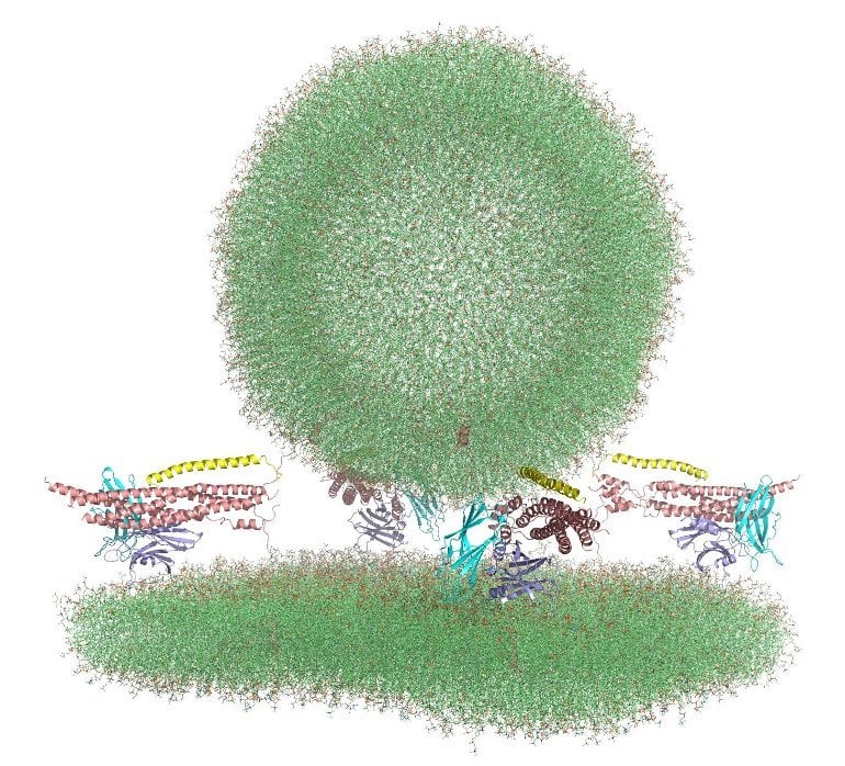
Recently, using the Frontera supercomputer at the Texas Advanced Computing Center (TACC), one of the most powerful systems in the world, Rizo-Rey has been exploring this process, creating a multi-million atom model of the proteins, the membranes, and their environment, and setting them in motion virtually to see what happens, a process known as molecular dynamics.

Writing in eLife in June 2022, Rizo-Rey and collaborators presented all-atom molecular dynamics simulations of synaptic vesicle fusion, providing a glimpse at the primed state. The research shows a system where several specialized proteins are “spring-loaded,” awaiting only the delivery of calcium ions to trigger fusion.

“It’s ready to release, but it doesn’t,” he explained. “Why doesn’t it? It’s waiting for the calcium signal. Neurotransmission is about controlling fusion. You want to have the system ready to fuse, so when calcium comes in, it can happen very fast, but it’s not fusing yet.”This shows a computer generated image of a synaptic vesicle 
 
Initial configuration of the molecular dynamics simulations designed to investigate the nature of the primed state of synaptic vesicles. Credit: Jose Rizo-Rey, UT Southwestern Medical Center

The study represents a return to computational approaches for Rizo-Rey, who recalls using the original Cray supercomputer at the University of Texas at Austin in the early 1990s. He went on to use primarily experimental methods like nuclear magnetic resonance spectroscopy over the past three decades to study the biophysics of the brain.“Supercomputers weren’t powerful enough to resolve this problem of how transmission was occurring in the brain. So for a long time, I used other methods,” he said. “However, with Frontera, I can model 6 million atoms and really get a picture of what’s going on with this system.”Rizo-Rey’s simulations only cover the first few microseconds of the fusion process, but his hypothesis is that the act of fusion should happen in that time. “If I see how it’s starting, the lipids starting to mix, then I’ll ask for 5 million hours [the maximum time available] on Frontera,” he said, to capture the snap of the spring-loaded proteins and the step-by-step process by which the fusion and transmission happens.Rizo-Rey says the sheer amount of computation that can be harnessed today is unbelievable. “We have a supercomputer system here at the University of Texas Southwestern Medical Center. I can use up to 16 nodes,” he said. “What I did on Frontera, instead of a few months, would have taken 10 years.”Investing in basic research—and in the computing systems that support this type of research—is fundamental to the health and well-being of our nation, Rizo-Rey says.“This country was very successful because of basic research. Translation is important, but if you don’t have the basic science, you have nothing to translate.”

Friday, April 1, 2022

'Younger' Brain Associated With Lower Risk of Post-Stroke Cognitive Disorders

 This doesn't matter at all. Your doctor is still responsible to get you 100% recovered regardless of your cognitive status.  Don't let your doctor weasel their way out of this responsibility by quoting the craptastic saying: 'All strokes are different, all stroke recoveries are different.' That quote means they know nothing about stroke and are out-of-date on research.  You don't want them treating you.

'Younger' Brain Associated With Lower Risk of Post-Stroke Cognitive Disorders


Dawn Elliott Knapp, PA-C, for Medscape

March 30, 2022

1

The study covered in this summary was published on MedRxiv.org as a preprint and has not yet been peer reviewed.

Key Takeaways

  • Patients with greater brain age gaps (BAG, the difference between predicted and chronological brain age) were more likely to have developed neurocognitive disorders (NCD) post-stroke.

  • "Younger" brains (lower BAG) appear to be more resilient to injury and are associated with lower risk for NCD up to 36 months after a stroke.

  • As BAG is sensitive to cognitive impairment after stroke, it may be used in order to predict cognitive outcome in stroke patients.

Why This Matters

  • Long-lasting mild to major cognitive impairments affect 35%–50% of stroke survivors and may not improve over time, a significant source of disability.

  • Brain age calculation could help identify which patients are particularly at risk for post-stroke NCD and could be used to guide treatment and rehabilitation to minimize impairment. 

Study Design

  • MRIs from 269 stroke survivors (55.4% women, mean age, 71 years) from the Nor-COAST study were assessed. MRIs were performed at baseline, 18 months, and 36 months post-stroke.

  • Neuropsychological data was collected at baseline, 3, 18, and 36 months post-stroke, consisting of standardized cognitive testing, Global Deterioration Scale, and classification of NCD using the DSM-5.

  • Predicted brain age was measured via machine learning models based on cortical thickness, surface area, and gray matter/white matter volume. Associations between cognitive status and longitudinal brain age were assessed using linear mixed effects models and survival analysis.

  • Statistical analyses were performed using R v. 3.3.3 and Stata v. 16 to test three hypotheses: 1) whether brain structural characteristics are associated with post-stroke NCD; 2) if a younger appearing brain among patients showing normal cognitive function at baseline is associated with lower risk of NCD at 18- and 36- months follow-up, and 3) whether patients showing preserved cognitive function from baseline onward show less evidence of brain aging over time compared with patients showing cognitive decline.

Key Results

  • "Older" brains (higher BAG) were associated with post-stroke cognitive impairment at 18 and 36 months, confirming that a “younger" brain may confer protective factors against cognitive decline after stroke.

  • There was no significant difference in BAG between the group with preserved brain function vs the group with NCD from baseline and across follow-up times.

  • Pre-stroke BAG may be a more reliable predictor for future cognitive decline than post-stroke BAG.

Sunday, March 28, 2021

Why is physical activity so important for health and wellbeing?

 It is your doctor's responsibility to get you recovered enough to do these physical activities. Don't let your doctor weasel out of that responsibility by quoting the crapola statement; 'All strokes are different, all stroke recoveries are different'. That is just their convenient excuse to shift blame from themselves to you for not recovering. Screaming in their face may be required.

Why is physical activity so important for health and wellbeing?

We know that staying active is one of the best ways to keep our bodies healthy. But did you know it can also improve your overall well-being and quality of life? 

Here are just a few of the ways physical activity can help you feel better, look better and live better. Because, why not?

It’s a natural mood lifter.

Regular physical activity can relieve stress, anxiety, depression and anger. You know that "feel good sensation" you get after doing something physical? Think of it as a happy pill with no side effects! Most people notice they feel better over time as physical activity becomes a regular part of their lives.

It keeps you physically fit and able.

Without regular activity, your body slowly loses its strength, stamina and ability to function properly. It’s like the old saying: you don’t stop moving from growing old, you grow old from stopping moving. Exercise increases muscle strength, which in turn increases your ability to do other physical activities. 

It helps keep the doctor away.

Stand up when you eat your apple a day! Too much sitting and other sedentary activities can increase your risk of heart disease and stroke. One study showed that adults who watch more than 4 hours of television a day had an 80% higher risk of death from cardiovascular disease.
 
Being more active can help you:

  • lower your blood pressure 
  • boost your levels of good cholesterol
  • improve blood flow (circulation)
  • keep your weight under control
  • prevent bone loss that can lead to osteoporosis

All of this can add up to fewer medical expenses, interventions and medications later in life!

It can help you live longer.

It’s true, 70 is the new 60… but only if you’re healthy. People who are physically active and at a healthy weight live about seven years longer than those who are not active and are obese. And the important part is that those extra years are generally healthier years! Staying active helps delay or prevent chronic illnesses and diseases associated with aging. So active adults maintain their quality of life and independence longer as they age.

Here are some other benefits you may get with regular physical activity: 

  • Helps you quit smoking and stay tobacco-free.
  • Boosts your energy level so you can get more done.
  • Helps you manage stress and tension.
  • Promotes a positive attitude and outlook.
  • Helps you fall asleep faster and sleep more soundly.
  • Improves your self-image and self-confidence.
  • Helps you spend more time outdoors.

The American Heart Association recommends at least 150 minutes of moderate-intensity aerobic activity each week. You can knock that out in just 30 minutes a day, 5 days a week. And every minute of moderate to vigorous activity counts toward your goal.

So, this is easy! Just move more, with more intensity, and sit less. You don’t have to make big life changes to see the benefits. Just start building more activity into your day, one step at a time.