This should obsolete all the million dollar mobile stroke units with scanners inside. But they don't say how fast it is. Can it beat these?
Hats off to Helmet of Hope - stroke diagnosis in 30 seconds February 2017
Microwave Imaging for Brain Stroke Detection and Monitoring using High Performance Computing in 94 seconds March 2017
New Device Quickly Assesses Brain Bleeding in Head Injuries - 5-10 minutes April 2017
The latest here:
Ski-Mask Design AIR Coil Offers Whole-Brain Imaging Without Claustrophobia
Imagine donning a balaclava – similar to the one you would put swish down a mountain on your skis – to undergo a brain MRI. With a prototype of a 16-channel head Adaptive Image Receive (AIR) radiofrequency coil, that could become a reality for some patients.
Based on performance results published in the American Journal of Roentgenology, this AIR coil, manufactured by GE Healthcare produced better results than a conventional 8-channel head coil for in vivo whole-brain imaging.
“This study shows the feasibility of the novel AIR coil technology for imaging the brain and provides insight for future coil design improvements,” said the study’s first author Petrice M. Cogswell, M.D., Ph.D., a radiologist with the Mayo Clinic in Rochester, Minn.

Even though the prototype did not perform as well as a conventional 32-channel head coil, it has a lightweight, flexible, open design, similar to a ski-mask has electrical characteristics that allow it to surpass the restrictions that limit traditional coil designs that are rigid.

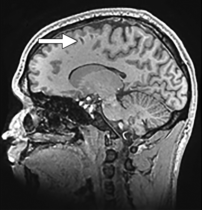
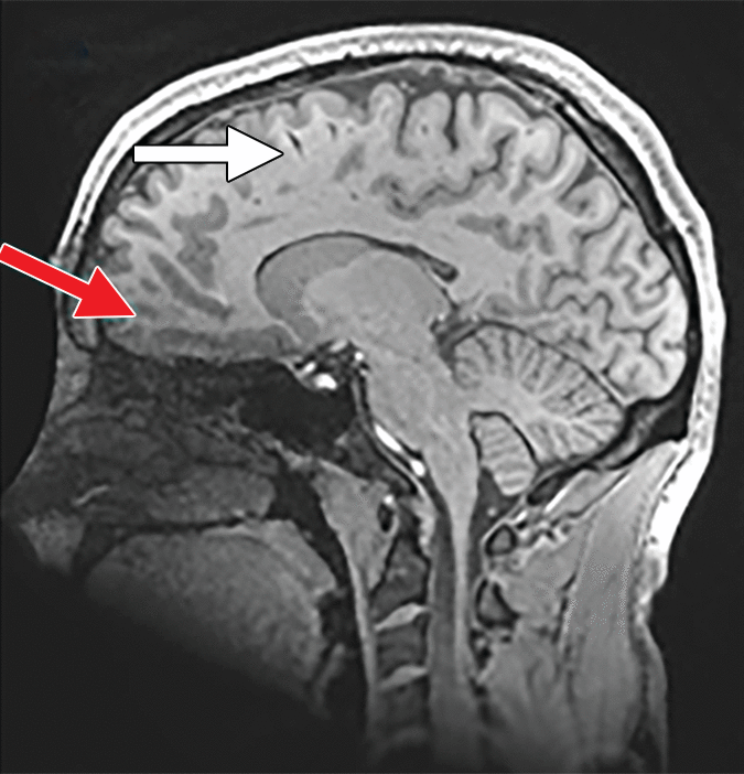
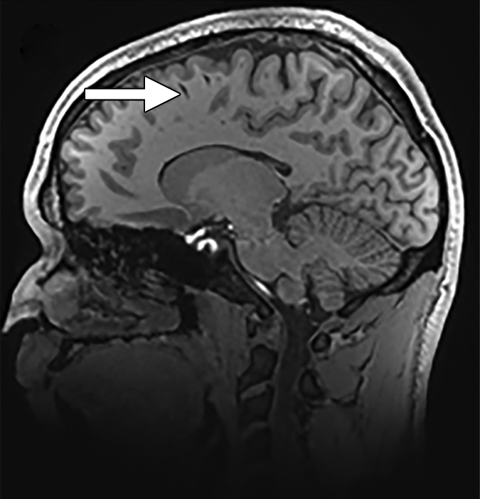
To determine whether the 16-coil prototype could provide images that were of high enough quality to be diagnostically useful, Cogswell’s team tested it on 15 healthy adult participants and a phantom, using clinically available MRI sequences. Two board-certified neuroradiologists scored the images on a five-point ordinal scale in multiple categories and compared the prototype’s results to those from the conventional 8-channel and 32-channel coils.

Based on their analysis, the signal-to-noise ratio, structural sharpness, and overall image quality scores gleaned from the 16-channel prototype outpaced the conventional 8-channel coil. Across all participants, the noise covariance matrices from the 16-channel AIR coil produced stable performance, and the media g-factors were less than those produced with the 8-channel coil. However, they were not less than the 32-channel.
According to the study findings, the 16-channel coil did have one inital drawback – the presence of artifacts. However, the team noted, adding foam padding to the ski-mask design kept the patient’s head more secure and reduced the chance of artifacts.
The outcome of the study is encourage, Cogswell said, and it presents multiple possibilities for improvements in whole-brain imaging.
“The advantages of the AIR coil technology for reduction of claustrophobia, improved airway access, and monitoring of patients under anesthesia, and overall better user comfort may be investigated in future studies,” the team said.
No comments:
Post a Comment