Changing stroke rehab and research worldwide now.Time is Brain! trillions and trillions of neurons that DIE each day because there are NO effective hyperacute therapies besides tPA(only 12% effective). I have 523 posts on hyperacute therapy, enough for researchers to spend decades proving them out. These are my personal ideas and blog on stroke rehabilitation and stroke research. Do not attempt any of these without checking with your medical provider. Unless you join me in agitating, when you need these therapies they won't be there.

What this blog is for:

My blog is not to help survivors recover, it is to have the 10 million yearly stroke survivors light fires underneath their doctors, stroke hospitals and stroke researchers to get stroke solved. 100% recovery. The stroke medical world is completely failing at that goal, they don't even have it as a goal. Shortly after getting out of the hospital and getting NO information on the process or protocols of stroke rehabilitation and recovery I started searching on the internet and found that no other survivor received useful information. This is an attempt to cover all stroke rehabilitation information that should be readily available to survivors so they can talk with informed knowledge to their medical staff. It lays out what needs to be done to get stroke survivors closer to 100% recovery. It's quite disgusting that this information is not available from every stroke association and doctors group.

Showing posts with label nerve stimulation. Show all posts

Monday, December 29, 2025

Audiovisual gamma stimulation restores hippocampal neurogenesis and neural circuit plasticity in aging mice

 Ask your competent? doctor how to EXACTLY do this even before human testing occurs! No ability to extrapolate; PURE INCOMPETENCE!

  • 40Hz sensory stimulation (3 posts to March 2021)
  • Audiovisual gamma stimulation restores hippocampal neurogenesis and neural circuit plasticity in aging mice


    Abstract

    Aging is the primary risk factor for cognitive decline and neurodegenerative disorders, characterized by impaired circuit plasticity and disrupted gamma oscillations. Non-invasive 40 Hz audiovisual stimulation (AuViS) has emerged as a promising strategy to restore cognition in models of Alzheimer’s disease and stroke. Yet, the mechanisms underlying these effects remain unclear. We found that AuViS increased gamma oscillations in the dentate gyrus of middle-aged mice. Control animals displayed scarce neurogenesis, and newborn neurons exhibited limited growth and remained functionally immature. Notably, AuViS triggered the proliferation of neural progenitor cells and shifted the balance from astrocytic towards neuronal differentiation. It also promoted neuronal maturation, leading to the development of complex dendritic trees and axons with large mossy terminals bearing filopodial extensions. These structural modifications were accompanied by increased spiking capacity and spontaneous synaptic activity, indicative of effective circuit integration. These effects were dependent on TrkB signaling, implicating neurotrophin pathways. Our findings demonstrate that AuViS reestablishes neurogenesis and promotes network remodeling in the healthy aging brain, which might aid to ameliorate neurological conditions.

    This is a preview of subscription content, access via your institution

    Sunday, December 15, 2024

    A Randomized Controlled Trial to Test the Effects of Repetitive Peripheral Magnetic Stimulation Versus Neuromuscular Electrical Stimulation in Patients with Spastic Hemiparesis After Stroke (REPMAST): Study Protocol

     Will this cure spasticity? That is the only endpoint for spasticity research! NOTHING LESS!

    A Randomized Controlled Trial to Test the Effects of Repetitive Peripheral Magnetic Stimulation Versus Neuromuscular Electrical Stimulation in Patients with Spastic Hemiparesis After Stroke (REPMAST): Study Protocol           

    Kristin Loreen Pohl 

     2, 2, 1 and 1,2,*
    1
    Section of Neurological Rehabilitation, Clinic of Neurology, Jena University Hospital, 07747 Jena, Germany
    2
    Department of Neurology, Gräfliche Kliniken Moritz Klinik GmbH, 07639 Bad Klosterlausnitz, Germany
    *
    Author to whom correspondence should be addressed.
    Brain Sci. 2024, 14(12), 1249; https://doi.org/10.3390/brainsci14121249
    Submission received: 28 October 2024 / Revised: 3 December 2024 / Accepted: 11 December 2024 / Published: 12 December 2024
    (This article belongs to the Special Issue New Studies on Stroke Care and Rehabilitation)

    Abstract

    Background/Objectives: 

    Innovative therapies are needed to reduce disability, facilitate activities of daily living, and improve the quality of life(Survivors want full recovery, NOT JUST IMPROVEMNT; Don't you ever talk to survivors without justifying your failures by parroting the tyranny of low expectations?)in patients with stroke. Non-invasive methods of stimulating the peripheral and central nervous system are increasingly being used to enhance the effects of existing therapies in stroke rehabilitation. One potentially relevant method for achieving greater improvement is repetitive peripheral magnetic stimulation (rPMS). This randomized controlled trial (RCT), the Peripheral MAgnetic stimulation in patients with spastic hemiparesis after Stroke Trial (REPMAST), will investigate whether rPMS improves upper extremity function, spasticity, and activities of daily living in patients with stroke compared with neuromuscular stimulation (NMS). 

    Methods: 

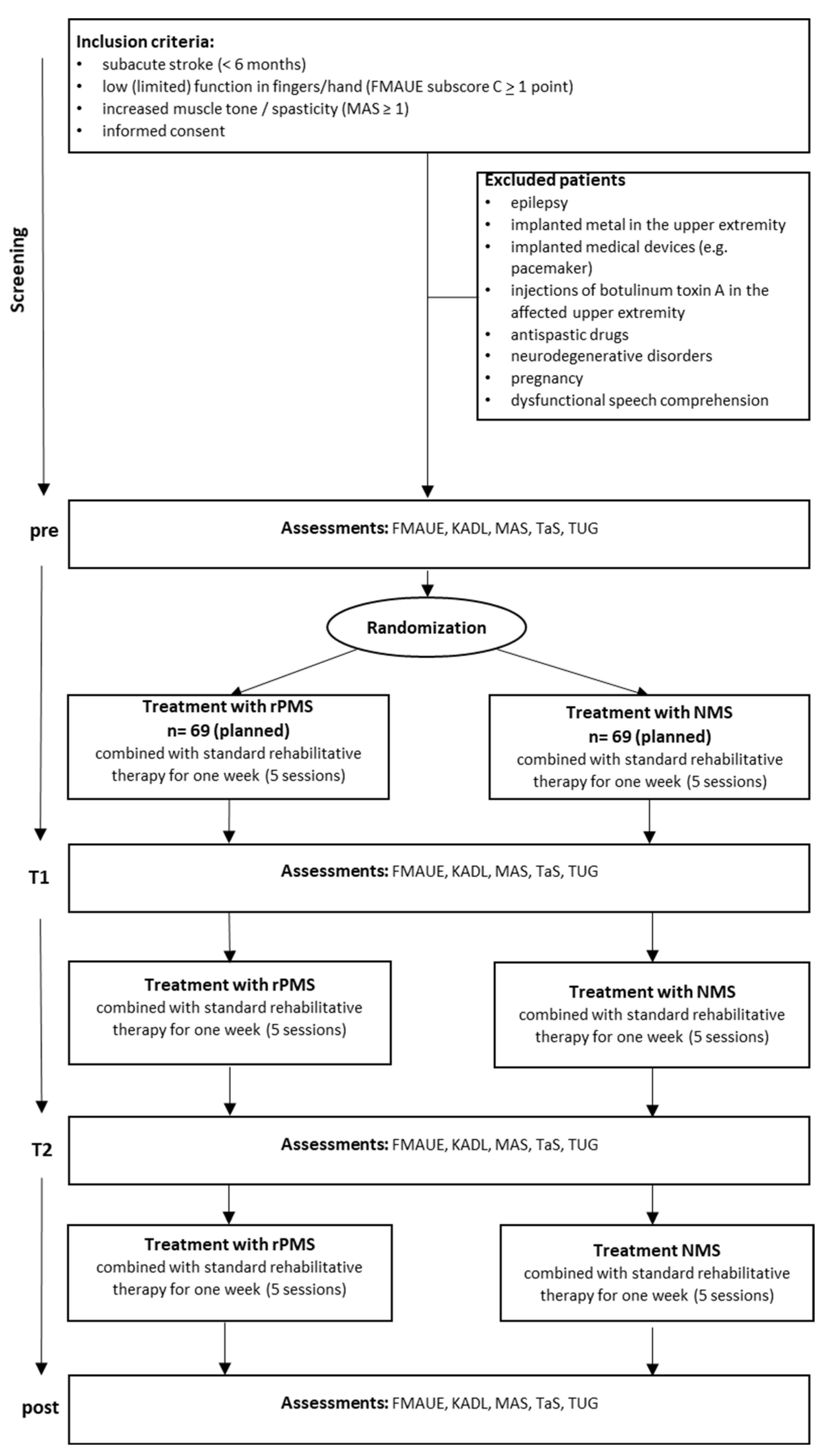
    REPMAST is an interventional, randomized controlled single-blinded study. Patients with subacute stroke are randomized to receive rPMS or NMS five days a week for three weeks in addition to standard rehabilitation therapy. The primary outcome is the change in the Fugl–Meyer Assessment for Upper Extremity between the beginning and end of the stimulation sessions. Secondary outcomes include changes in the Katz Index of Independence in Activities of Daily Living, the Timed Up and Go Test, the Modified Ashworth Scale, and the Tardieu Scale. A total sample size of 138 patients (69 in each group) is required to investigate the superiority of rPMS compared with NMS. 

    Conclusions: 

    The aim of this RCT is to provide evidence for an effective(Effective to a survivor is curing spasticty! Will this do that?) peripheral stimulation treatment for stroke recovery.

    1. Introduction

    Stroke is the third leading cause of death and disability combined worldwide [1]. Innovative therapies are needed to reduce disability, facilitate activities of daily living (ADL), and improve quality of life [2].
    In recent years, in addition to physiotherapy and occupational therapy, non-invasive brain stimulation methods such as transcranial direct current stimulation and repetitive transcranial magnetic stimulation (rTMS) have been increasingly used to modulate brain function in order to improve functional deficits after stroke.
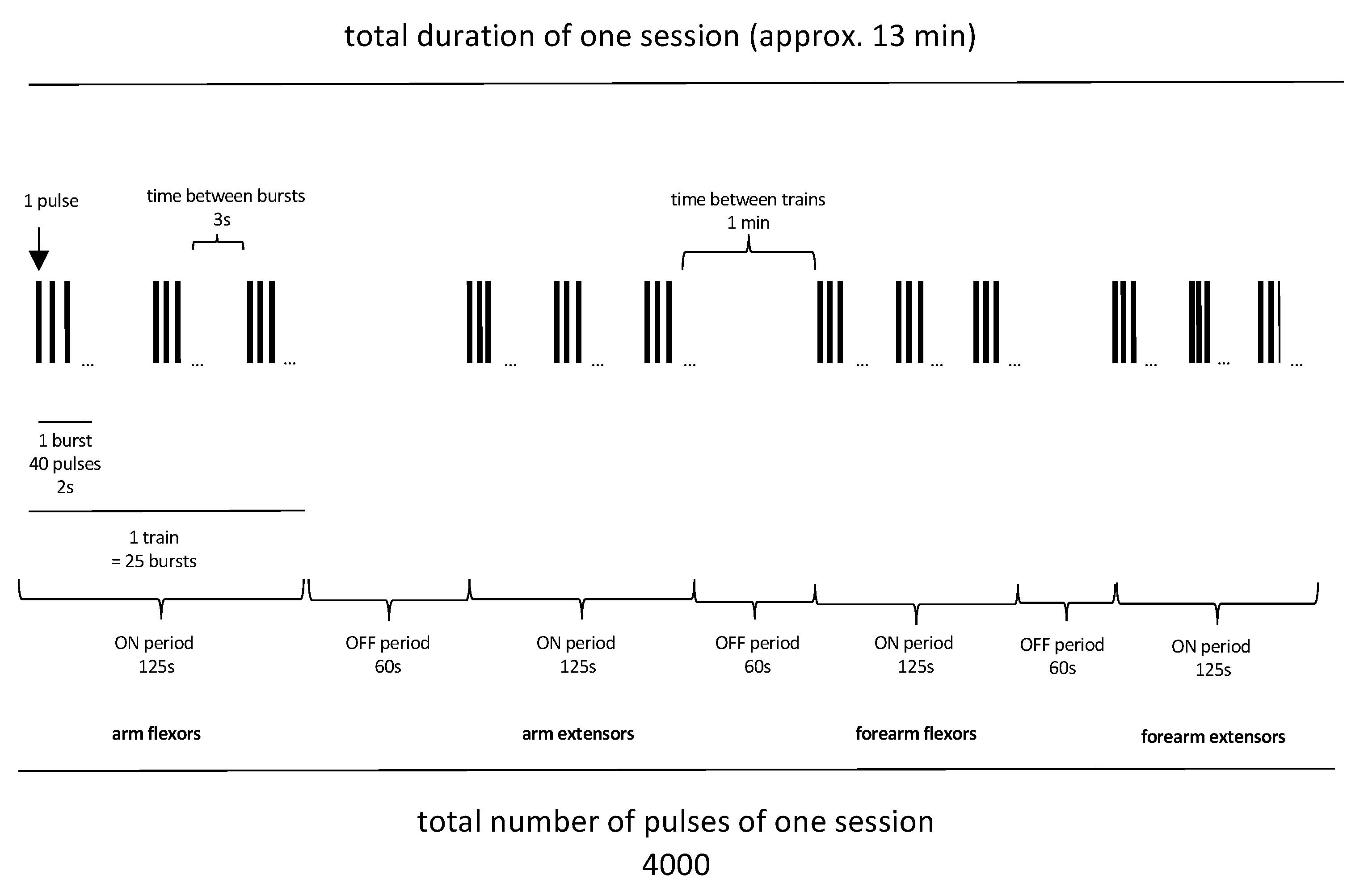
    Another possibility to modulate brain function is through peripheral application, such as peripheral electrical stimulation [3,4] or with the use of repetitive peripheral magnetic stimulation (rPMS) [5,6]. Repetitive peripheral magnetic stimulation (rPMS) is a painless stimulation method that uses rapidly changing magnetic fields to stimulate peripheral nerves and trigger repeated contractions of the skeletal muscles. Using rPMS is simple compared with other NIBS approaches. The stimulation coil is placed directly on the skin in the area of the target muscle to be stimulated. The magnetic field penetrates the tissue and causes depolarization due to the electric field that builds up, resulting in the induction of an action potential, which clinically leads to muscle contraction. The rPMS method has also been used to stimulate peripheral nerves or spinal nerve roots, with the stimulation coil placed paravertebrally or over the corresponding nerve [7,8,9,10]. The exact mechanism of how rPMS interacts with the central and peripheral nervous systems is not yet fully understood. Struppler and colleagues suggested that rPMS causes increased proprioceptive input to the brain by activating mechanoreceptors of the contracted muscle [11]. Another type of input has been suggested to come directly from the nerve fibers. Consequently, rPMS increases sensory input from the affected limb to the brain, initiating neuroplastic processes and leading to improved sensorimotor performance in patients.
    In the 1990s, the first studies by Struppler and colleagues reported improvements in perception, spasticity, and paresis after stroke and in patients with multiple sclerosis following the use of rPMS [12,13,14]. Over the past decade, the number of studies using rPMS has increased significantly [15]. Recent meta-analyses have reported that rPMS induces a better Fugl–Meyer Assessment for Upper Extremity (FMAUE) compared with control groups [16,17]. However, it has also been criticized that there is a lack of rPMS studies conducted as RCTs with large sample sizes [18,19,20]. Therefore, the current randomized controlled trial, the Repetitive Peripheral Magnetic stimulation in patients with spastic hemiparesis after Stroke Trial (REPMAST), will investigate the effect of rPMS compared to neuromuscular stimulation (NMS) in a large sample of patients with stroke. Because of the sensory influence of rPMS on the brain, the effect of rPMS in REPMAST will be compared with that of a control group that receives neuromuscular stimulation (NMS).
    Previous reports have compared the advantages and disadvantages of rPMS and NMS [5,6,21]. It is important to note that although both interventions stimulate some common peripheral structures [6], they nevertheless activate different networks. Repetitive PMS has been found to increase activation of the ipsilesional superior posterior parietal and premotor cortex [11]. By contrast, NMS has been described to increase activity in the ipsilesional sensorimotor cortex [22]. Recent clinical studies have compared rPMS over muscles with sham [23], standard care [21], conventional physiotherapy [24], or its combination with low-frequency rTMS [10]. REPMAST is a randomized controlled comparative interventional trial that aims to investigate the efficacy (superiority) of rPMS compared with NMS in a large number of patients with subacute stroke, as there is no direct comparison between rPMS and NMS.
    There are conflicting results regarding the effectiveness of rPMS in improving spasticity [14,16,18,19,20,23]. Therefore, this study will also analyze the effect of rPMS on spasticity.

    More at link.

    Monday, April 8, 2024

    Gamma oscillations induced by 40-Hz visual- auditory stimulation for the treatment of acute- phase limb motor rehabilitation after stroke: study protocol for a prospective randomized controlled trial

    40 Hz transcranial alternating current stimulation (tACS) has proven to be an effective treatment for improving cognition, a crucial factor in motor learning.If this statement is true, where is the protocol that your hospital is using? Or is your incompetent? hospital not using this?

    Gamma oscillations induced by 40-Hz visual-auditory stimulation for the treatment of acute-phase limb motor rehabilitation after stroke: study protocol for a prospective randomized controlled trial

    Wang Fu
    Seventh People's Hospital of Shanghai University of Traditional Chinese Medicine
    Xiaomi Yu
    Seventh People's Hospital of Shanghai University of Traditional Chinese Medicine
    Minhui Lai
    Seventh People's Hospital of Shanghai University of Traditional Chinese Medicine
    Yuanli Li
    Shanghai University of Traditional Chinese Medicine
    Yiting Yang
    Seventh People's Hospital of Shanghai University of Traditional Chinese Medicine
    Yong Qin
    Seventh People's Hospital of Shanghai University of Traditional Chinese Medicine
    Min Yu
    Seventh People's Hospital of Shanghai University of Traditional Chinese Medicine
    Feng Wang
    Seventh People's Hospital of Shanghai University of Traditional Chinese Medicine
    Cong Wang
    Seventh People's Hospital of Shanghai University of Traditional Chinese Medicine
    https://orcid.org/0000-0003-2289-2794

    https://doi.org/10.21203/rs.3.rs-3346592/v1

    This work is licensed under a CC BY 4.0 License

    Background: 

    40 Hz transcranial alternating current stimulation (tACS) has proven to be an effective treatment for improving cognition, a crucial factor in motor learning. However, current studies are predominantly focused on the motor cortex, and the potential brain mechanisms responsible for the therapeutic effects are still unclear. Given the interconnected nature of motor learning within the brain network, we have proposed a novel approach known as multitarget tACS. This study aims to ascertain whether multitarget tACS is more effective than single-target stimulation in stroke patients and to further explore the potential underlying brain mechanisms by using techniques such as transcranial magnetic stimulation (TMS) and magnetic resonance imaging (MRI).

    Methods:  

    This study employs a double-blind, sham-controlled, randomized controlled trial design with a 2-week intervention period. Both participants and outcome assessors will remain unaware of treatment allocation throughout the study. Thirty-nine stroke patients will be recruited and randomized into three distinct groups, including the sham tACS group (SS group), the single-target tACS group (ST group), and the multitarget tACS group (MT group), at a 1:1:1 ratio. The primary outcomes are series reaction time tests (SRTTs) combined with electroencephalograms (EEGs). The secondary outcomes include motor evoked potential (MEP), central motor conduction time (CMCT), short interval intracortical inhibition (SICI), intracortical facilitation (ICF), magnetic resonance imaging (MRI), box and block test (BBT) and blood sample RNA sequencing. The tACS interventions for all three groups will be administered over a 2-week period, with outcome assessments conducted at baseline (T0) and 1 day (T1), 7 days (T2), and 14 days (T3) of the intervention phase.

    Discussion: 

    The study’s findings will determine the potential of 40 Hz tACS to improve motor learning in stroke patients. Additionally, it will compare the effectiveness of multitarget and single-target approaches, shedding light on their respective improvement effects. Through the utilization of techniques such as TMS and MRI, the study aims to uncover the underlying brain mechanisms responsible for the therapeutic impact. Furthermore, the intervention has the potential to facilitate motor learning efficiency, thereby contributing to the advancement of future stroke rehabilitation treatment.

    Trial registration: Chinese Clinical Trial Registry ChiCTR2300073465. Registered on July 11, 2023.

    Wednesday, July 19, 2023

    New method could make post-stroke motor rehabilitation more effective and affordable - vagus nerve stimulation

    All this earlier research on vagus nerve stimulation and you still haven't written a protocol on it and distributed it worldwide.

    I'd fire you all for incompetence! Have you ever considered that survivors want to recover and are expecting you to deliver that recovery?

    The latest here:

    New method could make post-stroke motor rehabilitation more effective and affordable

    The longest nerve in the human body starts in the brain and meanders its way down the neck and into the chest, where it splits into separate branches, winding its twisting tendrils to touch each internal organ. Known as the "information superhighway" and aptly named from the Latin word meaning "wanders," the vagus nerve is a bundle of fibers responsible for the parasympathetic nervous system: digestion, heart rate, breathing.

    Sending electrical impulses down this tenth cranial nerve has proven effective in treating conditions like depression and epilepsy, and it has shown great success in amplifying the effects of motor rehabilitation after stroke. Implanting a device onto the vagus nerve in the neck provides direct stimulation to the information superhighway. And using this technology during post-stroke motor rehabilitation was approved by the Food and Drug Administration as a treatment option in 2021.

    But researchers at MUSC have found another method that accelerates treatment outcomes and improves motor function after stroke without this invasive and often-uninsured procedure.

    As described in a recent paper from Neurorehabilitation and Neural Repair, the research team placed sensors on the upper arm and in the ear and used the connected computer to send timed electrical impulses to the vagus nerve during motor rehabilitation. This noninvasive VNS method, known as motor activated auricular vagus nerve stimulation (MAAVNS), allows patients to gain the same amount of motor function improvement in 4 weeks that patients with the implanted device reached in 6 weeks – all without surgery.

    MAAVNS was developed and is currently pending patent at MUSC.

    Stroke is a leading cause of disability in the United States according to the American Stroke Association, and it leads to motor deficits and reduced mobility in almost half of stroke survivors over the age of 65. With such a large number affected, Bashar Badran, Ph.D., the director of the Neuro-X lab and Computational Brain Imaging Core at MUSC as well as the principal investigator on the paper, wanted to find a more accessible way to help patients recover.

    Motor rehabilitation is time-consuming and expensive, and oftentimes does not produce the results that patients want. Technology like the MAAVNS system can boost the effects of conventional motor rehabilitation and help patients get the most out of their therapy in a simple and relatively inexpensive manner. It's very exciting."

    Bashar Badran, Ph.D., Director of the Neuro-X lab and Computational Brain Imaging Core at MUSC

    With a small cart containing the computer and sticker-like sensors, the MAAVNS system can easily be incorporated into occupational therapy settings, with little change or impact to the current standard of care.

    With hundreds of repetitions in an hour, patients can focus on specific movements they want to improve, like sewing, cutting fabric or buttoning a shirt, while the MAAVNS system detects when the patient is moving and intricately delivers electrical stimulation to nerves in the ear.

    Badran says delivering the stimulation in conjunction with each movement is critical. "Our work shows that outcomes are much better when you time the stimulation in conjuction with movement, in a closed-loop approach," he said. "Interestingly, more stimulation is not better. In fact, less stimulation timed correctly produces the best outcomes."

    Since the vagus nerve elicits brain activity in areas responsible for the release of neurotransmitters like norepinephrine and serotonin which help the brain learn, stimulating it with electricity facilitates faster learning of motor skills.

    While the sample size for this pilot study was small, Badran and his team will be conducting a larger clinical trial next to further study the effects of this noninvasive VNS technique. "This is really promising technology," Badran said. "And the fact that the outcomes are mirroring what is already FDA approved is great. Not only do we believe this is an effective new technology for post-stroke motor rehabilitation, but it's cheaper and easier to incorporate into the standard of care than what is currently available."

    Source:
    Journal reference:

    Badran, B. W., et al. (2023) Motor Activated Auricular Vagus Nerve Stimulation as a Potential Neuromodulation Approach for Post-Stroke Motor Rehabilitation: A Pilot Study. Neurorehabilitation and Neural Repair. doi.org/10.1177/15459683231173357.

    Wednesday, May 24, 2023

    Gamma frequency tactile stimulation can reduce Alzheimer's disease pathology and symptoms

    Didn't your doctor already prescribe 40Hz for you?

    See this: Intermittent Light Exposures in Humans: A Case for Dual Entrainment in the Treatment of Alzheimer's Disease March 2021

    While this is a new area of research, preliminary evidence shows the potential of dual circadian and gamma-wave entrainment as an important therapy not only for those with AD, but for others with cognitive impairment.

    Google this: gamma 40hz light and sound stimulation device: and you get some examples. But you can't do that until your incompetent doctor in 50 years finally gets up-to-date on research. 

    Repeated Ketamine Anesthesia Restarts Plasticity in the Brain July 2021 This one talks about both 40 and 60 Hertz flickering lights. I have a 40Hz flickering light, not sure if I need to get a 60 Hz one. 


    The latest here:

    Gamma frequency tactile stimulation can reduce Alzheimer's disease pathology and symptoms

    Evidence that non-invasive sensory stimulation of 40 Hz gamma frequency brain rhythms can reduce Alzheimer's disease pathology and symptoms, already shown with light and sound by multiple research groups in mice and humans, now extends to tactile stimulation. A new study by MIT scientists shows that Alzheimer's model mice exposed to 40 Hz vibration an hour a day for several weeks showed improved brain health and motor function compared to untreated controls.

    The MIT group is not the first to show that gamma frequency tactile stimulation can affect brain activity and improve motor function, but they are the first to show that the stimulation can also reduce levels of the hallmark Alzheimer's protein phosphorylated tau, keep neurons from dying or losing their synapse circuit connections, and reduce neural DNA damage.

    This work demonstrates a third sensory modality that we can use to increase gamma power in the brain. We are very excited to see that 40 Hz tactile stimulation benefits motor abilities, which has not been shown with the other modalities. It would be interesting to see if tactile stimulation can benefit human subjects with impairment in motor function."

    Li-Huei Tsai, corresponding author of the study, director of The Picower Institute for Learning and Memory and the Aging Brain Initiative at MIT, and Picower Professor in the Department of Brain and Cognitive Sciences (BCS)

    Ho-Jun Suk, Nicole Buie, Guojie Xu and Arit Banerjee are lead authors of the study in Frontiers in Aging Neuroscience, and Ed Boyden, Y. Eva Tan Professor of Neurotechnology at MIT, is a co-senior author of the paper. Boyden, an affiliate member of The Picwoer Institute, is also appointed in BCS as well as the Departments of Bioengineering and Media Arts and Sciences, the McGovern Institute for Brain Research, and the K. Lisa Yang Cener for Bionics.

    Feeling the vibe

    In a series of papers starting in 2016, a collaboration led by Tsai's lab has demonstrated that light flickering and/or sound clicking at 40 Hz (a technology called GENUS for Gamma Entrainment Using Sensory stimuli), reduces levels of amyloid-beta and tau proteins, prevents neuron death and preserves synapses and even sustains learning and memory in a variety of Alzheimer's disease mouse models. Most recently in pilot clinical studies the team showed that 40 Hz light and sound stimulation was safe, successfully increased brain activity and connectivity and appeared to produce significant clinical benefits in a small cohort of human volunteers with early-stage Alzheimer's disease. Other groups have replicated and corroborated health benefits of 40 Hz sensory stimulation and an MIT spin-off company, Cognito Therapeutics, has launched stage III clinical trials of light and sound stimulation as an Alzheimer's treatment.

    The new study tested whether whole-body 40 Hz tactile stimulation produced meaningful benefits in two commonly used mouse models of Alzheimer's neurodegeneration, the Tau P301S mouse, which recapitulates the disease's tau pathology, and the CK-p25 mouse, which recapitulates the synapse loss and DNA damage seen in human disease. The team focused its analyses in two areas of the brain: the primary somatosensory cortex (SSp), where tactile sensations are processed, and the primary motor cortex (MOp), where the brain produces movement commands for the body.

    To produce the vibration stimulation, the researchers placed mouse cages over speakers playing 40 Hz sound, which vibrated the cages. Non-stimulated control mice were in cages interspersed in the same room so that all the mice heard the same 40 Hz sound. The differences measured between the stimulated and control mice were therefore made by the addition of tactile stimulation.

    First the researchers confirmed that 40 Hz vibration made a difference in neural activity in the brains of healthy (i.e. non-Alzheimer's) mice. As measured by expression of c-fos protein, activity increased two-fold in the SSp and more than 3-fold in the MOp, a statistically significant increase in the latter case.

    Once the researchers knew that 40 Hz tactile stimulation could increase neural activity, they assessed the impact on disease in the two mouse models. To ensure both sexes were represented, the team used male P301S mice and female CK-p25 mice.

    P301S mice stimulated for three weeks showed significant preservation of neurons compared to unstimulated controls in both brain regions. Stimulated mice also showed significant reductions in tau in the SSp by two measures, and exhibited similar trends in the MOp.

    CK-p25 mice received six weeks of vibration stimulation. These mice showed higher levels of synaptic protein markers in both brain regions compared to unvibrated control mice. They also showed reduced levels of DNA damage.

    Finally the team assessed the motor abilities of mice exposed to the vibration vs. not exposed. They found that both mouse models were able to stay on a rotating rod significantly longer. P301S mice also hung on to a wire mesh for significantly longer than control mice while CK-p25 mice showed a positive, though non-significant trend.

    "The current study, along with our previous studies using visual or auditory GENUS demonstrates the possibility of using non-invasive sensory stimulation as a novel therapeutic strategy for ameliorating pathology and improving behavioral performance in neurodegenerative diseases," the authors concluded.

    Support for the study came from The JPB Foundation, The Picower Institute for Learning and Memory, Eduardo Eurnekian, The DeGroof-VM Foundation, Halis Family Foundation, Melissa and Doug Ko Hahn, Lester Gimpelson, Eleanor Schwartz Charitable Foundation, The Dolby Family, Kathleen and Miguel Octavio, Jay and Carroll Miller, Anne Gao and Alex Hu and Charles Hieken.

    Source:
    Journal reference:

    Suk, H-J., et al. (2023) Vibrotactile stimulation at gamma frequency mitigates pathology related to neurodegeneration and improves motor function. Frontiers in Aging Neuroscience. doi.org/10.3389/fnagi.2023.1129510.

    Thursday, December 9, 2021

    Bilateral Transcutaneous Electrical Nerve Stimulation Improves Upper Limb Motor Recovery in Stroke: A Randomized Controlled Trial

     So still a failure. 'Improves', NOT 100% RECOVERY!

    Bilateral Transcutaneous Electrical Nerve Stimulation Improves Upper Limb Motor Recovery in Stroke: A Randomized Controlled Trial

    Originally publishedhttps://doi.org/10.1161/STROKEAHA.121.036895Stroke. 2021;0:STROKEAHA.121.036895

    Background and Purpose:

    Recent evidence has shown bilateral transcutaneous electrical nerve stimulation (Bi-TENS) combined with task-oriented training (TOT) to be superior to unilateral transcutaneous electrical nerve stimulation (Uni-TENS)+TOT in improving lower limb motor functioning following stroke. However, no research explored the effect of Bi-TENS+TOT in improving upper limb motor recovery. This study aimed to compare Bi-TENS+TOT with Uni-TENS+TOT, Placebo transcutaneous electrical nerve stimulation (Placebo-TENS)+TOT, and no treatment (Control) groups in upper limb motor recovery.

    Methods:

    This is a 4-group parallel design. One hundred and twenty subjects were given either Bi-TENS+TOT, Uni-TENS+TOT, Placebo-TENS+TOT, or Control without treatment in this randomized controlled trial. Twenty 60-minute sessions were administered 3× per week for 7 weeks. The outcome measure was the Fugl-Meyer Assessment of Upper Extremity, which was assessed at baseline, after 10 sessions (mid-intervention) and 20 sessions (post-intervention) of intervention, and at 1- and 3-month follow-up.

    Results:

    Patients in the Bi-TENS+TOT group showed greater improvement in the Fugl-Meyer Assessment of Upper Extremity scores than Uni-TENS+TOT (mean difference, 2.13; P=0.004), Placebo-TENS+TOT (mean difference, 2.63; P<0.001), and Control groups (mean difference, 3.11; P<0.001) at post-intervention. Both Bi-TENS+TOT (mean difference, 3.39; P<0.001) and Uni-TENS+TOT (mean difference, 1.26; P=0.018) showed significant within-group improvement in the Fugl-Meyer Assessment of Upper Extremity scores. Patients in the Bi-TENS+TOT group showed earlier within-group improvement in the Fugl-Meyer Assessment of Upper Extremity scores at mid-intervention than Uni-TENS+TOT. These improvements were maintained at the 3-month follow-up assessment.

    Conclusions:

    Bi-TENS combined with TOT is an effective therapy for improving upper limb motor recovery following stroke.

    Registration:

    URL: https://www.clinicaltrials.gov; Unique identifier: NCT03112473.


    Tuesday, November 23, 2021

    Stimulating the Facial Nerve to Treat Ischemic Stroke: A Systematic Review

    Interesting, but are these other methods to increase blood flow better? 

    Your doctor should be doing something from one of these. 

    Or is it more important to deliver more oxygen to your brain?

    Possible solutions: Obviously not vetted coming from me. Don't do them. 

    Normobaric oxygen (10)

    How to Improve Your Brain Function with An Oxygen Concentrator April 2018 

    Or is it more important to increase the loading ability of red blood cells to carry more oxygen? 

    Like this?

    University of Glasgow Study Demonstrates the Ability of Oxycyte® to Supply Oxygen to Critical Penumbral Tissue in Acute Ischemic Stroke  August 2012

    Or like this?

    chronic cannabis users have higher cerebral blood flow and extract more oxygen from brain blood flow than nonusers. August 2017  

    The latest here:

    Stimulating the Facial Nerve to Treat Ischemic Stroke: A Systematic Review

     
    Turner S. Baker1,2*, Justin Robeny1,2, Danna Cruz2,3, Alexis Bruhat1,2, Alfred-Marc Iloreta4, Anthony Costa1,2 and Thomas James Oxley1,2
    • 1Department of Neurosurgery, Icahn School of Medicine at Mount Sinai, New York, NY, United States
    • 2Sinai BioDesign, Icahn School of Medicine at Mount Sinai, New York, NY, United States
    • 3The Grove School of Engineering, The City College of New York, New York, NY, United States
    • 4Department of Otolaryngology, Icahn School of Medicine at Mount Sinai, New York, NY, United States

    Acute ischemic stroke (AIS) is a common devastating disease that has increased yearly in absolute number of cases since 1990. While mechanical thrombectomy and tissue plasminogen activator (tPA) have proven to be effective treatments, their window-of-efficacy time is very short, leaving many patients with no viable treatment option. Over recent years there has been a growing interest in stimulating the facial nerves or ganglions to treat AIS. Pre-clinical studies have consistently demonstrated an increase in collateral blood flow (CBF) following ganglion stimulation, with positive indications in infarct size and neurological scores. Extensive human trials have focused on trans-oral electrical stimulation of the sphenopalatine ganglion, but have suffered from operational limitations and non-significant clinical findings. Regardless, the potential of ganglion stimulation to treat AIS or elongate the window-of-efficacy for current stroke treatments remains extremely promising. This review aims to summarize results from recent trial publications, highlight current innovations, and discuss future directions for the field. Importantly, this review comes after the release of four important clinical trials that were published in mid 2019.

    Introduction

    Acute Ischemic Stroke

    Stroke is the leading cause of disability and the fifth leading cause of death in the United States (US). Approximately 795,000 people experience a new or recurrent stroke each year (1). Acute ischemic stroke (AIS) occurs when an obstruction within a blood vessel decreases cerebral blood flow, depriving nerve cells of oxygen and leading to severe metabolic failure and neural death (24). Immediately following stroke, a section of the brain referred to as the ischemic core is subject to extreme hypoxia, leading to irreversible brain damage (5). The area surrounding the ischemic core, the ischemic penumbra, is severely hypoperfused and non-functioning, yet can regain functionality if blood flow is restored to the area (5, 6). This recovery is highly time-dependent, as the penumbra rapidly evolves into the ischemic core (6, 7). The recovery of the penumbra has been demonstrated to have a significant effect on clinical outcomes; Meretoja et al. showed that for every 20-min reduction in time to reperfusion increases the average disability-free life span by 3 months (8).

    Current Treatments: Endovascular Thrombectomy and Tissue Plasminogen Activator

    Management for AIS relies on rapid treatment times to avoid penumbra evolution. The goal of modern stroke treatment facilities is to reperfuse the ischemic area via endovascular thrombectomy, mechanically removing the blood clot with catheter-based devices (9). The faster the patient achieves reperfusion, the more likely the patient will have excellent neurological outcomes at 90 days (10). The second treatment paradigm for AIS is the use of intravenous (IV) tissue plasminogen activator (tPA), which acts to chemically break down clots (1114). IV-tPA is commonly used prior to patient transport for thrombectomy.

    Both endovascular thrombectomy and IV-tPA suffer from a limited window-of-efficacy. Currently, stroke guidelines list the acceptable window of treatment for mechanical thrombectomy at 24 h (15, 16), and a recommended IV-tPA door-to-treatment time of 60 min (10, 17, 18). These windows are frequently missed, with fewer than a third of patients in the U.S. treated within the IV-tPA 60 min window (19). Mandatory neuroimaging, presence of a highly-trained neurointerventionalist, and hospital transfer times all reduce event-to-treatment times and result in reductions of successful functional outcomes following recanalization (16, 20, 21).

    Thrombectomy is a well-established treatment (15, 16, 22), but patients routinely fail to achieve functional independence (mRS ≤ 2) due to extensive event-to-treatment times (15, 16). There is a clear need for innovative approaches to extend the window-of-efficacy for endovascular thrombectomy and IV-tPA.

    Time Is Brain: Inhibiting the Evolution of the Ischemic Penumbra

    Researchers have recently sought to find new approaches to arrest the evolution of the ischemic penumbra and keep this susceptible region from becoming irreversibly damaged. Current approaches aim to either enhance oxygen delivery to the penumbra or reduce tissue oxygen demand (6). In addition to door-to-treatment time limitations, many patients also become ineligible for mechanical thrombectomy due to large ischemic core volumes. Inhibiting the evolution of the penumbra may help buy time by limiting core volume growth from reaching recommended exclusionary levels (23, 24). Inhibition of penumbra evolution could also be highly beneficial when combined with IV-tPA, potentially increasing its effectiveness and elongating its window-of-efficacy (6).

    Increased Collateral Blood Circulation Through Ganglion Stimulation

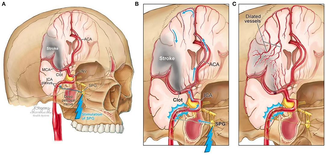
    Facial nerve-induced vasodilation of the cerebral arteries is an emerging therapeutic target that seeks to increase collateral blood flow in ischemic brain tissue, improve oxygen availability, and improve patient functional outcomes after stroke. Collateral circulation refers to alternative, pre-existing vascular pathways that deliver blood to target tissue when the primary vessel is occluded (25). Imaging of the brain and vessels has shown that collateral blood flow can preserve brain tissue for hours after major arteries to the brain are blocked (26). Figure 1 illustrates how facial nerve-induced increased collateral blood flow has the ability to ameliorate clot-induced tissue death by limiting ischemic penumbra evolution.

    FIGURE 1
    www.frontiersin.org

    Figure 1. Increased collateral blood circulation by sphenopalatine ganglion (SPG) stimulation. (A) Anatomical overview of stroke affected brain region before SPG stimulation. (B) The SPG contains parasympathetic fibers that synapse in the ganglion and innervate the internal carotid artery (ICA) through the deep petrosal nerve. (C) SPG stimulation induces vasodilation of blood vessels in the anterior circulation, increasing blood flow to the affected area limiting the evolution of the penumbra and reducing infarct volume.

    The sphenopalatine ganglion (SPG), also known as the pterygopalatine ganglion, is the largest and most superior ganglion of the sympathetic and parasympathetic nervous system and contains the largest collection of neurons in the calvarium outside of the brain (27). Humans have two SPGs, on each side of the midface, located within the viscerocranium in a space called the pterygopalatine fossa. This fossa has direct connections to the middle cranial fossa, nasal cavity, orbit, infratemporal fossa and oral cavity (28, 29). The SPG contains parasympathetic fibers that synapse in the ganglion and innervate the internal carotid artery (ICA) through the deep petrosal nerve (30). Electrical stimulation of the SPG activates the fibers, releasing several neurotransmitters such as acetylcholine, vasoactive intestinal polypeptide, peptide histidine isoleucine, and nitrous oxide that play a role in inducing vasodilation of blood vessels in the anterior circulation (26, 31).

    The geniculate ganglion is a small collection of somatosensory and gustatory ganglion cells (32) located within the temporal bone along the axis of the ear canal. Similar to the SPG, the geniculate ganglion has parasympathetic connections to cerebral arteries and has also been demonstrated to increase cerebral blood flow (33, 34). Its curved shape allows for greater susceptibility to be activated location makes it easier to access through non-invasive routes (33, 35).

    More at link.

    Tuesday, March 30, 2021

    Intermittent Light Exposures in Humans: A Case for Dual Entrainment in the Treatment of Alzheimer's Disease

     Sounds like it could be useful for stroke survivors. DEMAND YOUR DOCTOR CREATE A STROKE PROTOCOL ON THIS.

    Intermittent Light Exposures in Humans: A Case for Dual Entrainment in the Treatment of Alzheimer's Disease

    • 1Department of Population Health Science and Policy, Icahn School of Medicine at Mount Sinai, New York, NY, United States
    • 2Lighting Research Center, Rensselaer Polytechnic Institute, Troy, NY, United States
    • 3Department of Biological Sciences, Rensselaer Polytechnic Institute, Troy, NY, United States

    Circadian sleep disorders are common among American adults and can become especially acute among older adults, especially those living with Alzheimer's disease (AD) and mild cognitive impairment (MCI), leading to the exacerbation of symptoms and contributing to the development and advancement of the diseases. This review explores the connections between circadian sleep disorders, cognition, and neurodegenerative disease, offering insights on rapidly developing therapeutic interventions employing intermittent light stimuli for improving sleep and cognition in persons with AD and MCI. Light therapy has the potential to affect sleep and cognition via at least two pathways: (1) a regular and robust light-dark pattern reaching the retina that promotes circadian phase shifting, which can promote entrainment and (2) 40 Hz flickering light that promotes gamma-wave entrainment. While this is a new area of research, preliminary evidence shows the potential of dual circadian and gamma-wave entrainment as an important therapy not only for those with AD, but for others with cognitive impairment.

    Introduction

    Forty-five percent of Americans report sleep problems that affect their daily activities at least once per week, with 35% reporting poor or fair sleep quality and 20% reporting that they did not feel refreshed by sleep on any day of the past week (1, 2). Asynchrony between normal work and social schedules and the timing of the internal clock (3) can lead to sleep disorders and sleep deprivation, particularly if the asynchrony is prolonged for an extended period, which in turn can negatively affect task performance, cognition, and general health (46). Sleep disorders and their attendant decrements can become especially acute among older adults, especially those living with Alzheimer's disease (AD) and mild cognitive impairment (MCI), leading to the exacerbation of symptoms and contributing to the development and advancement of the diseases (79). In fact, of the estimated 5.8 million people in the United States living with AD and related dementias (ADRD) (10), at least one-third experience difficulty sleeping (11, 12) and approximately two-thirds of their estimated 18.5 million unpaid caregivers report sleep disturbances themselves (10, 13, 14).

    This review explores the connections between circadian sleep disorders, cognition, and neurodegenerative disease, offering insights on rapidly developing therapeutic interventions employing entraining light stimuli (both continuous and intermittent) for the treatment of sleep disorders, including in those with AD and MCI. Light therapy has the potential to affect sleep and cognition via at least two pathways: (1) a regular and robust light-dark pattern reaching the retina that promotes circadian phase shifting and thus, entrainment and (2) 40 Hz flickering light that promotes gamma wave entrainment. Both are discussed below.

    More at link.


    Saturday, March 27, 2021

    40Hz sensory stimulation induces gamma entrainment and affects brain structure, sleep and cognition in patients with Alzheimer's dementia

    Google this: gamma 40hz light and sound stimulation device: and you get some examples. But you can't do that until your incompetent doctor in 50 years finally gets up-to-date on research.

    40Hz sensory stimulation induces gamma entrainment and affects brain structure, sleep and cognition in patients with Alzheimer's dementia

    Diane Chan, Ho-Jun Suk, Brennan Jackson, Noah Pollak Milman, Danielle Stark, Elizabeth B. Klerman, Erin Kitchener, Vanesa S. Fernandez Avalos, Arit Banerjee, Sara D. Beach, Joel Blanchard, Colton Stearns, Aaron Boes, Brandt Uitermarkt, Phillip Gander, Matthew Howard III, Eliezer J. Sternberg, Alfonso Nieto-Castanon, Sheeba Anteraper, Susan Whitfield-Gabrieli, Emery N. Brown, Edward S. Boyden, Bradford Dickerson, Li-Huei Tsai

    ABSTRACT

    Non-invasive Gamma ENtrainment Using Sensory stimulation (GENUS) at 40Hz reduced Alzheimer’s disease (AD) pathology such as amyloid and tau levels, prevented cerebral atrophy and improved performance during behavioral testing in mouse models of AD. We report data from a randomized, placebo-controlled trial (n = 15) in volunteers with probable mild AD after 4 months of one-hour daily 40Hz sensory stimulation (NCT NCT04055376) to assess safety, compliance, entrainment and possible effects on brain structure, function, sleep activity and cognitive function. 40Hz light and sound GENUS was well-tolerated and compliance was high in both groups. Electroencephalography recordings show that our novel 40Hz GENUS device safely and effectively induced 40Hz entrainment in participants with mild AD. After 3 months of daily stimulation, the 40Hz GENUS group showed reduced ventricular dilation and stabilization of the hippocampal size compared to the control group. Functional connectivity was found to improve in the default mode network as well as with the medial visual network after 3 months of stimulation. Furthermore, actigraphy recordings show that circadian rhythmicity also improved with 40Hz stimulation. Compared to controls, the active group performed better on the face-name association delayed recall test. These results suggest that 40Hz GENUS can be used safely at home daily and shows favorable outcomes on cognitive function, structure and functional MRI biomarkers of AD-related degeneration. These results support further evaluation of GENUS in larger and longer clinical trials to evaluate its potential as a novel disease modifying therapeutic for Alzheimer’s dementia.

    ONE SENTENCE SUMMARY 40Hz sensory stimulation can safely and efficiently induce entrainment of neural oscillations in patients with mild probable Alzheimer’s disease and may be a novel therapeutic that can prevent brain atrophy while improving functional connectivity, sleep and cognition.