Changing stroke rehab and research worldwide now.Time is Brain! trillions and trillions of neurons that DIE each day because there are NO effective hyperacute therapies besides tPA(only 12% effective). I have 523 posts on hyperacute therapy, enough for researchers to spend decades proving them out. These are my personal ideas and blog on stroke rehabilitation and stroke research. Do not attempt any of these without checking with your medical provider. Unless you join me in agitating, when you need these therapies they won't be there.

What this blog is for:

My blog is not to help survivors recover, it is to have the 10 million yearly stroke survivors light fires underneath their doctors, stroke hospitals and stroke researchers to get stroke solved. 100% recovery. The stroke medical world is completely failing at that goal, they don't even have it as a goal. Shortly after getting out of the hospital and getting NO information on the process or protocols of stroke rehabilitation and recovery I started searching on the internet and found that no other survivor received useful information. This is an attempt to cover all stroke rehabilitation information that should be readily available to survivors so they can talk with informed knowledge to their medical staff. It lays out what needs to be done to get stroke survivors closer to 100% recovery. It's quite disgusting that this information is not available from every stroke association and doctors group.

Showing posts with label microglial inflammation. Show all posts

Thursday, August 27, 2026

Disrupted brain-immune signaling may help drive neurodegeneration

 Will your competent? doctor and hospital get followup research initiated that will create protocols that prevent neurodegeneration?

Do you prefer your doctor, hospital and board of director's incompetence NOT KNOWING? OR NOT DOING? Your choice; let them be incompetent or demand action!

Disrupted brain-immune signaling may help drive neurodegeneration

From gut-primed T cells to microglial signaling and persistent gene-regulatory states, researchers map an intricate immune network that connects the brain with the rest of the body.
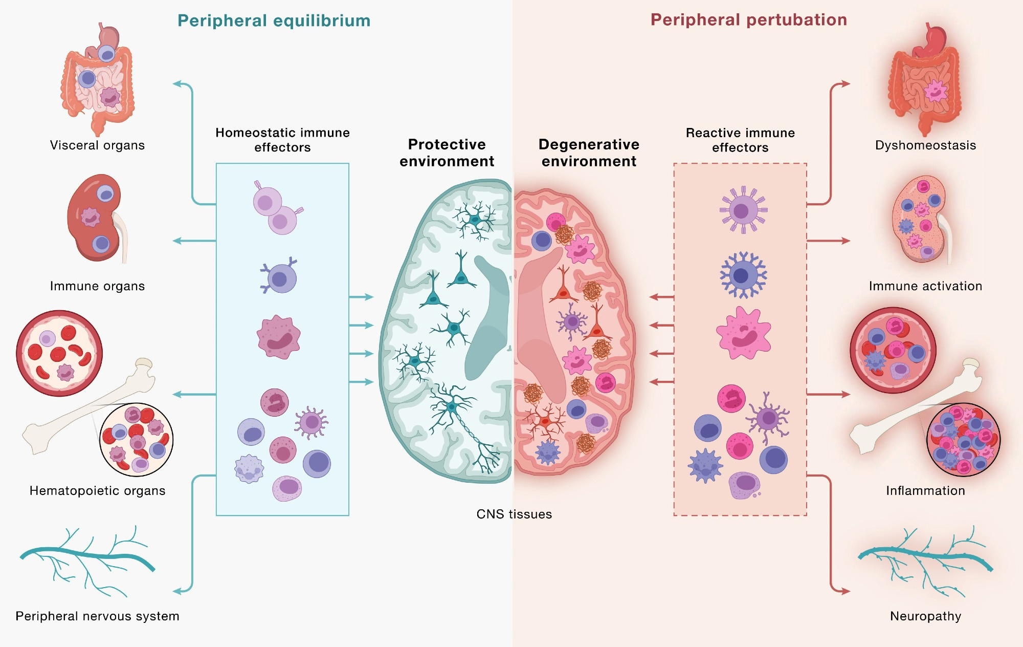
Immune cells communicate across a continuous, bidirectional circuit between the CNS and periphery. Immune cells from across the body engage CNS tissues and, in turn, are shaped by signals returning from the brain. (Left) Under peripheral equilibrium, immune effectors mobilized from visceral organs, secondary lymphoid organs, hematopoietic tissues, and the peripheral nervous system (PNS) - including CD4+ and CD8+ T cells, B cells, monocytes (top 3 cell types), NK cells, dendritic cells, mast cells, neutrophils, and innate lymphoid cells (bottom cluster of cells) - engage the CNS in a homeostatic dialogue that supports neural cell health, plasticity, and repair, sustaining a protective environment. (Right) Age-associated dysfunction across the same peripheral compartments reshapes this circuit: altered immune effectors enter the CNS and contribute to shifting the local environment toward a degenerative state in which microglia adopt disease-associated programs, astrocytes become reactive, and neurons accumulate pathology. This model underscores how the same circuitry can yield protective or pathogenic outcomes and that neurodegeneration is downstream of dysfunctional brain-immune crosstalk. Study: Neurodegeneration as a dysregulation of neuroimmune crosstalk

Immune cells communicate across a continuous, bidirectional circuit between the CNS and periphery. Immune cells from across the body engage CNS tissues and, in turn, are shaped by signals returning from the brain. (Left) Under peripheral equilibrium, immune effectors mobilized from visceral organs, secondary lymphoid organs, hematopoietic tissues, and the peripheral nervous system (PNS) - including CD4+ and CD8+ T cells, B cells, monocytes (top 3 cell types), NK cells, dendritic cells, mast cells, neutrophils, and innate lymphoid cells (bottom cluster of cells) - engage the CNS in a homeostatic dialogue that supports neural cell health, plasticity, and repair, sustaining a protective environment. (Right) Age-associated dysfunction across the same peripheral compartments reshapes this circuit: altered immune effectors enter the CNS and contribute to shifting the local environment toward a degenerative state in which microglia adopt disease-associated programs, astrocytes become reactive, and neurons accumulate pathology. This model underscores how the same circuitry can yield protective or pathogenic outcomes and that neurodegeneration is downstream of dysfunctional brain-immune crosstalk. Study: Neurodegeneration as a dysregulation of neuroimmune crosstalk

A recent perspective published in the journal Cell synthesizes scientific evidence suggesting that neurodegeneration involves intricate crosstalk between neurons and immune cells, linking the brain to peripheral immunity through bidirectional exchange. Strategies that restore immune homeostasis or recalibrate neuroimmune signaling may potentially slow neurodegeneration and promote recovery.

Historically, immune dysregulation has often been considered a consequence of neurodegenerative disorders. Recent studies, however, are beginning to change this scientific mindset, suggesting that disordered communication between the brain and immune cells may also contribute to disease onset and progression. The authors describe immune dysfunction as a “concause” of neurodegeneration, meaning it may interact with neuronal and glial vulnerabilities without necessarily being the initial trigger. It is essential to advance understanding of the pathophysiology of neurodegenerative diseases to inform therapeutic development and the development of immune-based strategies.

In this perspective, researchers examined brain-immune interactions and their potential role in neurodegeneration. They organized emerging evidence into three frameworks: “outside-in” effects driven by peripheral immunity, “inside-out” signaling coordinated by brain-resident microglia, and “locked-in” gene regulatory programs that can stabilize maladaptive neuroimmune states.

The brain-immune communication network

The brain continuously communicates with peripheral immune networks. Components of the CNS, including the choroid plexus, meninges, and lymphatic and vascular structures, interact with immune cells to relay signals related to neural needs.

Helper and cytotoxic T cells can enter CNS border regions and, under defined conditions, the brain parenchyma. Brain-immune communication supports neural integrity but can promote pathology when dysregulated. Microglia and BAMs provide surveillance, while lymphocytes confer antigen specificity and immunological memory.

Cytokines, complement, and MHC-I are traditionally linked to immunity, but CNS cells also produce or sense these molecules during neural activity. Innate lymphoid cells in the dura can respond to injury, while the choroid plexus helps regulate inflammatory signaling. In mice, increased neuronal activity may draw antibody-secreting B-lineage cells into the hippocampus during synaptic remodeling.

The gut also influences brain immunity. T cells educated in gut-associated immune tissues can subsequently traffic to the borders of the CNS and, under certain conditions, into the brain, while plasma cells secreting IgA antibodies protect blood vessels in the meninges. In addition, changes in the gut microbiome could influence immune activity and microglial function. Through the GBA, the gut and brain are in constant dialogue with each other. The vagus nerve conveys immunity-related information from the intestines to the brain. Reward-related neural pathways can, in turn, influence peripheral immune activity.

Brain-immune interactions in neurodegenerative disease

T cell activity has been implicated in PD, AD, ALS, and dementia with Lewy bodies (DLB). In ALS4, an inherited form of ALS, cytotoxic T cells are detected early in the blood and brain and expand as the disease progresses, consistent with antigen-driven responses.

Monday, April 14, 2025

Aerobic Exercise Restores Hippocampal Neurogenesis and Cognitive Function by Decreasing Microglia Inflammasome Formation Through Irisin/NLRP3 Pathway

You'll want your competent? doctor to get you recovered enough to do these exercises to reduce this part of your Parkinson's risk.  

 Did your incompetent doctors and hospital do anything about getting NLRP3 inflammasone research done after this earlier research came out in February 2020? NO? Then you don't have a functioning stroke doctor or hospital.  I expect a competent doctor to know more than I do!

Molecular 'switch' reverses chronic inflammation and aging

 February 2020 

And this; March 2023

Berberine ameliorates depression-like behaviors in mice via inhibiting NLRP3 inflammasome-mediated neuroinflammation and preventing neuroplasticity disruption

The latest here:

 Aerobic Exercise Restores Hippocampal Neurogenesis and Cognitive Function by Decreasing Microglia Inflammasome Formation Through Irisin/NLRP3 Pathway

1 of 17Aging Cell, 2025; 0:e70061
https://doi.org/10.1111/acel.70061
Aging Cell
RESEARCH ARTICLE OPEN ACCESS
Aerobic Exercise Restores Hippocampal
Neurogenesis and Cognitive Function by Decreasing
Microglia Inflammasome Formation Through
Irisin/NLRP3 Pathway
Renqing Zhao | Xin Tian | Haocheng Xu | Yuanxin Wang | Junjie Lin | Bin Wang
College of Physical Education, Yangzhou University, Yangzhou, China
Correspondence: Renqing Zhao (zhrenqing@gmail.com)
Received: 29 November 2024 | Revised: 26 February 2025 | Accepted: 14 March 2025
Funding: This work was supported by the Natural Science Foundation of Jiangsu Province (BK20201435).
Keywords: aerobic exercise | cognition | FNDC5/irisin | hippocampal neurogenesis | NLRP3 inflammasome | Parkinson disease

ABSTRACT

Persistent microglial inflammation is a detrimental contributor to the progression of Parkinson disease (PD) pathology and related issues such as impaired adult hippocampal neurogenesis (AHN) and cognition. We conducted a 10-week exercise program with MPTP-treated mice to determine whether neuroinflammation can be addressed by aerobic exercise and elucidate its underlying regulatory mechanisms. Ten weeks of exercise significantly reduced PD-related pathology and enhanced AHN and
memory. These changes were linked to a reduction in neuronal apoptosis, microglial inflammation, and NLRP3 inflammasome activation. In cultured microglia, fibril α-synuclein reduced FNDC5/irisin protein levels and induced NLRP3 inflammasome formation and IL-1β production, which could be diminished by recombinant irisin treatment. Interestingly, “runner serum” isolated from exercising rodents enhanced FNDC5/irisin expression and reduced NLRP3 inflammasome components and IL-1β
secretion in α-synuclein-treated microglia. These effects could be diminished by blocking irisin signaling with cyclo RGDyk or NLRP3 agonist, nigericin sodium salt. Exercise-induced neuroprotective effects were weakened by treatment of MPTP-treated mice with cyclo RGDyk. In contrast, systematic administration of irisin partially replicated the beneficial effects of exercise
on PD pathology, AHN, and memory function. As a nonpharmacological strategy, aerobic exercise effectively addresses PD pathology and preserves adult neurogenesis and cognition by mitigating microglial inflammation via mediating irisin/NLRP3 inflammasome pathways.